Comparative analysis of adhesion virulence protein FadA from gut-associated bacteria of colorectal cancer patients (F. nucleatum) and healthy individuals (E. cloacae)
Nadia Hussain, Fatima Muccee, Naeem Mahmood Ashraf, Tayyaba Afsar, Fohad Mabood Husain, Arslan Hamid, Suhail Razak

TL;DR
This study compares the FadA protein from gut bacteria in colorectal cancer patients and healthy individuals to understand its role in disease and potential treatment strategies.
Contribution
The study reveals structural and functional differences in FadA proteins from F. nucleatum and E. cloacae, offering insights for targeted CRC therapies.
Findings
FadA from F. nucleatum shows helical configuration and hydrophilic properties, differing from E. cloacae's globular and hydrophobic FadA.
F. nucleatum FadA has fewer antigenic epitopes and binds strongly to COX2 drugs, suggesting a role in virulence.
Structural differences in FadA could inform vaccine design and drug development to inhibit CRC-related bacterial virulence.
Abstract
Background: Colorectal cancer (CRC) is a gastrointestinal disease linked with GIT microbial dysbiosis. The present study has targeted the comparative analysis of virulent factor FadA from gut-associated bacteria of CRC patients (F. nucleatum) and healthy individuals (E. cloacae). Methods: For this purpose, FadA protein sequences of fifteen strains of F. nucleatum and four strains of E. cloacae, were retrieved from the UniProt database. These sequences were analysed through VirulentPred, PSLpred, ProtParam, PFP-FunDSeqE, PROTEUS Structure Prediction Server, SWISS-MODEL, SAVES validation server, MEME suite 5.5.0, CAVER Web tool, Webserver VaxinPAD, HPEPDOCK and HDOCK servers. Results: FadA protein from F. nucleatum was found to exhibit significant differences as compared to E. nucleatum i.e. it exhibited helical configuration, cytoplasmic, periplasmic, outer-membrane and extracellular…
Genes, proteins, chemicals, diseases, species, mutations and cell lines named across the full text — each resolved to its canonical identifier and authoritative record.
Click any figure to enlarge with its caption.
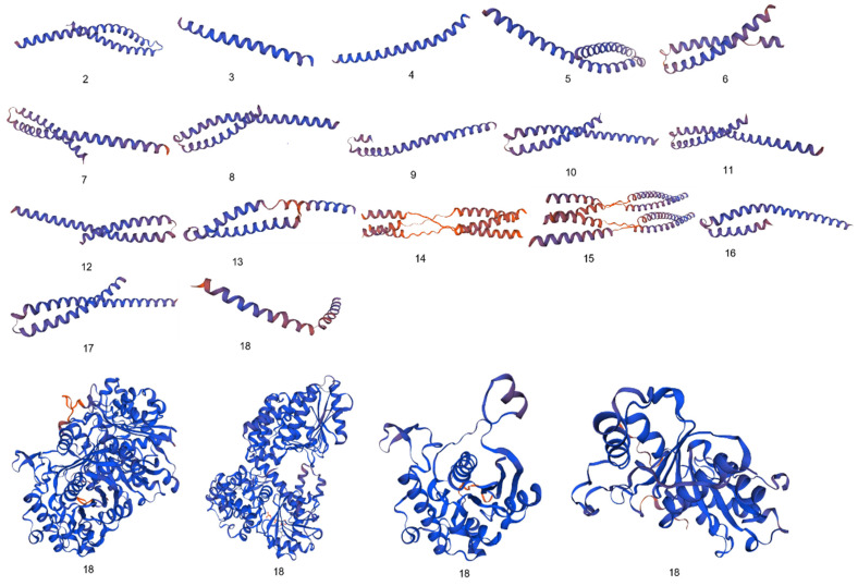 Figure 1
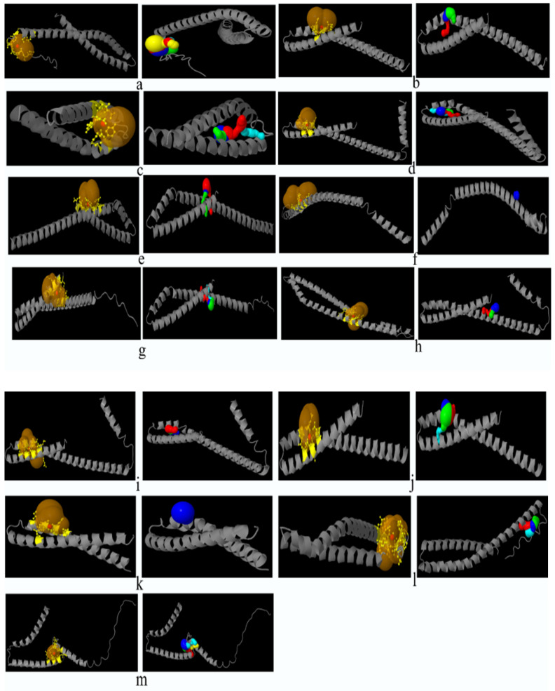 Figure 2
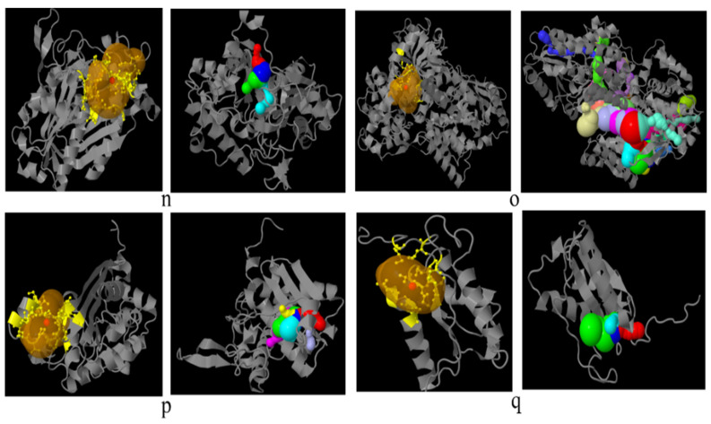 Figure 3
 Figure 4
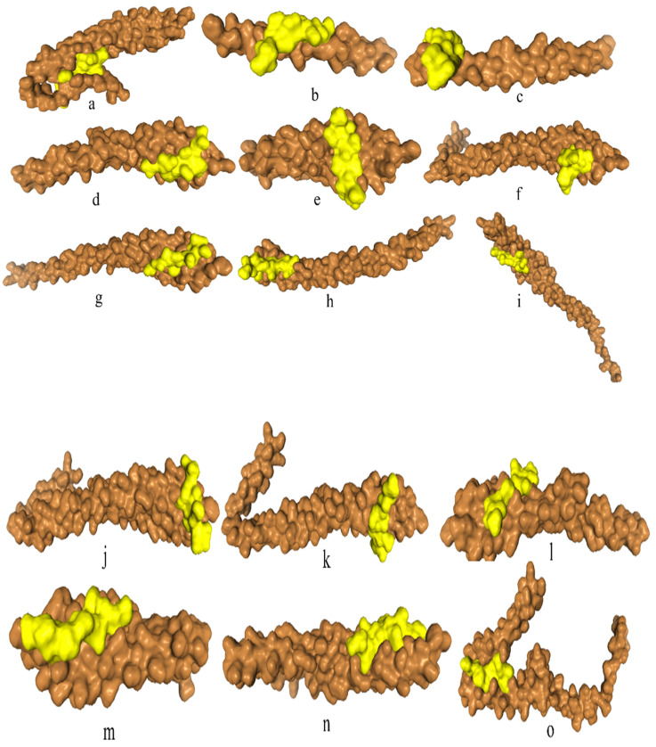 Figure 5
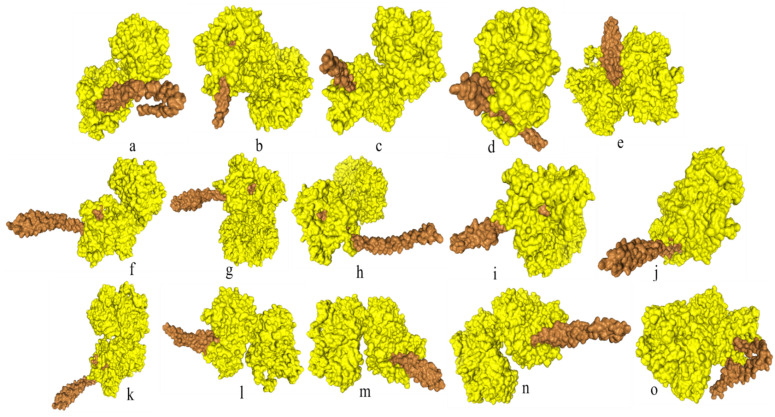 Figure 6
Peer Reviews
No public reviews on file for this paper yet. If you reviewed it on a platform where reviews are public (OpenReview, ICLR, NeurIPS, ICML), you can paste yours below so the community can read it here.
Videos
No videos yet. Explain this paper in a talk, walkthrough, or lecture? Add one.
Taxonomy
TopicsGut microbiota and health · Probiotics and Fermented Foods · Enterobacteriaceae and Cronobacter Research
Background
Colorectal cancer (CRC) is the second and third most abundant cancer concerning morbidity (9.2%) and diagnosis (6.1%) as per the research of the Centers for Disease Control and Prevention (CDC). According to a study, by 2030 CRC is expected to increase by 60% with 2.2 million new cases and 1.1 million deaths 1. It has contributed to 9, 35000 deaths caused by cancer. It is characterized by polyps in the rectum, change in bowel habits, constipation, diarrhoea, rectal bleeding, change in stool colour, shape, abdominal cramps, gas, pain, changed stool consistency, tenesmus, anaemia, loss of appetite and weight, nausea, vomiting, jaundice and fatigue 1. The risk factors include smoking, physical inactivity, obesity, unhealthy and improper diet routine, family history of diabetes, inflammatory bowel diseases, colon polyps, genetics, race, gender, age and gastrointestinal tract (GIT) microbiome 2, 3.
The composition of the GIT-associated microbial population always varies with oncogenesis. This dysbiosis always involves a shift toward pathogenic microbes including Fusobacterium nucleatum, Escherichia coli, Enterococcus faecalis, Bacteroides fragilis, Salmonella enterica, Streptococcus, Rothia, Faecalibacterium, Porphyromonas, Collinsella, Slackia, Alistipes, Porphyromonas, Prevotella, Methanobrevibacter, Gemella, Mogibacterium, Parvimonas, Peptostreptococcus, Solobacterium, Helicobacter, Klebsiella, Akkermansia and Thermanaerovibrio 4-14. Following bacteria have been reported to reduce in CRC patients i.e. Ruminococcus, Roseburia, Lactobacillus, Eubacterium, Clostridium, Anaerostipes, Bacteroides, Bifidobacterium, Citrobacter, Treponema, Serratia, Kluyvera and Cronobacter 10-20.
E. cloacae is reported as a harmless GIT commensal microbe normally found in healthy individuals 21. On the contrary, F. nucleatum is a commensal-turned and obligate pathogen associated with various GIT disorders 22. It potentiates the development and progression of CRC due to its enrichment in colorectal malignant tumour tissue 23-25. Being asaccharolytic, it metabolizes the peptides and amino acids into short-chain fatty acids and formyl-methionyl-leucyl-phenylalanine 26. These metabolites attract myeloid cells in the tumour microenvironment thus contributing to tumour enrichment with myeloid cells. Additionally, metabolic products also promote vascularization and infiltration of immune cells in tumours 9. Being slightly capable of respiring aerobically, Fusobacterium successfully survives hypoxic tumour conditions 27. It increases oncogenic micro RNAs 28-30. It damages DNA and induces single nucleotide polymorphisms (SNPs) by producing reactive oxygen species via its metabolite hydrogen sulfide 31. It suppresses tumour immune microenvironment via interference with functions of T-cells, macrophages, dendritic cells, neutrophils and natural killer cells (NKCs) 9, 32. Therefore, it can be considered one of the major non-invasive prognostic, diagnostic and therapy response biomarkers of CRC 9, 33-42.
F. nucleatum being an adhesive pathogen has multiple virulence factors contributing to its colonization of colorectal tissues and CRC incidence. The known virulence factors of F. nucleatum promote adhesion to intestinal epithelial cells via FadA 8. This factor has a strong connection with CRC due to its association with NF-Kb inflammatory response and cell adhesion and invasion of Fusobacterium 43. It enables the bacterium to get attached to fibroblasts, and endothelial and epithelial cells 22. To cause disease, FadA binds the EC5 domain (ASANWTLOYNDP) of the E-cadherin protein leading to its phosphorylation. This activates the Wnt signal in the Wnt/β- cadherin pathway. Wnt binds receptors and disrupts the destruction complex comprising of Axin, APC, GSK-3β, PP2A and Ck1α. This complex under normal circumstances targets β-catenin for ubiquitination and digestion by proteasome. Disruption of this complex inhibits phosphorylation of β-catenin resulting in its stability. The β-catenin gets accumulated in the cytosol and translocated to the nucleus. In the nucleus, β-catenin binds with TCF/LEF transcription factors and activates the transcription of genes involved in inflammation and other cancer-initiating activities 44.
The present study focused on comparative analysis of the FadA protein in F. nucleatum and E. cloacae at the levels of physicochemical properties, sub-cellular localization, conserved domains, antigenic epitopes, functional domains, 2D and 3D structures, the number of catalytic sites and tunnels that might help design strategies for inhibition of tumorigenic roles of F. nucleatum FadA protein.
Methodology
Retrieving the sequences of FadA protein in F. nucleatum and E. cloacae
Sequences of FadA protein in F. nucleatum and E. cloacae were retrieved from the UniProt database (https://www.uniprot.org, accessed on 11 Nov. 2022) 18. Sequences were retrieved for a total of fifteen strains of F. nucleatum and four strains of E. cloacae (Supplementary data Table 1).
Evaluation of virulence potential
To predict the virulence status of the FadA protein in bacteria documented in the present study VirulentPred tool (http://bioinfo.icgeb.res.in/virulent/, accessed on 13 Nov. 2022) has been consulted 45. This is a Support Vector Machine (SVM) based method. It was determined based on prediction scores. A positive score indicates virulence while a negative score shows the non-virulent nature of protein.
Phylogenetic tree construction
To analyze the phylogenetic relationship among present study bacteria, the Clustal Omega Multiple Sequence Alignment Tool was used for multiple sequence alignment 46. Alignment was followed by tree construction using MEGA version 7. Trees were inferred by the neighbour-joining method 47, 48. The evolutionary distances were computed using the Poisson correction method and are in the units of the number of amino acid substitutions per site 49.
Evaluation of sub-cellular localization
To predict the sub-cellular localization of proteins, the PSLpred tool (webs.iiitd.edu.in/raghava/pslpred/submit.html, accessed on 26 Nov. 2022) was used 50. The prediction approach used was based on a hybrid approach.
Evaluation of physicochemical properties
To determine the differences between FadA proteins of bacteria documented in the present study, the ProtParam tool (https://web.expasy.org/protparam/, accessed on 12 Dec. 2022) was used 51. Properties compared include the number of amino acids, molecular weight, theoretical isoelectric point (pI), half-life, instability index, aliphatic index, extinction coefficient and grand average of hydropathy (GRAVY) 51. The pI value helps in prediction of the acidity or alkalinity of the protein.
Prediction of functional domains
To predict the functional domains in FadA protein sequences of bacteria inhabiting normal and cancerous patients GIT, Functional domain or motif prediction (PFP-FunDSeqE) tool (www.csbio.sjtu.edu.cn/bioinf/PFP-FunDSeqE/#, accessed on 25 Nov. 2022) was used 52.
Prediction of 2D configuration
To predict the 2D configuration of the present study proteins, PROTEUS Structure Prediction Server 2.0 (www.proteus2.ca/proteus2/, accessed on 20 Dec. 2022) was used 53. This server helped us to determine the helix, beta sheet, coil content, signal peptide and membrane content of proteins 54.
Prediction of 3D configuration
To predict the 3D structure of bacterial proteins, a homology modelling server SWISS-MODEL (https://swissmodel.expasy.org, accessed on 12 Dec. 2022) was used 55.
Validation of 3D structures
To validate the 3D structures of FadA proteins predicted using the SWISS-MODEL, the SAVES validation server (https://saves.mbi.ucla.edu, accessed on 20 Dec. 2022) was used. Two programs of this server i.e. ERRAT and PROCHECK were used.
Prediction of conserved protein motifs
To predict the conserved protein motifs, Multiple Em for Motif Elicitation (MEME) suite 5.5.0 (https://meme.suite.org/meme/tools/meme, accessed on 21 Dec 2022) was used. A total of ten motifs were found using default values of all parameters. For ontology analysis of predicted motifs, Lambda Predict Protein (ƛPP) (https://embed.predictprotein.org/0, accessed on 23 Dec 2022) was used.
Assessment of catalytic pockets and tunnels
To assess the catalytic pockets and tunnels in FadA proteins of bacteria documented in the present study, the CAVER Web tool was used (https://loschmidt.chemi.muni.cz/caverweb, accessed on 20-21 Dec. 2022) 56.
Evaluation of immunomodulatory A-cell epitopes
Webserver VaxinPAD (https://webs.iiitd.edu.in/raghava/vaxinpad/batch.php, accessed on 25 - 27 Dec 2022) was used to explore the immunomodulatory A-cell epitopes in the FadA protein of F. nucleatum and* E. cloacae* 57.
Assessment of binding affinity of FadA with inhibitor peptide
To determine the binding affinity of FadA protein from virulent F. nucleatum bacteria with a previously reported inhibitor peptide ASANWTIQYND, peptide-protein docking web-server, HPEPDOCK (huanglab.phys.hust.edu.cn/hpepdock/, accessed on 26 & 27 Dec. 2022) was used 8, 58. To dock FadA protein with cyclooxygenase-2 (COX2), the HDOCK server (http://hdock.phys.hust.edu.cn/, accessed on 31 Dec, 2022) was used 59.
Results
Virulence status prediction
Sequences of the FadA protein retrieved from the UniProt database were analyzed for virulence status. All the sequences from E. cloacae were found to be non-virulent with negative scores while those from F. nucleatum showed virulent status with positive scores (Table 1).
Phylogeny
The optimal tree for the F. nucleatum strains with the sum of branch length 11.14917307 is shown in Supplementary Data Figure 1 (a). According to this tree, F. nucleatum IV and V, F. nucleatum II and III, F. nucleatum I and subsp. polymorphum 3 and F. nucleatum subsp. animalis 7_1 and animalis D11 (I) were closely related as compared to others as they share the same clade with each other.
The optimal tree for the E. cloacae strains with the sum of branch length 3.79674773 is shown in Supplementary Data Figure 1 (b). According to this tree, E. cloacae I and IV are more closely related to each other as compared to others as they are originating from the same branch point. While E. cloacae III and II are distantly related to each other and from E. cloacae I and IV.
Sub-cellular localization prediction
Assessment of sub-cellular localization of FadA protein using PSLPred analysis tool revealed FadA protein to be localized in the cytoplasm in the case of all four strains of E. cloacae. in the case of colorectal cancer GIT bacteria, in addition to the cytoplasm (F. nucleatum (IV), F. nucleatum (VI), F. nucleatum 13_3C, F. nucleatum CTI-5, F. nucleatum subsp. Vincentii, F. nucleatum subsp. animalis 7_1, F. nucleatum subsp. animalis D11 (I), F. nucleatum subsp. animalis D11 (II) and polymorphum 2) the protein was also found to be localized in periplasmic space (F. nucleatum (II) and F. nucleatum (III)), outer-membrane (F. nucleatum (I) and polymorphum 3) and extracellular membrane (F. nucleatum (V) and F. nucleatum subsp. animalis 11_3_2) (Supplementary data Table 2).
Physicochemical properties prediction
Analysis of the physicochemical properties of FadA proteins revealed a wide range of variations in proteins of F. nucleatum from E. cloacae. In E. cloacae, the pI was observed in the range of 5.97 to 6.90 while in the case of F. nucleatum, the minimum and highest values of pI were observed to be 4.06 and 6.94, respectively. The highest deviation was observed in the case of F. nucleatum I, F. nucleatum II, F. nucleatum III, F. nucleatum IV, F. nucleatum subsp. polymorphum 2 and F. nucleatum subsp. polymorphum 3 (Table 2). F. nucleatum II, F. nucleatum III and F. nucleatum IV showed a very unusual value of half-life i.e. 2 min., as compared to all other bacteria documented in the present study. High deviation of instability index from E. cloacae, was observed in the case of F. nucleatum II (40.2), F. nucleatum V (46.80), F. nucleatum VI (48.33), F. nucleatum 13_3C (52.95), F. nucleatum CTI-5 (49.32), F. nucleatum subsp. vincentii (51.94), F. nucleatum subsp. animalis 7_1 (47.92), F. nucleatum subsp. animalis D11 I (50.36), F. nucleatum subsp. animalis D11 II (50.37), F. nucleatum subsp. animalis 11_3_2 (46.13), F. nucleatum subsp. polymorphum 2 (43.71) and F. nucleatum subsp. polymorphum 3 (69.24). As far as the aliphatic index is concerned, F. nucleatum I (82.79), F. nucleatum II (77.45), F. nucleatum III and F. nucleatum IV (75.79), F. nucleatum V (81.41) and F. nucleatum subsp. polymorphum 3 (75.32) exhibited variation from that of E. cloacae. F. nucleatum III and F. nucleatum IV (2980), F. nucleatum V and F. nucleatum subsp. animalis 11_3_2 (11460), F. nucleatum subsp. animalis D11 II (12950), polymorphum 2 (4470) and polymorphum 3 (1490) was observed to exhibit variation in extinction coefficient values than the GIT inhabiting bacteria from healthy individuals. All the bacteria from CRC patients' gut were found to have very different values of GRAVY i.e. ranging from -0.440 to -1.460 as compared to bacteria from healthy individuals (Table 2).
Functional domains prediction
Functional domain analyses also revealed a large degree of variation of FadA proteins between F. nucleatum and E. cloacae strains. Two fold types were found in E. cloacae protein i.e. NAD(P)-binding Rossmann-fold and (TIM)-barrel. On the other hand, four different types of fold were observed in F. nucleatum strains i.e. 4 helical up and down bundle, DNA-binding 3-helical bundle, EF-hand and 4-helical cytokines (Table 3).
Two-dimensional structures prediction
The 2D structure of F. nucleatum FadA protein was markedly different from that of E. cloacae. i. e. the α-helix content was significantly higher in F. nucleatum (70-96%) as compared to E. cloacae (30-56%). No beta sheets were observed in F. nucleatum while E. cloacae secondary structure exhibited 12-21% beata sheets. Coil content was extremely lower in F. nucleatum (4-16%) than E. cloacae (33-50%). Only E. cloacae III exhibited signal peptide (17%) content while the majority of the F. nucleatum strains were found to have signal peptide content i.e. 14-19% (Table 4, Supplementary data Figure 2).
Three-dimensional structures prediction
Significant diversity was found between the FadA protein of F. nucleatum and E. cloacae at the level of 3D configuration as is evident from Figure 1. The globular structure was found in E. cloacae proteins while only a helical folding pattern was observed in the case of F. nucleatum. Validation of these structures using Procheck and ERRAT scores is described in detail in Supplementary Data Table 3.
Catalytic pockets and tunnels prediction
All the bacteria except F. nucleatum II and F. nucleatum III were found to exhibit catalytic pockets and tunnels. The highest number of tunnels was observed in E. cloacae II (Table 5, Figure 2).
Prediction of conserved motifs
Conserved motif analysis revealed that all the bacteria documented in the present study except F. nucleatum II, F. nucleatum III, F. nucleatum IV and F. nucleatum V exhibited the conserved regions (Figure 3, Supplementary data Table 4).
A-cell epitopes prediction
Epitope assessment revealed that a sufficient number of antigenic peptides were present in FadA protein from healthy individuals' GIT. i.e. E. cloacae I (52), II (40), III (28) and IV (18). However, in most of the bacteria from CRC patients GIT, FadA was found to have very few epitopes like F. nucleatum subsp. polymorphum 2 (1), F. nucleatum subsp. polymorphum 3 (6) and F. nucleatum I (4) or no epitopes i.e. F. nucleatum II and III. Others were found to have epitopes in the range of 12-20 (Supplementary data Table 5).
Docking of FadA protein inhibitor peptide
Docking analysis of F. nucleatum FadA proteins with inhibitor peptide showed that all the proteins from virulent bacteria tend to bind with this peptide. Good binding affinity is reflected by the docking energy scores ranging from -136.574 for F. nucleatum II to -209.754 for F. nucleatum 13_3C (Figure 4, Table 6).
Docking of FadA protein with COX2
Docking analysis of F. nucleatum FadA protein with FDA-approved anti-inflammatory drug COX2 revealed a high binding affinity of FadA with this drug with good energy scores ranging from -204.87 to -286.12 (Figure 5).
Discussion
In present study, FadA protein is compared among the two groups of GIT bacteria. In one group (F. nucleatum), this protein causes pathogenicity while in other (E. cloacae) it is non-virulent. F. nucleatum is reported to occur abundantly in gut of CRC patients. Present research analyzed the distinguishing characteristics of FadA which might contribute to its virulent character in F. nucleatum.
FadA has been found virulent in all the strains of F. nucleatum documented in the present study as compared to that of E. cloacae which is consistent to previous findings 21, 22.
According to the present study,* E. cloacae-associated* FadA protein is localized in the cytoplasm while in six strains of F. nucleatum, the localization is different. Marked deviation in half-life was observed in F. nucleatum II-IV i.e. 2 min. as compared to > 10 hours in other cases. In cases of E. cloacae, an instability index below 40 suggests their poor in-vitro stability as compared to F. nucleatum 60. An aliphatic index is a measure of protein thermostability 61. In the present work, FadA in the bacteria from GIT of both the normal and diseased individuals was found to be thermostable. GRAVY values indicated the hydrophilic nature of F. nucleatum FadA as compared to most of the E. cloacae which were hydrophobic with GRAVY score > 0 62. In most cases of F. nucleatum, the pI of the FadA virulence factor was observed strongly acidic as compared to slightly acidic pI in cases of E. cloacae-associated FadA.
In the present study, healthy individuals associated with bacterial FadA protein have been found to comprise NAD(P)-binding Rossmann-fold and (TIM)-barrel domains. On the other hand, CRC patients' associated bacteria FadA factors were found to consist of four helical up and down bundles, EF-hand and DNA-binding 3-helical bundle. This finding of the helical domain was consistent with literature where the FadA protein monomer of F. nucleatum has been reported as alpha-helical which gives rise to a hair-pin-like structure 63.
As far as the 2D configuration is concerned, no beta-sheet content, large number of helix residues and low coil content was observed in F. nucleatum FadA protein as compared to that of E. cloacae. Eight strains of F. nucleatum versus only one strain of E. cloacae were found to contain signal peptides. The length of signal peptides ranged between 17-20 residues in F. nucleatum protein. Signal peptide comprising eighteen amino acids i.e. MKKFLLLAVLAVSASAFA has been reported in the FadA protein 64.
Differences between 3D configurations of FadA proteins of two groups of bacteria documented in the present study were understandable. F. nucleatum FadA contained only a helical configuration, while from E. cloacae the globular structure was observed.
The number of tunnels was comparable between FadA proteins of two types of bacteria in the present study, however, only E. cloacae (II) exhibited twenty-six tunnels. Different parameters of catalytic pockets were also analyzed. i. e. druggability, length and bottleneck radius. The binding tendency of a catalytic site of protein for a drug is referred to as druggability. FadA in case of F. nucleatum subsp.* polymorphum* 2, F. nucleatum subsp. polymorphum 3 and E. cloacae (I), were found to a good therapeutic target drugs with druggability scores closer to 1 65. In all other cases, the FadA was not found to be a druggable protein. Tunnel length and curvature reflected the substrate specificity of the protein. Parameters of tunnel and curvature might be used to reduce FadA activity through the inhibition of its substrate binding and catalysis.
Being virulent, FadA from F. nucleatum can be the most appropriate vaccine target against this pathogenic bacterium. Identification of antigenic epitopes could be proven helpful in this regard. In the present study, A-cell epitopes were found to be higher in number in E. cloacae i.e. ranging from 18-52 as compared to F. nucleatum i.e. 0-20. No epitopes were observed in F. nucleatum II and III. Overall, the presence of a variety of epitopes in cases of F. nucleatum VI, F. nucleatum subsp. Vincentii, F. nucleatum subsp.* animalis 7_1*, F. nucleatum subsp.* animalis* D11 (I) AND (II) and F. nucleatum subsp. animalis 11_3_2 is consistent with earlier literature reporting antigenic heterogeneity in different strains of F. nucleatum 66. A protein containing a large number of A-cell epitopes is capable of stimulating the immune system in the host. So, FadA in E. cloacae I, II and III with antigenic peptides 52, 40 and 28, respectively might boost the immune system. The epitopes predicted in virulent FadA from F. nucleatum strains might also be used for designing peptide-based vaccine adjuvants after getting insight into their antigenic potential 57.
According to the literature, the FadA gene is highly conserved not only among different strains of F. nucleatum but also among other oral species of Fusobacterium. Hence, our findings are in agreements with previous reports 64, 67, 69.
Inhibitory peptides might be the promising candidates as immunotherapeutic agents. In the present study, FadA protein from F. nucleatum has been docked with an already reported peptide comprising of eleven amino acid residues i.e. ASANWTIQYND. This peptide has been derived from the EC5 protein and is designated as the inhibitory one 8. docking analysis revealed the strong binding affinity of FadA proteins with this peptide. Keeping in view, the involvement of FadA protein in inflammation, this protein has been evaluated for binding tendency with an FDA-approved anti-inflammatory drug COX2 68. The docking analysis results with energy scores of -224.60 to -286.12 reflected the high binding affinity of FadA virulent factor with COX2. Hence, this drug might be used for successful inhibition of F. nucleatum-associated FadA protein.
This study is limited to in-silico comparison of FadA protein among the healthy and CRC patients gut bacteria. Its findings are significant because they highlighted the pathogenicity associated properties of protein in F. nucleatum. However, validity of these findings should be confirmed in future via experimental studies. For this purpose, fecal samples of healthy and CRC patients will be collected and used for isolation of gut associated bacteria 70. Bacteria will be targeted for FadA protein extraction and analysis.
Conclusion
Wide range of differences observed in FadA protein not only justifies the virulence associated with *F. nucleatum-*derived FadA but also suggests multiple strategies to inhibit the oncogenic potential of this protein. Present study revealed that due to the high instability index, F. nucleatum FadA protein tends to remain stable in-vitro, so these proteins can be easily studied and mutated in the laboratory. Tunnels and different attributes of catalytic sites explored might be used to design the drugs targeting FadA virulence factors. Antigenic peptides of virulent FadA proteins might be targeted for peptide-based vaccine designing. Divergence of 2D and 3D structure between virulent and non-virulent FadA protein might help to inactivate the virulency form of this protein through site-directed mutation induction leading to configuration alterations in active regions.
The reference list from the paper itself. Each links out to its DOI / PubMed record.
- 1Paschke S Jafarov S Staib L Kreuser ED Maulbecker-Armstrong C Roitman M Are colon and rectal cancer two different tumor entities?. A proposal to abandon the term colorectal cancer International journal of molecular sciences 20181925773020021510.3390/ijms 19092577 PMC 6165083 · doi ↗ · pubmed ↗
- 2Bray F Ferlay J Soerjomataram I Siegel RL Torre LA Jemal A Global cancer statistics 2018: GLOBOCAN estimates of incidence and mortality worldwide for 36 cancers in 185 countries CA: a cancer journal for clinicians 2018683944243020759310.3322/caac.21492 · doi ↗ · pubmed ↗
- 3Wong MCS Huang J Lok V Wang J Fung F Ding H Differences in incidence and mortality trends of colorectal cancer worldwide based on sex, age, and anatomic location Clinical Gastroenterology and Hepatology 20211995566 e 613208830010.1016/j.cgh.2020.02.026 · doi ↗ · pubmed ↗
- 4Sobhani I Tap J Roudot-Thoraval F Roperch JP Letulle S Langella P Microbial dysbiosis in colorectal cancer (CRC) patients Plo S one 20116 e 163932129799810.1371/journal.pone.0016393 PMC 3029306 · doi ↗ · pubmed ↗
- 5Zhang S Kong C Yang Y Cai S Li X Cai G Human oral microbiome dysbiosis as a novel non-invasive biomarker in detection of colorectal cancer Theranostics 202010115953305223510.7150/thno.49515 PMC 7545992 · doi ↗ · pubmed ↗
- 6Kalasabail S Engelman J Zhang LY El-Omar E Yim HCHA Perspective on the Role of Microbiome for Colorectal Cancer Treatment Cancers 20211346233457285010.3390/cancers 13184623 PMC 8468110 · doi ↗ · pubmed ↗
- 7Hernández-Luna MA Lopez-Briones S Luria-Pérez R The four horsemen in colon cancer Journal of Oncology 2019201956362723166275210.1155/2019/5636272 PMC 6791268 · doi ↗ · pubmed ↗
- 8Rubinstein MR Wang X Liu W Hao Y Cai G Han YW Fusobacterium nucleatum promotes colorectal carcinogenesis by modulating E-cadherin/β-catenin signaling via its Fad A adhesin Cell host & microbe 2013141952062395415810.1016/j.chom.2013.07.012PMC 3770529 · doi ↗ · pubmed ↗