Identification of high-performing antibodies for SPARC-related modular calcium-binding protein 1 (SMOC-1) for use in Western Blot and immunoprecipitation
Riham Ayoubi, Sara González Bolívar, Michael Nicouleau, Kathleen Southern, Carl Laflamme, Christian Tiede, Kathleen Southern, Deborah Moshinsky

TL;DR
This paper identifies reliable antibodies for SMOC-1, a protein linked to Alzheimer's disease, to help improve research consistency and diagnostic exploration.
Contribution
The study evaluates and identifies high-performing commercial antibodies for SMOC-1 suitable for Western blot and immunoprecipitation.
Findings
Seven commercial SMOC-1 antibodies were tested using a standardized protocol.
Successful antibodies for Western blot and immunoprecipitation were identified.
The findings aim to guide researchers in selecting appropriate antibodies for their studies.
Abstract
SPARC-related modular calcium-binding protein 1, otherwise known as SMOC-1, is a secreted glycoprotein involved in various cell biological processes including cell-matrix interactions, osteoblast differentiation, embryonic development, and homeostasis. SMOC-1 was found to be elevated in asymptomatic Alzheimer’s disease (AD) patient cortex as well as being enriched in amyloid plaques and in AD patientcerebrospinal fluid, arguing for SMOC-1 as a promising biomarker for AD. Having access to high-quality SMOC-1 antibodies is crucial for the scientific community. It can ensure the consistency and reliability of SMOC-1 research, and further the exploration of its potential as both a therapeutic target or diagnostic marker.. In this study, we characterized seven SMOC-1 commercial antibodies for Western blot and immunoprecipitation, using a standardized experimental protocol based on comparing…
Genes, proteins, chemicals, diseases, species, mutations and cell lines named across the full text — each resolved to its canonical identifier and authoritative record.
Click any figure to enlarge with its caption.
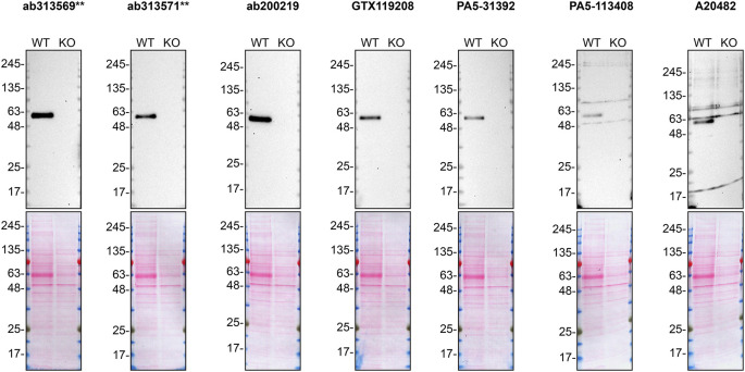 Figure 1
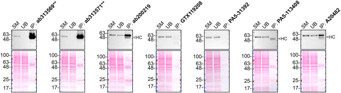 Figure 2
| Institution | Catalog number | RRID (Cellosaurus) | Cell line | Genotype |
|---|---|---|---|---|
| ATCC | CCL-2 |
| HeLa | WT |
| Montreal Neurological Institute | - |
| HeLa |
|
| Company | Catalog number | Lot number | RRID (Antibody Registry) | Clonality | Clone ID | Host | Concentration (μg/μL) | Vendors recommended applications |
|---|---|---|---|---|---|---|---|---|
| Abcam | ab313569
| 3101091230 | AB_2941846
| recombinant-mono | EPR26922-29 | rabbit | 0.50 | Wb, IP, IF |
| Abcam | ab313571
| 3101065175 | AB_2941847
| recombinant-mono | EPR26922-31 | rabbit | 0.50 | WB, IP |
| Abcam | ab200219 | GR3370372-1 |
| polyclonal | - | rabbit | 0.50 | Wb |
| GeneTex | GTX119208 | 40331 |
| polyclonal | - | rabbit | 0.90 | Wb |
| Thermo Fisher Scientific | PA5-31392 | 130141931 |
| polyclonal | - | rabbit | 0.90 | Wb |
| Thermo Fisher Scientific | PA5-113408 | WL3463969 |
| polyclonal | - | rabbit | 3.50 | Wb |
| ABclonal | A20482 | 125410101 |
| polyclonal | - | rabbit | 2.65 | Wb |
- —Genome Quebec
- —Genome Canada
- —Ontario Genomics
- —Canadian Institutes of Health Research Foundation
- —National Institute on Aging
- —Mitacs
Peer Reviews
No public reviews on file for this paper yet. If you reviewed it on a platform where reviews are public (OpenReview, ICLR, NeurIPS, ICML), you can paste yours below so the community can read it here.
Videos
No videos yet. Explain this paper in a talk, walkthrough, or lecture? Add one.
Taxonomy
TopicsBone and Dental Protein Studies · Oral and gingival health research · Protease and Inhibitor Mechanisms
Introduction
The SMOC1 gene encodes the SPARC (secreted protein acidic and rich in cysteine)-related calcium-binding protein 1 (SMOC-1), a secreted glycoprotein involved in numerous extracellular processes. ^ 1 ^ ^–^ ^ 3 ^ Expressed in various tissues with localization to the basement membrane and extracellular matrix, SMOC-1 regulates cell-matrix interactions through its ability to bind cell-surface receptors, growth factors, extracellular matrix and cytokines. ^ 2 ^ ^,^ ^ 4 ^ Through its binding to receptors on the cells surface, SMOC-1 modulates growth factor signalling involved in osteoblast differentiation. ^ 5 ^ In addition to being a critical regulator of various biological processes, SMOC-1 plays a role in the pathophysiology of diverse diseases, including cancer development and progression. ^ 1 ^
Proteomic studies have uncovered SMOC-1 to be highly enriched in a subpopulation of amyloid plaques, in AD patients and to be elevated in asymptomatic AD cortex. ^ 6 ^ Recently, SMOC-1 was shown to be elevated in cerebrospinal fluid from AD patients. ^ 7 ^ Although it remains unknown why SMOC-1 co-localizes with only some amyloid plaques, it is hypothesized that SMOC-1 may interact with amyloid-beta (Aβ) species that have been subjected to post-translational modifications. ^ 6 ^ More comprehensive research is required to examine the mechanistic role of SMOC-1 in AD.
Mechanistic studies would be greatly facilitated with the availability of high-quality antibodies. Here, we compared the performance of a range of seven commercially-available antibodies for SMOC-1, and validated several high-quality antibodies for Western blot and immunoprecipitation, enabling experts in the field to select the most appropriate antibodies for their experimental needs. In turn, allow a reproducible approach to the biochemical and cellular assessment of SMOC-1 properties and function, in both a healthy and diseased state.
Results and discussion
Our standard protocol involves comparing readouts from parental and knockout cells. ^ 8 ^ ^–^ ^ 12 ^ To identify a cell line that expresses adequate levels of SMOC-1 protein to provide sufficient signal to noise, we examined public proteomics databases, namely PaxDB ^ 13 ^ and DepMap. ^ 14 ^ HeLa was identified as a suitable cell line and thus HeLa was modified with CRISPR/Cas9 to knockout the corresponding SMOC1 gene ( Table 1).
SMOC-1 is predicted to be a secreted protein. Accordingly, we collected concentrated culture media from both parental and SMOC1 KO cells and used the conditioned media to probe the performance of the antibodies ( Table 2) side-by-side by Western blot and immunoprecipitation. The profiles of the tested antibodies are shown in Figures 1 and 2.
*SMOC-1 antibody screening by Western blot on culture media.HeLa WT and SMOC1 KO were cultured in serum free media, and 30 μg of protein from concentrated culture media were processed for Western blot with the indicated SMOC-1, antibodies. The Ponceau stained transfers of each blot are shown. Antibody dilutions were chosen according to the recommendations of the antibody supplier. All antibodies were tested at 1/2000. Predicted band size: 48 kDa. *= recombinant antibody.
SMOC-1 antibody screening by immunoprecipitation on culture media.Immunoprecipitation was performed on concentrate culture media from HeLa WT, and using 2.0 μg of the indicated SMOC-1, antibodies pre-coupled to Dynabeads protein A. Samples were washed and processed for Western Blot with the indicated SMOC-1, antibody. For Western blot, ab313569* was used at 1/1000. The Ponceau stained transfers of each blot are shown for similar reasons as in Figure 1. SM=8% starting material; UB=8% unbound fraction; IP=immunoprecipitated, HC= antibody heavy chain, *= recombinant antibody.
In conclusion, we have screened seven SMOC-1 commercial antibodies by Western blot and immunoprecipitation. Under our standardized experimental conditions, several high-quality antibodies were identified, however, the authors do not engage in result analysis or offer explicit antibody recommendations. A limitation of this study is the use of universal protocols - any conclusions remain relevant within the confines of the experimental setup and cell line used in this study. Our primary aim is to deliver top-tier data to the scientific community, grounded in Open Science principles. This empowers experts to interpret the characterization data independently, enabling them to make informed choices regarding the most suitable antibodies for their specific experimental needs.
The underlying data can be found on Zenodo, an open-access repository. ^ 15 ^ ^,^ ^ 16 ^
Methods
Antibodies
All SMOC-1, antibodies are listed in Table 2, together with their corresponding Research Resource Identifiers (RRID), to ensure the antibodies are cited properly. ^ 17 ^ Peroxidase-conjugated goat anti-rabbit is from Thermo Fisher Scientific (cat. number 65-6120).
CRISPR/Cas9 genome editing
HeLa SMOC1 KO clone was generated with low passage cells using an open-access protocol available on Zenodo.org. The guide RNA used to knockout the SMOC1 gene is CUCGUAGGACCUGCCAUCAG.
Cell culture
Both HeLa WT and SMOC1 KO cell lines used are listed in Table 1, together with their corresponding RRID, to ensure the cell lines are cited properly. ^ 18 ^ Cells were cultured in DMEM high-glucose (GE Healthcare cat. number SH30081.01) containing 10% fetal bovine serum (Wisent, cat. number 080450), 2 mM L-glutamate (Wisent cat. number 609065), 100 IU penicillin and 100 μg/mL streptomycin (Wisent cat. number 450201). Cells were starved in DMEM high-glucose containing L-glutamate and penicillin/streptomycin.
Antibody screening by Western blot on culture media
HeLa cells WT and SMOC1 KO were washed three times with PBS 1x and starved for ~18 hrs. Culture media were collected and centrifuged for 10 min at 500 x g to eliminate cells and larger contaminants, then for 10 min at 4500 x g to eliminate smaller contaminants. Culture media were concentrated by centrifuging at 4000 x g for 30 min using Amicon Ultra-15 Centrifugal Filter Units with a membrane NMWL of 10 kDa (MilliporeSigma cat. number UFC901024). Culture media were supplemented with 1x protease inhibitor cocktail mix (MilliporeSigma, cat. number P8340).
Western blots were performed as described in our standard operating procedure. ^ 10 ^ ^–^ ^ 12 ^ ^,^ ^ 19 ^ Western blots were performed with precast midi 4-20% Tris-Glycine polyacrylamide gels from Thermo Fisher Scientific (cat. number WXP42012BOX) ran with Tris/Glycine/SDS buffer from Bio-Rad (cat. number 1610772), loaded in Laemmli loading sample buffer from Thermo Fisher Scientific (cat. number AAJ61337AD) and transferred on nitrocellulose membranes. BLUelf prestained protein ladder from GeneDireX (cat. number PM008-0500) was used. Proteins on the blots were visualized with Ponceau S staining (Thermo Fisher Scientific, cat. number BP103-10) which is scanned to show together with individual Western blot. Blots were blocked with 5% milk for 1 hr, and antibodies were incubated overnight at 4°C with 5% milk in TBS with 0,1% Tween 20 (TBST) (Cell Signalling Technology, cat. number 9997). Following three washes with TBST, the peroxidase conjugated secondary antibody was incubated at a dilution of ~0.2 μg/ml in TBST with 5% milk for 1 hr at room temperature followed by three washes with TBST. Membranes were incubated with Pierce ECL from Thermo Fisher Scientific (cat. number 32106) prior to detection with the iBright™ CL1500 Imaging System from Thermo Fisher Scientific (cat. number A44240).
Antibody screening by immunoprecipitation on culture media
Immunoprecipitation was performed as described in our standard operating procedure. ^ 10 ^ ^–^ ^ 12 ^ ^,^ ^ 20 ^ Antibody-bead conjugates were prepared by adding 2 μg of antibody to 500 μL of Pierce IP Lysis Buffer from Thermo Fisher Scientific (cat. number 87788) in a 1.5 mL microcentrifuge tube, together with 30 μL of Dynabeads protein A- (for rabbit antibodies) from Thermo Fisher Scientific (cat. number 10002D). Tubes were rocked for ~1 hr at 4°C followed by two washes to remove unbound antibodies.
Starved HeLa WT culture media were concentrated as described above and supplemented with protease inhibitor. 0.3 mL aliquots at 1.6 mg/mL of protein were incubated with an antibody-bead conjugate for ~1 hr at 4°C. The unbound fractions were collected, and beads were subsequently washed three times with 1.0 mL of IP lysis buffer and processed for SDS-PAGE and Western blot on a precast midi 4-20% Tris-Glycine polyacrylamide gels. VeriBlot for IP Detection Reagent:HRP from Abcam (cat. number ab131366) was used as a secondary detection system at a concentration of 0.3 μg/mL.
The reference list from the paper itself. Each links out to its DOI / PubMed record.
- 1Gao Q Mok HP Zhuang J : Secreted modular calcium-binding proteins in pathophysiological processes and embryonic development. Chin. Med. J. 2019;132(20):2476–2484. 10.1097/CM 9.0000000000000472 31613820 PMC 6831058 · doi ↗ · pubmed ↗
- 2Vannahme C Smyth N Miosge N : Characterization of SMOC-1, a novel modular calcium-binding protein in basement membranes. J. Biol. Chem. 2002;277(41):37977–37986. 10.1074/jbc.M 203830200 12130637 · doi ↗ · pubmed ↗
- 3Huang XQ Zhou ZQ Zhang XF : Overexpression of SMOC 2 Attenuates the Tumorigenicity of Hepatocellular Carcinoma Cells and Is Associated With a Positive Postoperative Prognosis in Human Hepatocellular Carcinoma. J. Cancer. 2017;8(18):3812–3827. 10.7150/jca.20775 29151969 PMC 5688935 · doi ↗ · pubmed ↗
- 4Bornstein P Sage EH : Matricellular proteins: extracellular modulators of cell function. Curr. Opin. Cell Biol. 2002;14(5):608–616. 10.1016/S 0955-0674(02)00361-7 12231357 · doi ↗ · pubmed ↗
- 5Choi YA Lim J Kim KM : Secretome analysis of human BMS Cs and identification of SMOC 1 as an important ECM protein in osteoblast differentiation. J. Proteome Res. 2010;9(6):2946–2956. 10.1021/pr 901110 q 20359165 · doi ↗ · pubmed ↗
- 6Drummond E Kavanagh T Pires G : The amyloid plaque proteome in early onset Alzheimer's disease and Down syndrome. Acta Neuropathol. Commun. 2022;10(1):53. 10.1186/s 40478-022-01356-1 35418158 PMC 9008934 · doi ↗ · pubmed ↗
- 7Johnson ECB Bian S Haque RU : Cerebrospinal fluid proteomics define the natural history of autosomal dominant Alzheimer's disease. Nat. Med. 2023;29(8):1979–1988. 10.1038/s 41591-023-02476-4 37550416 PMC 10427428 · doi ↗ · pubmed ↗
- 8Laflamme C Mc Keever PM Kumar R : Implementation of an antibody characterization procedure and application to the major ALS/FTD disease gene C 9ORF 72. elife. 2019;8:8. 10.7554/e Life.48363 PMC 679409231612854 · doi ↗ · pubmed ↗