Oculomic stratification of COVID-19 patients’ intensive therapy unit admission status and mortality by retinal morphological findings
Ella Courtie, Matthew Taylor, Dominic Danks, Animesh Acharjee, Thomas Jackson, Ann Logan, Tonny Veenith, Richard J. Blanch

TL;DR
This study shows that retinal thickness measured by OCT can predict hospital length of stay and long-term mortality in COVID-19 patients.
Contribution
The study introduces retinal thickness as a potential biomarker for predicting disease severity and mortality in COVID-19 patients.
Findings
Retinal thickness was significantly associated with long-term mortality in COVID-19 patients (p = 0.015).
Retinal and GCL+IPL thicknesses were significantly linked to hospital length of stay in COVID-19 patients (p = 0.006).
These associations were not observed in pneumonia patients from before 2020.
Abstract
To investigate if retinal thickness has predictive utility in COVID-19 outcomes by evaluating the statistical association between retinal thickness using OCT and of COVID-19-related mortality. Secondary outcomes included associations between retinal thickness and length of stay (LoS) in hospital. In this retrospective cohort study, OCT scans from 230 COVID-19 patients admitted to the Intensive Care Unit (ITU) were compared with age and gender-matched patients with pneumonia from before March 2020. Total retinal, GCL + IPL, and RNFL thicknesses were recorded, and analysed with systemic measures collected at the time of admission and mortality outcomes, using linear regression models, Pearson’s R correlation, and Principal Component Analysis. Retinal thickness was significantly associated with all-time mortality on follow up in the COVID-19 group (p = 0.015), but not 28-day mortality (p =…
Genes, proteins, chemicals, diseases, species, mutations and cell lines named across the full text — each resolved to its canonical identifier and authoritative record.
Click any figure to enlarge with its caption.
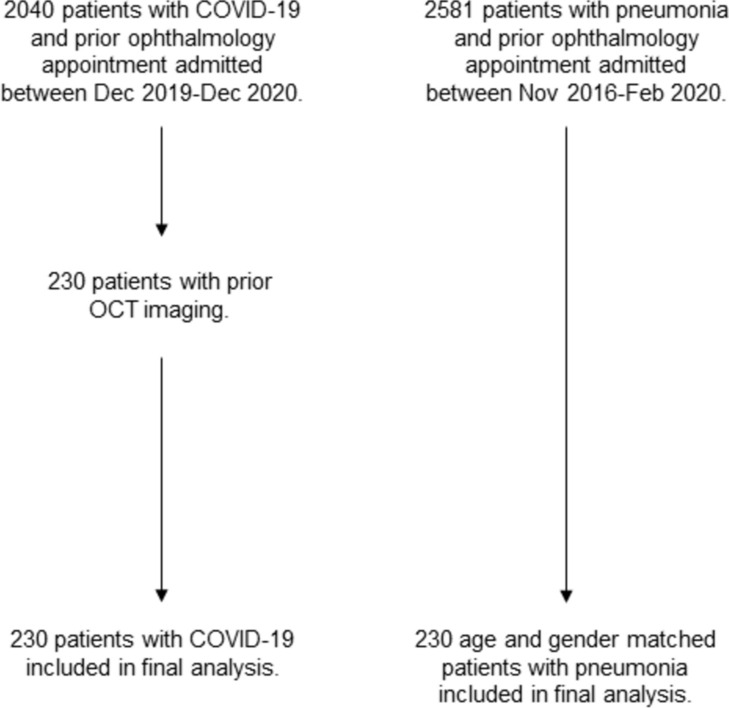 Figure 1
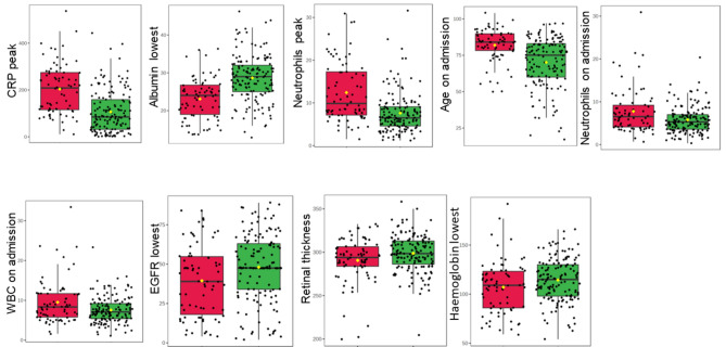 Figure 2
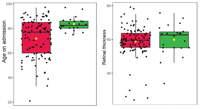 Figure 3
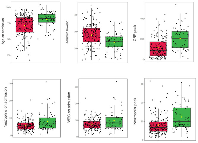 Figure 4
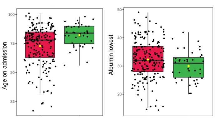 Figure 5
- —http://dx.doi.org/10.13039/501100013629Surgical Reconstruction and Microbiology Research Centre
Peer Reviews
No public reviews on file for this paper yet. If you reviewed it on a platform where reviews are public (OpenReview, ICLR, NeurIPS, ICML), you can paste yours below so the community can read it here.
Videos
No videos yet. Explain this paper in a talk, walkthrough, or lecture? Add one.
Taxonomy
TopicsRetinal and Optic Conditions · Retinal Imaging and Analysis · Retinal Diseases and Treatments
Introduction
The novel coronavirus COVID-19 (SARS-CoV-2) causes respiratory failure, with 4.9 million deaths globally, and severely affected patients requiring intensive treatment unit (ITU) support^1^. Multiorgan dysfunction and an exaggerated inflammatory response are associated with poor prognosis and mortality after COVID-19^2–4^.
Critically ill patients with COVID-19 and multiorgan dysfunction have an extensive injury to vascular endothelium with microcirculatory thrombus, manifesting as reduced functional capillary density^5^ and a functional reduction in oxygen delivery, propagating organ dysfunction^6^.
The retinal structure and microvasculature can be sequentially monitored non-invasively using optical coherence tomography (OCT). This laser imaging modality generates high-resolution cross-sectional retinal images, including of the macula and retinal nerve fibre layer (RNFL)^7,8^. Retinal neuronal and microvascular changes mirror systemic and cerebral pathology in health and diseases such as Parkinson’s and Alzheimer’s disease^6,9–11^. As retinal and cerebral circulations share similar mechanisms of autoregulation^12^, the retina may act as a biomarker for cerebral perfusion^8,13^.
A previous study involving 12 symptomatic COVID-19 patients used OCT to suggest retinal microvascular findings may be associated with COVID-19 neurological events, with results showing cotton wool spots, microhaemorrhages, and hyper-reflective lesions at the ganglion cell and inner plexiform layers in the participants^14^. However, this study did not detail the presence of pre-existing medical conditions, and the interpretation of the results was disputed^15^. A larger study involving 108 COVID-19 positive patients showed one in nine had retinal microvascular signs on colour fundus imaging and OCT, including microhaemorrhages, retinal vascular tortuosity, cotton wool spots, and hyper-reflectivity, which could be related to underlying cardiovascular and thrombotic alterations associated with COVID-19^3^.
The ability to image the retina non-invasively to detect ocular biomarkers of systemic disease is termed “oculomics”, which could predict disease progression by retinal microvascular and structural changes reflecting systemic change^16^. This study aimed to investigate retinal degenerative changes, as a potential biomarker for frailty, to predict health outcomes, including mortality after COVID-19. Thicknesses of the retinal ganglion cell layer (GCL) and RNFL—the neuronal layers most closely associated with cerebral structure and most sensitive to retinal ischaemia—were analysed to determine whether alterations in the retinal layer thickness correlated to outcomes of COVID-19 assessed by length of stay (LoS) in hospital and mortality after admission.
Methods
Study design and setting
This was a retrospective cross-sectional study investigating predictors of outcome after COVID-19 by retinal thickness analysis of previously acquired OCT images. This work uses data provided by patients and collected by the National Health Service (NHS) as part of their care and support at University Hospitals Birmingham NHS Foundation Trust and was approved by UHB Trust governance institutional review and permissions through the DeCOVID under application number [UHB-COV166]. This was entirely hospital electronic patient record-based, analysing results from the Queen Elizabeth Hospital Birmingham (QEHB) only, therefore no patient interaction was required. As only retrospective data were accessed, the determination by the DeCOVID ethics and institutional review process was that patient consent was not required. The study was conducted between December 2020 and August 2022, and we complied with the Declaration of Helsinki and the Data Protection Act 2018.
Key eligibility criteria were: (1) patients admitted to either the QEHB wards or ITU who tested COVID-19 positive and had at least one of the following investigations in the QEHB Ophthalmology Department within the past 10 years: “Fast Macula”, “Posterior Pole” (p-pole) or “RNFL” OCT scans and; (2) patients diagnosed with pneumonia from other causes before March 2020 who had the same OCT investigations in the QEHB Ophthalmology Department between 2010–2020 to act as age and gender-matched controls.
Ophthalmic imaging
OCT Fast Macula (25 B-scans over an area of 5.7 mm^2^ at an automatic real time (ART) setting of 9 A-scans, averaged) and p-pole (61 B-scans over an area of 8.9 mm × 7.4 mm at an ART of 9) scans were exported from the Heidelberg EyeExplorer program (Heidelberg Engineering, Heidelberg, Germany). All scans had been obtained using the SPECTRALIS^®^ Heidelberg OCT2 table-top module (Heidelberg Engineering, Heidelberg, Germany).
Health measures obtained
The following underlying diagnoses and health measures were collected for each patient included in the study: diabetes, ischaemic heart disease, chronic renal failure, chronic obstructive pulmonary disorder (COPD), National Early Warning Score (NEWS), glucose, blood pressure, C-reactive protein (CRP) levels, haematocrit, haemoglobin, eosinophil count, while blood cell (WBC) count, neutrophils, lymphocytes, albumin, estimated glomerular filtration rate (eGFR), and time to mortality following admission. Patient LoS in hospital is the number of days that an in-patient remains in hospital during a single admission event^17^. All measures were retrieved from the hospital database.
OCT thickness extraction
The Fast Macula and p-pole scans were exported from Heidelberg EyeExplorer as E2E files for each patient and anonymised. The E2E files were converted to .VOL files using a research license and run through OCTseg—a custom made code—which automatically segmented and documented the total retinal thickness, combined GCL-plus-inner plexiform layer (GCL + IPL) thickness, and RNFL thickness^18^. Outliers with retinal thickness values more than 3 standard deviations from the mean were excluded.
OCT thickness analysis
Patients were grouped by eye diagnosis (before thickness analysis was completed. Diagnoses included neovascular/dry/wet age-related macular degeneration, diabetic retinopathy, primary open angle glaucoma, diabetic maculopathy, and macula oedema.
Statistics
We used univariate t test with false discovery rate correction (Benjamini-Hochberg, BH Procedure). A significance label was considered as p < 0.05. Associations with length of stay in COVID-19 patients and pneumonia control patients were estimated using linear regression, with corrected p values and variation explained (R^2^) reported. Correlation between measures was calculated using Pearson’s R correlation. In addition, Principal Component Analysis (PCA) was performed to study the variation in both cohorts. For PCA, data were normalised (standardised) using auto scaling. All analyses were performed in R (v4.2.3; R Core Team 2023)^19^.
Informed consent statement
Informed consent requirement was waived by the DeCOVID institutional ethics committee for this retrospective study.
Results
2040 COVID-19 positive patients were admitted to either the QEHB wards or ITU between December 2019 and December 2020, of which 230 patients had OCT imaging in the prior 10 years and were included in the final analysis. 2581 pneumonia patients who had attended a previous ophthalmology appointment were admitted to either the QEHB wards or ITU between November 2016 and February 2020 and from this group 230 age and gender-matched patients were included in the final analysis (all admitted before March 2020) (Fig. 1, Table 1). Mortality was assessed as “all time” at any point in follow up from the date of admission (0 to 1700 days, Table 1), as well as 28-day mortality after admission.Figure 1. Study flowchart.Table 1. Patient demographics table for COVID-19 and pneumonia groups.VariablePatients with COVID-19Patients with pneumoniaP valueMale, N (%)1141130.93Female, N (%)1161170.93Age on admission (SD)74.5 (16.5)74.5 (16.4)1.00Comorbidities, N (%) Diabetes mellitus86 (37.4)74 (32.2)0.28 Ischaemic heart disease11 (4.8)18 (7.8)0.25 Chronic renal failure41 (17.8)19 (8.3)0.03 COPD22 (9.6%)35 (15.2)0.09Eye disorders, N (%) Dry age-related macular degeneration17 (7%)18 (8%)0.87 Diabetic macular oedema13 (6%)13 (6%)1.00 Diabetic retinopathy37 (16%)20 (9%)0.02 Glaucoma24 (10%)15 (7%)0.15 None (no retinal diagnosis)103 (45%)131 (57%)0.06 Other23 (10%)12 (5%)0.06 Wet age-related macular degeneration13 (6%)21 (9%)0.17Mortality, N (%)77 (23.3)147 (35.5) < 0.01Time to mortality, daysRange (mean)0–143 (20.5)0–1700 (489)0.016NEWS on admission (SD)4.16 (3.3)4.38 (3.0)0.56CRP peak, mg/L (SD)139.2 (110.3)162.1 (124.6)0.04LoS, days11.8 (14.4)11.2 (12.1)0.63HCT lowest (SD)0.34 (0.1)0.32 (0.1)0.01HGB lowest, g/L (SD)111.9 (23.8)107.2 (22.9)0.03WBC on admission, × 10^9^/L (SD)8.36 (4.7)12.6 (6.1) < 0.001Neutrophils on admission, × 10^9^/L (SD)6.57 (4.4)10.3 (5.8) < 0.001Neutrophils peak, × 10^9^/L (SD)9.30 (6.2)12.4 (6.6) < 0.001ALB lowest, g/L (SD)26.9 (6.6)31.9 (6.8) < 0.001EGFR lowest, ml/min/1.73 m^2^ (SD)44.7 (23.9)48.5 (22.7)0.06Demographics and clinical characteristics of the participants included in the study given as mean (standard deviation). P-values of < 0.05 are considered significant.N number, SD standard deviation, COPD chronic obstructive pulmonary disorder, CRP C-reactive protein, HCT haematocrit, HGB haemoglobin, WBC white blood cells, ALB albumin, EGFR estimated glomerular filtration rate.
LoS in hospital associated with retinal thickness in patients with COVID-19
Retinal and GCL + IPL layer thicknesses were both strongly associated with LoS in patients with COVID-19 (p = 0.006 for both; Table 2, Supplementary Fig. 1), but not in patients with pneumonia (p = 0.706 and 0.989 respectively; Table 2). RNFL thickness was weakly associated with LoS in hospital in the COVID-19 group (p = 0.097) but not in patients with pneumonia (p = 0.692). Table 2. Associations with length of stay in patients with COVID-19 and patients with pneumonia.GroupMeasureMean ± SDP-value% Variation R^2^COVID-19Retinal thickness298.4 ± 28.130.0063.385**GCL + IPL thickness61.39 ± 13.930.0063.469RNFL thickness47.79 ± 13.340.0971.267Albumin lowest26.85 ± 6.609 < 0.00121.45**Neutrophils peak9.299 ± 6.175** < 0.001*****19.96Haemoglobin lowest**111.9 ± 23.81*** < 0.00116.37Haematocrit lowest0.340 ± 0.070**** < 0.00116.24CRP peak139.2 ± 110.3**** < 0.0019.961WBC on admission8.356 ± 4.661****0.0014.670Neutrophils on admission6.573 ± 4.3750.004*****3.800EGFR lowest44.67 ± 23.870.0083.229Age on admission74.47 ± 16.480.1131.151PneumoniaRetinal thickness295.4 ± 19.340.7060.153GCL + IPL thickness56.92 ± 12.060.9890.000RNFL thickness47.84 ± 12.740.6920.168Albumin lowest32.61 ± 6.7180.0038.916Neutrophils peak12.36 ± 7.111****0.0038.842**Haemoglobin lowest106.5 ± 22.32** < 0.001*****17.10Haematocrit lowest**0.320 ± 0.067*** < 0.00116.68CRP peak154.7 ± 114.70.1142.639WBC on admission12.18 ± 5.7890.6550.213Neutrophils on admission9.995 ± 5.4600.6590.208EGFR lowest47.46 ± 22.660.0404.412Age on admission75.78 ± 14.710.9890.000Rows in bold text represent statistically significant differences in measures, where p ≤ 0.05.SD standard deviation, GCL ganglion cell layer, IPL inner plexiform layer, RNFL retinal nerve fibre layer, CRP C-reactive protein, WBC white blood cells, EGFR estimated glomerular filtration rate. * p < 0.05.
LoS associated with acute inflammatory blood markers in patients with COVID-19
The LoS in hospital for patients with COVID-19 was significantly associated with blood inflammatory markers, with the strongest association for levels of peak neutrophils, WBC, and CRP levels (p < 0.001 for all measures) (Table 2, Supplementary Fig. 1), while only peak neutrophil levels were significantly associated with LoS for patients with pneumonia but not WBC or CRP levels (p = 0.003, 0.655, 0.144 respectively; Supplementary Fig. 2). NEWS was not significantly different between patients with pneumonia and patients with COVID-19 (4.38 and 4.16 respectively, p = 0.56; Table 1), suggesting similar levels of disease severity. LoS in hospital was also significantly associated with low haemoglobin and haematocrit both in patients with COVID-19 (p < 0.001) and patients with pneumonia (p < 0.001; Table 2).
Retinal thickness associated with all-time mortality in patients with COVID-19 and pneumonia, but not 28-day mortality
Most blood markers were significantly associated with all-time mortality in COVID-19 patients, with the strongest association with peak CRP level, CRP on admission, and lowest albumin level (p < 0.001 for all; Fig. 2, Table 3). Total retinal thickness was also associated with all-time mortality (p = 0.015), although GCL + IPL and RNFL thickness were not (Fig. 2, Table 3). No blood markers were significantly associated with all-time mortality in pneumonia patients (Fig. 3, Table 3), but similar to the COVID-19 group, retinal thickness was associated with all-time mortality in pneumonia patients (p = 0.024; Fig. 3, Table 3).Figure 2. Boxplots of significant associations with all time mortality in patients with COVID-19. The green boxplots represent patients who survived, the red boxplots represent patients who died. Each black dot represents a patient with COVID-19. The yellow rhombus represents the mean value of the distribution. CRP C-reactive protein, WBC white blood cells, EGFR estimated glomerular filtration rate.Table 3. Associations with all-time mortality.GroupMeasureP-valueFDRCOVID-19Retinal thickness****0.0150.033GCL + IPL thickness0.6940.671RNFL thickness0.4520.502**Albumin lowest*** < 0.001***** < 0.001Neutrophils peak < 0.001***** < 0.001Haemoglobin lowest0.0320.065Haematocrit lowest0.0650.114CRP peak*** < 0.001***** < 0.001WBC on admission0.0010.004Neutrophils on admission0.0010.002EGFR lowest0.0060.013Age on admission < 0.001 < 0.001PneumoniaRetinal thickness0.0070.094GCL + IPL thickness0.1750.474RNFL thickness0.9810.993Albumin lowest0.7100.959Neutrophils peak0.3230.726Haemoglobin lowest0.0820.345Haematocrit lowest0.1700.474CRP peak0.6810.959WBC on admission0.9520.993Neutrophils on admission0.8830.993EGFR lowest0.0540.345Age on admission* < 0.001*0.002Rows in bold text represent statistically significant differences in measures, where p ≤ 0.05.FDR false discovery rate, GCL ganglion cell layer, IPL inner plexiform layer, RNFL retinal nerve fibre layer, CRP C-reactive protein, WBC white blood cells, EGFR estimated glomerular filtration rate.Figure 3. Boxplots to show associations with all time mortality of patients with pneumonia and significant markers. The green boxplots represent patients who survived, the red boxplots represent patients who died. Each black dot represents a patient with pneumonia. The yellow rhombus represents the mean value of the distribution.
No retinal thickness measurements were associated with 28 day mortality in patients with either COVID-19 (retinal thickness p = 0.151, GCL + IPL p = 0.610, RNFL p = 0.480) or pneumonia (GCL + IPL p = 0.344, RNFL p = 0.813) except for total retinal thickness in patients with pneumonia, which was weakly associated with 28 day mortality (p = 0.067; Fig. 4, Table 4). The blood markers most strongly associated with 28-day mortality in COVID-19 patients were lowest albumin level, peak neutrophil count, and peak CRP (p < 0.001), while the strongest markers for pneumonia patients were lowest albumin level and age on admission (p = 0.043 and p = 0.001 respectively, Fig. 5, Table 4).Figure 4. Boxplots to show associations with 28-day mortality of patients with COVID-19 and significant markers. The green boxplots represent patients who survived, the red boxplots represent patients who died. Each black dot represents a patient with COVID-19. The yellow rhombus represents the mean value of the distribution. CRP C-reactive protein, WBC white blood cells.Table 4. Associations with 28-day mortality.GroupMeasureP-valueFDRCOVID-19Retinal thickness0.1510.244GCL + IPL thickness0.6100.610RNFL thickness0.4800.533Albumin lowest**** < 0.001*** < 0.001Neutrophils peak < 0.001***** < 0.001Haemoglobin lowest0.2000.286Haematocrit lowest0.3280.410CRP peak**** < 0.001***** < 0.001WBC on admission0.0010.004Neutrophils on admission < 0.001****0.001EGFR lowest0.0980.184Age on admission*** < 0.001***** < 0.001PneumoniaRetinal thickness0.0670.347GCL + IPL thickness0.3440.813RNFL thickness0.5790.947Albumin lowest0.0430.347Neutrophils peak0.2070.617Haemoglobin lowest0.5610.947Haematocrit lowest0.7130.947CRP peak0.6240.947WBC on admission0.6180.947Neutrophils on admission0.6300.947EGFR lowest0.0600.347Age on admission****0.001***0.021Rows in bold text represent statistically significant differences in measures, where p ≤ 0.05.FDR false discovery rate, GCL ganglion cell layer, IPL inner plexiform layer, RNFL retinal nerve fibre layer, CRP C-reactive protein, WBC white blood cells, EGFR estimated glomerular filtration rate.Figure 5. Boxplots to show associations with 28-day mortality of patients with pneumonia and significant markers. The green boxplots represent patients who survived, the red boxplots represent patients who died. Each black dot represents a patient with pneumonia. The yellow rhombus represents the mean value of the distribution.
Associations with retinal layer thicknesses in retinal diagnostic subgroups
The retinal diagnoses for which the included patients attended the ophthalmology clinic are reported in Table 1, with comparable proportions of patients with diabetic macular oedema and wet age-related macular degeneration, but a higher proportion of patients with diabetic retinopathy and glaucoma in the COVID-19 group compared to the pneumonia group. Subgroup analysis of the association between LoS and GCL+ thickness in the different retinal diagnostic groups in patients with COVID-19 revealed the same directionality of association in the diagnostic groups: other (p = 0.04), glaucoma (p = 0.05), none (p = 0.11), dry age-related macular degeneration (p = 0.57), and diabetic macular oedema (p = 0.64); but not in: diabetic retinopathy (p = 0.92) or wet age-related macular degeneration (p = 0.83). Similarly, for the association between LoS and retinal thickness, subgroup analysis revealed the same directionality of association in the diagnostic groups: none (p < 0.001), glaucoma (p = 0.009), diabetic retinopathy (p = 0.85), and wet age-related macular degeneration (p = 0.43) but not the other groups. For the association with mortality in patients with COVID-19, retinal thickness retained the same (non-significant) association in all retinal diagnostic groups except diabetic macular oedema, whilst a model could not be fit for the same subgroup analysis in patients with pneumonia.
Discussion
When analysing previously acquired structural OCT images from patients admitted to hospital with COVID-19, retinal thickness and GCL + IPL layer thickness were significantly associated with the LoS in hospital in this group of patients but not in patients with pneumonia, whilst retinal thickness was predictive of all-time but not 28 day mortality for patients with both COVID-19 and pneumonia. These findings suggest that retinal thickness could be a predictor of COVID-19 and pneumonia severity and potentially represent a marker for frailty, supporting wider associations between systemic disease and retinal signs^20–23^.
Frailty is a medical syndrome which increases an individual’s vulnerability to and risk of adverse health outcomes when exposed to a stressor^24^, which considers weight loss, exhaustion, low activity, slowness, and weakness in the screening process^24^. Frailty is an independent predictor of mortality in COVID-19 patients^25^, with an increased risk of mortality in frail patients who were under 65 years old as well as an increased incidence of ITU admission^26^. Frailty is a risk factor for increased susceptibility to and severity of pneumonia in adults ≥ 65 years old^27^. The severity of physical frailty assessed by the Fried phenotype (which includes physical inactivity, exhaustion, weakness, weight loss, and slow walking speed) correlated with elevated total white matter hyperintensity and lower grey matter volume^28^. The neuroretina contains central nervous system neurons and retinal perfusion and structural changes may reflect cerebral changes in illnesses from stroke to multiple sclerosis in addition to systemic diseases such as sepsis and cardiac disease^6,29^. An association between retinal changes and frailty could therefore reflect the manifestation of multiple neurodegenerative and cardiovascular diseases. We found that retinal thickness was associated with all-time mortality in both patients with COVID-19 and pneumonia, though not 28-day mortality, which could support the ability of retinal thickness to be a marker of long-term frailty, consistent with the stronger findings in patients with COVID-19 and LoS. We were unable to control for frailty, as frailty was only recorded routinely in the COVID-19 group.
Both retinal thickness and GCL + IPL thickness significantly associated with LoS in COVID-19 patients, but not in pneumonia patients. LoS is not just dependent on the medical reason for admission and can be affected by patient demographics, past medical history, treatment complexity, and complications^17^. Log regression statistics, machine learning, and data mining are all used to predict LoS with the aim to predict the level of care that may be required for the patient, although it remains a complicated area to model due to external factors^17^. As retinal thickness is associated with COVID-19 LoS, retinal assessments may be able to contribute to predictive markers for patient LoS in this group. However, retinal thickness was not associated with LoS in pneumonia patients, which may be because this group of patients was less severely ill (although NEWS was equal between the groups) or the fact that we were not able to separate community acquired and hospital acquired pneumonia, so the pneumonia patient group may be more heterogenous. During the pandemic, hospital admissions were under greater pressure than before, with the consequence that only the most severely ill and vulnerable patients were admitted^30^. The difference between COVID-19 and pneumonia patients could relate to the frailty of the patient prior to admission, with a stronger signal from retinal thickness in the frailer COVID-19 group but not in the less frail pneumonia group. Blood infection markers were also associated with LoS and all-time mortality, and therefore associate with illness severity, although these results came from blood collected during admission. Because retinal layer thicknesses showed associations with these outcomes, and the OCT images were taken before admission, retinal thickness may predict these outcomes and susceptibility to severe disease.
There was a higher number of patients with COVID-19 who had chronic renal failure compared to patients with pneumonia. A recent study found that patients with chronic kidney disease had retinal and choroidal thinning on OCT compared with healthy volunteers^31^, with similar results shown in late-stage chronic kidney disease in another study^32^. Renal failure also represents a risk for severe COVID-19, which may also cause acute kidney injury. It is therefore unsurprising that renal failure would be more common and EGFR lower in patients with COVID-19 than in patients with pneumonia. Many other systemic diseases may also manfest in retinal thickness or perfusion changes^6^. The PCA analysis examines the contribution of each included predictor independently, and it would be informative in future larger studies to examine the interaction between renal failure, retinal thickness and disease susceptibility.
The associations between retinal thickness, LoS and mortality were present across most diagnostic groups, although the study was most likely not powered for these subgroup analyses. In particular retinal thickness associated with LoS and mortality in patients with no retinal diagnosis, and glaucoma, but not in patients with age-related macular degeneration or diabetic maculopathy, which may reflect greater variability in the data caused by retinal oedema in these cohorts.
Conclusions
The associations between retinal thickness and LoS in hospital in COVID-19 patients, and retinal thickness and all-time mortality in both patients with COVID-19 and those with pneumonia, suggests that retinal structure could be a marker of frailty and a predictor of disease severity in this group.
Supplementary Information
Supplementary Figures.
The reference list from the paper itself. Each links out to its DOI / PubMed record.
- 1World Health Organisation. Weekly epidemiological update on COVID-19. https://www.who.int/publications/m/item/weekly-epidemiological-update-on-covid-19---21-september-2021 (2020).
- 2Mayer, M. OCTSEG (Optical Coherence Tomography Segmentation and Evaluation GUI). (https://www.mathworks.com/matlabcentral/fileexchange/66873-octseg-optical-coherence-tomography-segmentation-andevaluation-gui) (MATLAB Central File Exchange, 2024).
- 3SCIE. Social Care Institute for Excellence. https://www.scie.org.uk/care-providers/coronavirus-covid-19/commissioning/hospital-discharge-admissions (2022).