Microhardness and characterization of human dental tissue after application of enzymatic chemical agents: In vitro study
Caio-Luiz Lins-Candeiro, Wender Batista-de-Souza, Murilo Navarro-de-Oliveira, Paulo-César-Freitas Santos-Filho, Luiz-Renato Paranhos

TL;DR
This study examines how enzymatic agents affect the hardness of human dental tissue in an in vitro setting.
Contribution
The study evaluates the impact of enzymatic chemical agents on dentin and enamel hardness, providing insights into their conservative dental applications.
Findings
Enzymatic chemical agents did not significantly alter enamel hardness compared to a water-soluble gel.
Phosphoric acid showed a statistically significant difference in dentin hardness compared to other groups.
Brix 3000 had lower pH values than Papacárie Duo, indicating higher acidity.
Abstract
The use of enzymatic chemical agents are papain-based materials used in the selective removal of decayed dentin tissue, assisting in conservative techniques and reducing the chances of accidental pulp exposure. A research protocol was subjected to and approved by an Ethics Committee. Using a reporting guide for laboratory studies (CRIS). Healthy human teeth comprised the produced dentin discs subjected to polishing and washing in an ultrasonic bath. Next, the discs received material application according to the experimental groups: water-soluble gel for two minutes, 37% phosphoric acid for 15 seconds in dentin and 30 seconds in enamel, Papacárie Duo (PD) for 30 seconds and two minutes, and Brix 3000 (BX) for two minutes and 30 seconds. The measurement of material pH used solutions at concentrations of 0.1 ml and 2.7 ml prepared for each enzymatic agent. Then, a bench pH meter (n=10)…
Genes, proteins, chemicals, diseases, species, mutations and cell lines named across the full text — each resolved to its canonical identifier and authoritative record.
Click any figure to enlarge with its caption.
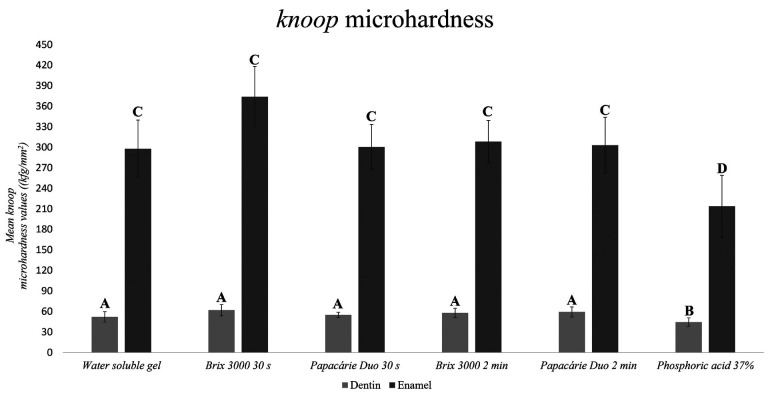 Figure 1
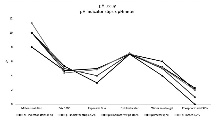 Figure 2
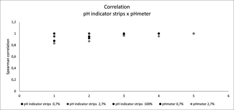 Figure 3
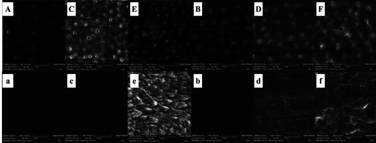 Figure 4
Peer Reviews
No public reviews on file for this paper yet. If you reviewed it on a platform where reviews are public (OpenReview, ICLR, NeurIPS, ICML), you can paste yours below so the community can read it here.
Videos
No videos yet. Explain this paper in a talk, walkthrough, or lecture? Add one.
Taxonomy
TopicsDental materials and restorations · Dental Erosion and Treatment · Dental Implant Techniques and Outcomes
Introduction
Dental caries is a multifactorial and non-communicable disease mediated by biofilm and modulated by diet, causing mineral losses from hard dental tissues (1). It usually originates from the action of microorganisms in oral microbiota and simple carbohydrate fermentation, mainly sucrose (2).
As for the treatment of cavitated carious lesions, the non-selective removal of carious tissues has long been advocated, completely removing the affected and contaminated tissue in surrounding and pulpal walls, and this is the technique most used by dentists (3,4). However, selective caries removal significantly reduced the risk of pulp exposure, symptoms, and postoperative pulpal complications (5). Thus, selectively removing the carious tissues showed a higher cost-effectiveness than non-selective removal, favoring the vitality maintenance of deeply decayed teeth (6,7).
Brix 3000 is a new papain-based material with higher enzyme concentrations that presents favorable characteristics, such as lower cytotoxicity and genotoxicity levels and faster carious tissue removal (8,9). The amount of papain in its formulation (3000 U/mg at 10%) distinguishes this material. It also uses the EBE (Encapsulating Buffer Emulsion) technology with encapsulated papain, increasing enzymatic activity and providing higher stability.
Numerous studies have been developed after introducing enzymatic chemical agents (ECAs) for selective caries removal. The publications include studies assessing the activity of these ECAs on cells (10), especially pulp cells, analyzing their direct (9) and indirect (11) cytotoxicity, genotoxicity (8), bioactivity (12), and comfort in controlling the painful experience without local anesthetics (13). Other studies assessed the effect of dentin deproteinization caused by chemo-mechanical removers on self-etch adhesive sealing (14,15).
Therefore, the topic has been investigated, increasingly promoting discussions in the literature. However, data regarding ECA action in mineralized dental tissues, such as enamel and dentin, are still scarce. Therefore, the present study assessed surface structure, enamel and dentin hardness, and the pH behavior of these materials after applying enzymatic chemical removers.
Material and Methods
-Protocol and ethical criteria
It is a controlled in vitro study. The researchers complied with ethical criteria related to studies in human beings (CAAE 28490619.3.0000.5152) and followed the CRIS (Checklist for Reporting In-vitro Studies) reporting guide.
-Sample calculation
Gpower 3.1.9.2 software assisted sample calculation using the parameters α = 0.05, 0.63 effect, and 95% power. Therefore, the sample size included 60 samples (n=10). Pilot trials determined sample parameters. The sample size calculation was based on the study outcomes, namely the Knoop microhardness of dental tissues.
-Sample preparations
The study used healthy human third molars donated after exodontia with clinical and radiographic indication. Immediately after exodontia, the teeth were cleaned with periodontal curettes and stored in distilled water under refrigeration in a glass recipient hermetically sealed.
The teeth were sectioned in a cutting machine (Isomet1000; Buehler Ltd, Lake Bluff, IL) with a diamond disc (Diamond Wafering Blade. #3041201 - 4” x.012 X 1/2” - 102 mm x 0.3 mm x 127 mm Odeme Dental Research, Miami, FL, USA) in the buccolingual direction, leveling out the occlusal surface. Later, a new section was made, producing dentin and enamel discs with 2 mm of thickness. Seventy-eight discs were obtained (only one per tooth) so that all would maintain the occlusal-cervical thickness dimensions. The study did not use teeth with carious lesions, anomalies, cracks, fractures, restorations, and discs with pulp horn exposure. The discs were stored in microtubes for up to 30 days in distilled water and constant refrigeration.
The discs underwent a surface polishing protocol with 400, 600, 1200, and 1500-grit sandpapers for 10 seconds each, with a metallic device (50 g) in constant water refrigeration. Next, the discs were washed in an ultrasonic bath (Cristófoli Equipamentos de Biossegurança LTDA, Campo Mourão – PR, Brazil) with distilled water for two five-minute cycles, leaving the polished surface of the disc upward (11).
-Material application protocol
Regarding material application, the samples were randomly distributed into six groups according to the materials: water-soluble gel for two minutes, 37% phosphoric acid for 15 seconds in dentin and 30 seconds in enamel, Papacárie Duo (PD) for 30 seconds and two minutes, and Brix 3000 (BX) for two minutes and 30 seconds. Insulin syringes applied the materials to standardize their quantity. After material application according to the protocol, the discs were washed in distilled water aided by a triple syringe and dried in sterile cotton.
-Knoop hardness test
The discs were included with crystal resin, and the matrix comprised a polyvinyl chloride pipe with 15 mm of height and 21 mm with utility wax at the base. Tilt during indentations was prevented by leveling out the base with 400-grit sandpapers, and the samples underwent the microhardness test (n=10) with a microhardness tester (FM – 700, Future-Tech Corp., Tokyo, Japan). Three indentations were made per sample with a load of 50 g for 15 seconds, respecting a minimum distance of 100 µm between each indentation for dentin and enamel. The mean length of the two diagonals produced by the indenter calculated hardness values. The results correspond to the means of the three indentations for each sample.
-pH test
Two solutions helped measure hydrogen potential by dissolving 0.1 ml and 0.4 ml of each enzymatic remover into 14 ml of distilled water, obtaining two final solutions with 0.7% and 2.7% concentrations, respectively. Solution homogenization used a sterile Falcon tube where the material was poured with distilled water in a multi-platform agitator (Kasvi® K40-10208) at 1900 rpm. Next, a 24-well plate received the solution, using six wells for each material, with 2 ml of solution per well. Control solutions comprised the Milton solution, distilled water, pure water-soluble gel, and 0.7% phosphoric acid (n=10). A bench pH meter (MS Tecnopon Equip. Especiais LTDA) assessed the pH individually for each well in triplicate. Then, a new assessment was performed using pH indicator strips (Merck KGaA, Darmstadt, Germany). Three operators performed all analyses, tabulating the mean values of each evaluation.
-Scanning electron microscopy
The samples received the material application protocol (n=3) and were dehydrated in alcoholic solutions in an increasing sequence of 50%, 70%, 80%, and 90% for ten minutes in each concentration and absolute alcohol three times for ten minutes each. Then, the discs were fixed in stubs, dried at room temperature in a chapel for 24 hours, maintained for 15 days in a desiccator, and metalized before scanning electron microscopy.
-Statistical analysis
The findings were described and tabulated in Excel software, version 2016 Microsoft Office®. Next, the values underwent descriptive analysis, the Shapiro-Wilk normality test, one-way ANOVA supplemented by Tukey’s test at 5% significance, and the Spearman correlation test in JAMOVI statistical software, version 2.3.21. Qualitative results were described and inserted in the study, complementing the quantitative data.
Results
-Knoop hardness test
The dentinal hardness analysis (Graph 1) showed that samples receiving phosphoric acid differed statistically from the other groups (p<0.05). The knoop microhardness values of tooth enamel (Fig. 1) indicates that, after material application, the samples receiving enzymatic chemical agents (ECAs) did not statistically differ from those receiving the water-soluble gel (p<0.05).
Figure 1. Mean knoop hardness values of human dentin and enamel after applying the experimental materials (n=10). One-way ANOVA supplemented by Tukey’s test at 5% significance, Shapiro-Wilk p=0.513. Different letters indicate statistical differences.
-pH test
In the pH test, BX values were lower (4.37 ± 0.01) than PD (4.85 ± 0.06) (Fig. 2). The pH values did not present normal data distribution p<0.001) (Table 1).
Figure 2. Mean pH values obtained through different techniques: pH indicator strips and bench pH meter.
The Shapiro-Wilk normality analysis showed that groups did not present a normal distribution (p<0.001); hence, the Spearman test was used. Spearman correlation coefficient demonstrated significance between all groups (p=0.001), with positive correlations with a minimum of 0.831 (Fig. 3).
Figure 3. Spearman correlation between different techniques and concentrations (p=0.001).
-Scanning electron microscopy
Scanning electron microscopy showed that the longer the contact time of ECAs with dentin (Fig. 4), the higher the exposure of dentinal tubules, although changes in inter- and peritubular dentin are not visible. Enamel images (Fig. 4) demonstrate that Papacárie Duo and Brix 3000 did not significantly change enamel prisms as phosphoric acid. Water-soluble gel images present an irregular surface, possibly due to oily compounds.
Figure 4SEM of dentin after the material application protocol. (A) Papacárie Duo for 30 seconds; (B) Papacárie Duo for two minutes; (C) Brix 3000 for 30 seconds; (D) Brix 3000 for two minutes; (E) Phosphoric acid for 15 seconds; (F) Water-soluble gel for two minutes. The SEM of enamel (a) Papacárie Duo for 30 seconds; (b) Papacárie Duo for two minutes; (c) Brix 3000 for 30 seconds; (d) Brix 3000 for two minutes; (e) Phosphoric acid for 15 seconds; (f) Water-soluble gel for two minutes. Magnification of 5000x.
Discussion
Understanding material behavior in dental tissues is essential for clinical practice. This knowledge aids dentists and researchers in direct or indirect operative and rehabilitating decision-making, preserving the dental structure to improve adhesion for the success of the proposed treatment and promoting comfort to patients. The literature has been discussing the application of these materials to people with phobias, challenging behavioral management, and young permanent teeth, showing higher patient acceptance (16).
Chemo-mechanical removal methods have promoted less stressful and painful treatments (17). Methods using enzymatic chemical agents (ECAs) for selectively removing dentinal carious tissue promote lower anxiety than treatments with rotary instruments, as therapies with ECAs were less painful when assessing pain experience (18). Thus, treatments with ECAs are a viable therapeutic proposal, considering patient benefits during treatment associated with the positive effect on healthy dental tissues and the maintenance of viable dental tissues (6).
ECAs did not show significant differences in hardness values compared to the control group, indicating that healthy dentin and enamel presented good quality after applying the materials. The quality of tooth remnants is directly associated with adhesion (19,20). Thus, new studies must be performed to assess more parameters affecting the quantity of tooth remnants after applying these materials.
The microscopy images showed differences in enamel surface between the groups using ECAs and the control, and the former presented an erosion pattern. Systematic reviews with meta-analyses found that erosion did not significantly affect enamel bond strength (21,22). However, erosion causes a rough surface similar to conditions after phosphoric acid etching, potentially benefiting adhesive systems that favor surface erosion (23). Studies assessing dental substrate adhesion after using ECAs are relevant and necessary to substantiate clinical decision-making and restorative surgical protocol.
After ECA applications at different times, an intratubular smear plug appeared with intertubular dentin exposure, while the group that received phosphoric acid showed a complete smear layer removal. Smear layer removal may increase fluid flow on the exposed dentin surface, potentially interfering with postoperative sensitivity and the adhesion process, and moisture control and adhesive system selection will be essential for clinical success (24).
This study is not free of limitations. Considering it is an in vitro study, the findings may not be considered for clinical practice because the tests occurred in a controlled environment. Thus, studies assessing the quality of tissues with different adhesive systems and clinical studies evaluating postoperative sensitivity, restoration survival, and restorative materials are relevant to explain ECA use and expand the discussions about applying these protocols to the clinical routine.
Conclusions
ECAs for carious dentinal tissue removal did not significantly change the hardness of healthy human enamel and dentin despite being acidic. The pH measurement technique was a viable alternative to materials commercially presented in gel, especially when the composition of these materials has pigments.
The reference list from the paper itself. Each links out to its DOI / PubMed record.
- 1Pitts NB Zero DT Marsh PD Ekstrand K Weintraub JA Ramos-Gomez F Dental caries Nat Rev Dis Primers 20173170302854093710.1038/nrdp.2017.30 · doi ↗ · pubmed ↗
- 2Mathur VP Dhillon JK Dental Caries: A Disease Which Needs Attention Indian J Pediatr 201885(3)2022062864316210.1007/s 12098-017-2381-6 · doi ↗ · pubmed ↗
- 3Bjørndal L Simon S Tomson PL Duncan HF Management of deep caries and the exposed pulp Int Endod J 201952(7)9499733098594410.1111/iej.13128 · doi ↗ · pubmed ↗
- 4Casagrande L Seminario AT Correa MB Werle SB Maltz M Demarco FF Longevity and associated risk factors in adhesive restorations of young permanent teeth after complete and selective caries removal: a retrospective study Clin Oral Investig 201721(3)8478552710358710.1007/s 00784-016-1832-1 · doi ↗ · pubmed ↗
- 5Li T Zhai X Song F Zhu H Selective versus non-selective removal for dental caries: a systematic review and meta-analysis Acta Odontol Scand 201876(2)1351402907381410.1080/00016357.2017.1392602 · doi ↗ · pubmed ↗
- 6Barros MMAF De Queiroz Rodrigues MI Muniz FWMG Rodrigues LKA Selective, stepwise, or nonselective removal of carious tissue: which technique offers lower risk for the treatment of dental caries in permanent teeth? A systematic review and meta-analysis Clin Oral Investig 202024(2)5215323177337110.1007/s 00784-019-03114-5 · doi ↗ · pubmed ↗
- 7Schwendicke F Stolpe M Meyer-Lueckel H Paris SDörfer CE Cost-effectiveness of one- and two-step incomplete and complete excavations J Dent Res 201392(10)88072394597510.1177/0022034513500792 · doi ↗ · pubmed ↗
- 8Santos TML Bresciani E Matos FS Camargo SEA Hidalgo APT Rivera LML Comparison between conventional and chemomechanical approaches for the removal of carious dentin: an in vitro study Sci Rep 202010(1)81273241519010.1038/s 41598-020-65159-x PMC 7229020 · doi ↗ · pubmed ↗