Semi-automated analysis of HER2 immunohistochemistry in invasive breast carcinoma using whole slide images: utility for interpretation in clinical practice
Chiu-Hsiang Connie Liao, Nilay Bakoglu, Emine Cesmecioglu, Matthew Hanna, Fresia Pareja, Hannah Y. Wen, Timothy M. D’Alfonso, Edi Brogi, Yukako Yagi, Dara S. Ross

TL;DR
This study evaluates a semi-automated tool for analyzing HER2 immunohistochemistry in breast cancer, showing it can help provide objective and accurate results for clinical treatment decisions.
Contribution
The study introduces a semi-automated HER2 IHC analysis tool that demonstrates high concordance with manual scoring and potential for clinical use.
Findings
Semi-automated HER2 IHC scoring showed a Kappa value of 0.77 in the calibration dataset, indicating substantial agreement with manual scoring.
The tool achieved 100% sensitivity and 56-61% specificity in detecting HER2 amplification.
Only 17% of validation cases had discordant results between manual and semi-automated analysis.
Abstract
Human epidermal growth factor receptor 2 (HER2) gene amplification and subsequent protein overexpression is a strong prognostic and predictive biomarker in invasive breast carcinoma (IBC). ASCO/CAP recommended tests for HER2 assessment include immunohistochemistry (IHC) and/or in situ hybridization (ISH). Accurate HER2 IHC scoring (0, 1+, 2+, 3+) is key for appropriate classification and treatment of IBC. HER2-targeted therapies, including anti-HER2 monoclonal antibodies and antibody drug conjugates (ADC), have revolutionized the treatment of HER2-positive IBC. Recently, ADC have also been approved for treatment of HER2-low (IHC 1+, IHC 2+/ISH-) advanced breast carcinoma, making a distinction between IHC 0 and 1+ crucial. In this focused study, 32 IBC with HER2 IHC scores from 0 to 3+ and HER2 FISH results formed a calibration dataset, and 77 IBC with HER2 IHC score 2+ and paired FISH…
Genes, proteins, chemicals, diseases, species, mutations and cell lines named across the full text — each resolved to its canonical identifier and authoritative record.
Click any figure to enlarge with its caption.
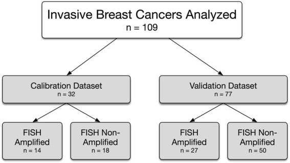 Figure 1
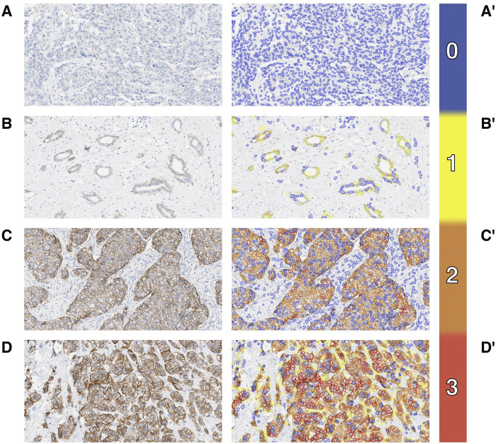 Figure 2
 Figure 3
 Figure 4
Peer Reviews
No public reviews on file for this paper yet. If you reviewed it on a platform where reviews are public (OpenReview, ICLR, NeurIPS, ICML), you can paste yours below so the community can read it here.
Videos
No videos yet. Explain this paper in a talk, walkthrough, or lecture? Add one.
Taxonomy
TopicsHER2/EGFR in Cancer Research · Monoclonal and Polyclonal Antibodies Research · Nanofabrication and Lithography Techniques