Complete genome sequence of Bacillus subtilis phage fHSPT3
Tushar Midha, Aaina Choudhary, Somesh Baranwal

TL;DR
This paper reports the full genome sequence of a new bacteriophage, fHSPT3, isolated from sewage and infecting Bacillus subtilis.
Contribution
The novel contribution is the complete genome characterization of phage fHSPT3, including its coding potential and life cycle prediction.
Findings
The phage fHSPT3 has a genome of 150,187 bp with 221 protein-coding sequences.
Phage fHSPT3 belongs to the genus Siophivirus and lacks virulence or antibiotic resistance genes.
The phage is predicted to have a virulent life cycle using PhageScope and PhageAI.
Abstract
Here, we describe a Bacillus subtilis bacteriophage isolated from sewage water. The phage fHSPT3 was isolated against the host Bacillus subtilis 168 and has a genome size of 150,187 bp with 221 protein-coding sequences. The fHSPT3 belongs to the genus Siophivirus, lacks virulence or antibiotic resistance genes, and shows a virulent life cycle predicted through PhageScope and PhageAI.
Genes, proteins, chemicals, diseases, species, mutations and cell lines named across the full text — each resolved to its canonical identifier and authoritative record.
Click any figure to enlarge with its caption.
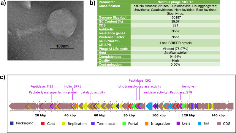 Fig 1
Fig 1- —Council of Scientific and Industrial Research, India (CSIR)
- —University Grants Commission (UGC)
- —Department of Science and Technology, Ministry of Science and Technology, India (DST)
Peer Reviews
No public reviews on file for this paper yet. If you reviewed it on a platform where reviews are public (OpenReview, ICLR, NeurIPS, ICML), you can paste yours below so the community can read it here.
Videos
No videos yet. Explain this paper in a talk, walkthrough, or lecture? Add one.
Taxonomy
TopicsLogic, Reasoning, and Knowledge · Logic, programming, and type systems · Semantic Web and Ontologies
ANNOUNCEMENT
Bacillus subtilis is a Gram-positive, rod-shaped, spore-forming model organism with applications in food fermentation, enzyme production, feed additive, and plant biocontrol (1, 2). We report a complete genome of Bacillus subtilis phage fHSPT3 belonging to the genus Siophivirus isolated from influent sewage water located at Dabwali, Haryana, India (N29°56′18.56″, E74°41′5.497″).
The phage was isolated after enrichment with host Bacillus subtilis 168 (BEI resources, NIAID, NIH) and purified with three rounds of double-layer agar overlay method to get monophage (3). Briefly, 2 mL water sample was filtered with a 0.22 µm filter (HiMedia) and incubated at 37°C with 200 µL overnight host bacterial culture in 3 mL double strength tryptic soy broth for 6 h with continuous shaking at 120 rpm. Then, it was centrifuged at 8,000 g for 5 minutes; 500 µL supernatant was used for a double-layer plate approach, mixed with 500 µL of log-phase Bacillus subtilis 168 and 5 mL of molten top agar, and plated on tryptic soy agar (HiMedia) plate and left to incubate overnight at 37°C. A plaque with clear, brilliant, and smooth edges was inoculated into 500 µL of SM buffer pH 7.5. For transmission electron microscopy, fHCPT3 phage lysate was stained with 2% phosphotungstic acid (pH 7.0) for 15 seconds after treatment with ammonium acetate (4). The stained grid was observed with Tecnai G20 HR-TEM at SAIF, AIIMS, New Delhi. The length of the phage was determined to be 275 ± 01 nm with a head width of 133 ± 2 nm using Image J 1.54 software (NIH, Bethesda, MD, USA) (Fig. 1a). Phenol-chloroform-based extraction was used for DNA isolation from phage lysate following treatment with DNase to remove the host DNA contamination (3).
Details of Bacillus subtilis phage fHSPT3. (a) Morphology of phage observed through transmission electron microscopy. (b) Genomic details, and (c) genomic map of phage fHSPT3 depicting various protein-coding regions.
The purified phage DNA was outsourced to the National Institute for Biomedical Genomics, Kalyani (India) for library preparation through NexteraXT (Illumina) and Illumina pair-based whole genome sequencing through NovoSeq 6000-2 × 250 bp SP v1.5-150x (Illumina Inc.). FastQC 0.11.7 was performed to check the quality. A de novo genome assembly was performed with Q.C. pass reads through Unicycler version 0.4.8, functioning as SPAdes-optimiser 3.13.0 (5). PhageTerm 1.0.12 and QUAST 5.2.0 were used to determine phage termini and assembly quality. A single contig with 94% mapping reads was obtained for phage fHSPT3 (6, 7). Then, the genome was annotated with Prokka 1.14.6 (8) and further with PhageScope to determine host and completeness (9). Phagescope uses CheckV version 1.0.1 to determine contamination and completeness. The phage fHSPT3 genome is a complete linear genome of 150,187 bp with 39.07% GC content and is of high quality. A total of 221 protein-coding sequences were obtained without any tRNA, antibiotic-resistant gene, virulence factor, or CRISPR elements. The phage life cycle was determined to be virulent with 78.97% confidence with PhageAI 1.0.2 tool (10) (Fig. 1b). A genomic map was prepared depicting the lysis proteins with Proksee version 1.0.0a6 (Fig. 1c) (11). The NCBI BLASTn (12) showed that phage fHSPT3 is closely related to genus Siophivirus and family Herelleviridae phages, Bacillus phage vB_BspH_Mawwa (MW749002.1) and Bacillus phage vB_BspH_TimeGriffin (MW749007.1) with a percentage identity of 98.03% and 97.75%, respectively, and query cover of 92% and 90%, respectively.
The reference list from the paper itself. Each links out to its DOI / PubMed record.
- 1Su Y, Liu C, Fang H, Zhang D. 2020. Bacillus subtilis: a universal cell factory for industry, agriculture, biomaterials and medicine. Microb Cell Fact 19:173. doi:10.1186/s 12934-020-01436-832883293 PMC 7650271 · doi ↗ · pubmed ↗
- 2Kovács ÁT. 2019. Bacillus subtilis. Trends Microbiol 27:724–725. doi:10.1016/j.tim.2019.03.00831000489 · doi ↗ · pubmed ↗
- 3Choudhary A, Midha T, Gulati I, Baranwal S. 2024. Isolation, genomic characterization of Shigella prophage f PSFA that effectively infects multi-drug resistant Shigella isolates from the Indian poultry sector. Microb Pathog 188:106538. doi:10.1016/j.micpath.2024.10653838184177 · doi ↗ · pubmed ↗
- 4Rathor N, Thakur CK, Das BK, Chaudhry R. 2022. An insight into the therapeutic potential of a novel lytic Pseudomonas phage isolated from the river Ganga. J Appl Microbiol 133:1353–1362. doi:10.1111/jam.1563935616159 · doi ↗ · pubmed ↗
- 5Wick RR, Judd LM, Gorrie CL, Holt KE. 2017. Unicycler: resolving bacterial genome assemblies from short and long sequencing reads. P Lo S Comput Biol 13:e 1005595. doi:10.1371/journal.pcbi.100559528594827 PMC 5481147 · doi ↗ · pubmed ↗
- 6Gurevich A, Saveliev V, Vyahhi N, Tesler G. 2013. QUAST: quality assessment tool for genome assemblies. Bioinformatics 29:1072–1075. doi:10.1093/bioinformatics/btt 08623422339 PMC 3624806 · doi ↗ · pubmed ↗
- 7Garneau JR, Depardieu F, Fortier L-C, Bikard D, Monot M. 2017. Phage Term: a tool for fast and accurate determination of phage termini and packaging mechanism using next-generation sequencing data. Sci Rep 7:8292. doi:10.1038/s 41598-017-07910-528811656 PMC 5557969 · doi ↗ · pubmed ↗
- 8Seemann T. 2014. Prokka: rapid prokaryotic genome annotation. Bioinformatics 30:2068–2069. doi:10.1093/bioinformatics/btu 15324642063 · doi ↗ · pubmed ↗