Double chamber-like left ventricle
Mikio Shiba, Yoshiharu Higuchi

Abstract
Genes, proteins, chemicals, diseases, species, mutations and cell lines named across the full text — each resolved to its canonical identifier and authoritative record.
Click any figure to enlarge with its caption.
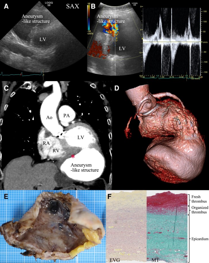 Figure 1
Peer Reviews
No public reviews on file for this paper yet. If you reviewed it on a platform where reviews are public (OpenReview, ICLR, NeurIPS, ICML), you can paste yours below so the community can read it here.
Videos
No videos yet. Explain this paper in a talk, walkthrough, or lecture? Add one.
Taxonomy
TopicsCongenital Heart Disease Studies · Cardiac Structural Anomalies and Repair · Vascular anomalies and interventions
An 89-year-old woman who had undergone surgical aortic valve replacement 25 years ago was admitted to our hospital with deficient articulation and diagnosed with a cerebrovascular accident. An echocardiogram revealed what appeared to be a ventricular aneurysm (Panel A and see Supplementary material online, Movie S1), with a to-and-fro flow observed between it and the left ventricle (Panel B and see Supplementary material online, Movie S2). A contrast-enhanced thoracic computed tomography (CT) scan indicated a left ventricular aneurysm 90 mm in diameter, with a mural thrombus that was very thin and stratified (Panels C and D). On history taking, it was revealed that 4 months ago, she had received conservative treatment for a ventricular rupture secondary to acute myocardial infarction due to the occlusion of the right coronary artery. The aneurysmal structure was surgically resected (Panel E), and the heart was repaired with a bovine pericardial patch. Histological examination led to a diagnosis of a pseudoaneurysm (Panel F).
The patient was discharged without complications after a month of hospital stay. Pericardial adhesions resulting from her previous open-heart surgery may have provided structural support, preventing rupture and allowing the pseudoaneurysm to enlarge over time. Thrombus formation in a pseudoaneurysm is one of the causes of embolic stroke. In addition to imaging diagnostics, such as echocardiography and CT, a detailed medical history provides key insights into the mechanism of anatomical deformation and subsequent thrombus formation.
Supplementary Material
ytae459_Supplementary_Data