Further notes on the genus Eurhaphidophora Gorochov, 1999 (Orthoptera, Rhaphidophoridae) with description of a new species from China

Abstract
Genes, proteins, chemicals, diseases, species, mutations and cell lines named across the full text — each resolved to its canonical identifier and authoritative record.
Click any figure to enlarge with its caption.
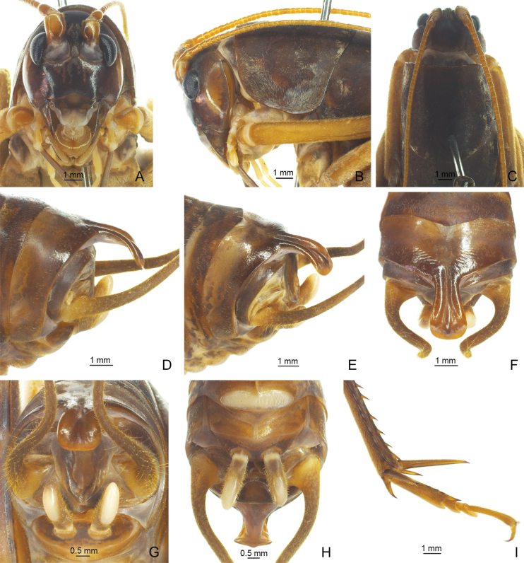 Figure 1
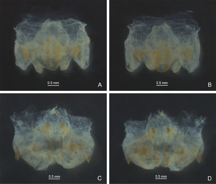 Figure 2
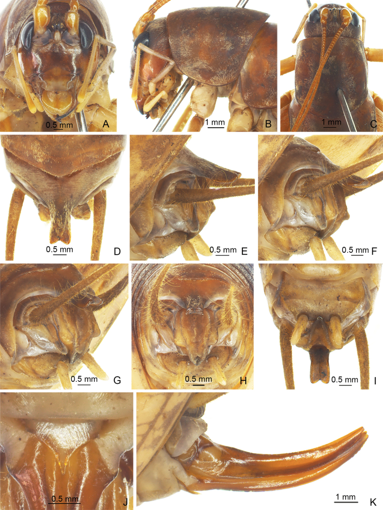 Figure 3
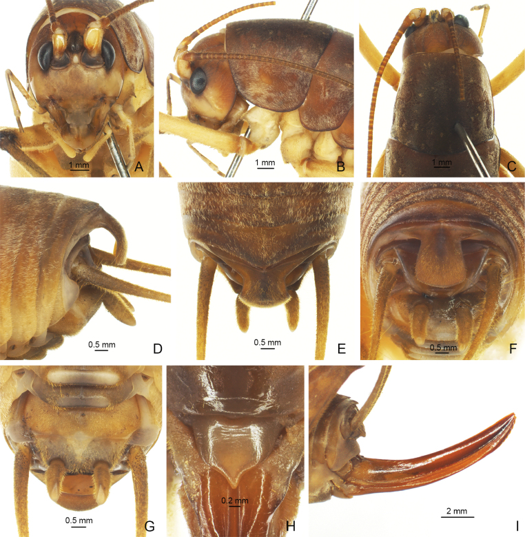 Figure 4
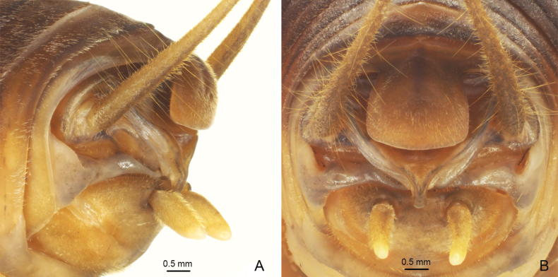 Figure 5
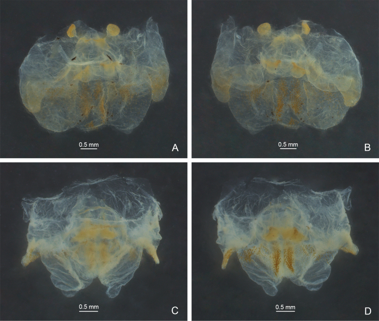 Figure 6
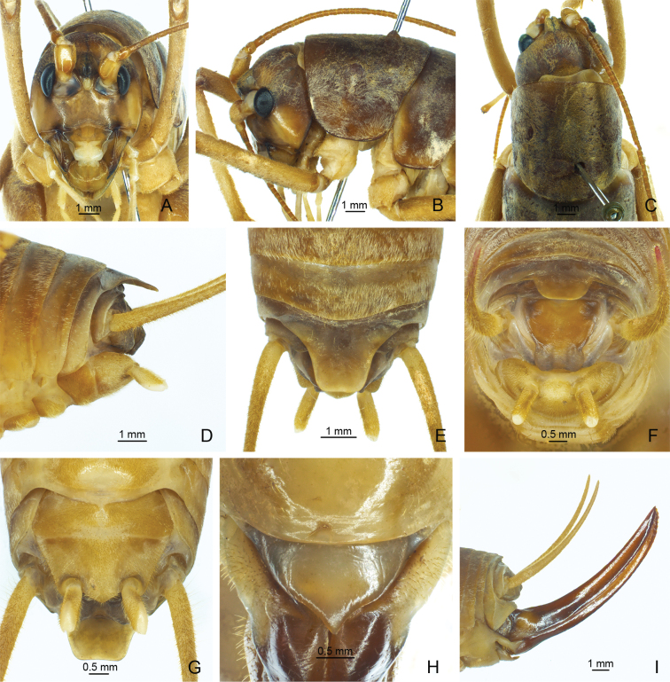 Figure 7
Peer Reviews
No public reviews on file for this paper yet. If you reviewed it on a platform where reviews are public (OpenReview, ICLR, NeurIPS, ICML), you can paste yours below so the community can read it here.
Videos
No videos yet. Explain this paper in a talk, walkthrough, or lecture? Add one.
Taxonomy
TopicsOrthoptera Research and Taxonomy · Fossil Insects in Amber · Ecology and Vegetation Dynamics Studies
Introduction
Gorochov (1999) established the genus Eurhaphidophora Gorochov, 1999 and assigned Eurhaphidophoranataliae Gorochov, 1999 from Vietnam as type species. Thereafter, nine species were described from China, Vietnam, Laos, Thailand and Malaysia (Gorochov 2010, 2011, 2012). Later, E.truncata Bian & Shi, 2016, E.curvata Lu, Huang & Bian, 2022 and E.fossa Lu, Huang & Bian, 2022 were published from China (Bian and Shi 2016; Lu et al. 2022), while E.pawangkhananti Dawwrueng, Gorochov & Suwannapoom, 2020, E.tarasovidoitungensis Dawwrueng, Gorochov & Suwannapoom, 2020 and E.apicoexcisa Dawwrueng, Gorochov, Pinkaew & Vitheepradit, 2023 were discovered from Thailand (Dawwrueng et al. 2020, 2023).
Up to now, the genus Eurhaphidophora includes fifteen species, four of which are recorded from China. Here, we describe a new species E.dulongjiangensis Zhu & Shi, sp. nov. from China, describe the females of E.tarasovidoitungensis Dawwrueng, Gorochov & Suwannapoom, 2020 and E.fossa Lu, Huang & Bian, 2022 for the first time, and propose E.curvata Lu, Huang & Bian, 2022, syn. nov. to become a new synonym of Eurhaphidophorapawangkhananti Dawwrueng, Gorochov & Suwannapoom, 2020.
Materials and methods
Specimens were collected by hand at night and preserved in 75% ethanol. The genitalia were dissected with an insect needle and then put in 10% KOH solution to clean the tissue. Images were taken with a Zeiss AxioCam ICc5 digital camera attached to a Zeiss Stereo Discovery V12 microscope and edited with ADOBE PHOTOSHOP 2022. With regard to the scheme of arrangement of spines on the tibiae and hind basitarsus we follow Gorochov and Storozhenko (2015) and for measurements we follow Zhu et al. (2022). The type specimen is deposited in the Museum of Hebei University, Baoding, China (HBU).
Results
Eurhaphidophora
Taxon classificationAnimaliaOrthopteraRhaphidophoridae
Genus
Gorochov, 1999
14A15F23-622D-5260-9E2B-27E80F443FD5
Type species.
Eurhaphidophoranataliae Gorochov, 1999, by original designation.
Diagnosis.
Body medium-sized in Rhaphidophorinae. Seventh and eighth abdominal tergites of male with a small posterior median projection that is nearly rounded or angular. Posterior margin of ninth abdominal tergite of male provided with a large median process. Male epiproct simple. Male genitalia membranous. Lateral lobes of dorso-median blade large, almost entirely covering central lobe of this blade.
Eurhaphidophoradulongjiangensis Zhu & Shi, sp. nov. A–I ♂ A–C head and pronotum A frontal view B lateral view C dorsal view D–H apex of abdomen D lateral view E apico-lateral view F dorsal view G apical view H ventral view I hind tarsus in lateral view.
Distribution.
China, Laos, Malaysia, Thailand and Vietnam.
Eurhaphidophora
dulongjiangensis
Taxon classificationAnimaliaOrthopteraRhaphidophoridae
Zhu & Shi sp. nov.
C8523007-929C-5C26-8C59-01197BF62653
https://zoobank.org/FAE6DAB0-1028-40DD-A5A2-1F8BA6D2411A
Type material.
Holotype. ♂, China: Yunnan Province, Gongshan County, Dulongjiang Town, Bapo Village, 27.7418°N, 98.3561°E, alt. 1610 m, 9.VII.2021, Shengchuan Yang leg.
Diagnosis.
The new species can be distinguished from other congeneric species by the shape of the male epiproct and the ninth abdominal tergite. The ninth abdominal tergite of the male has a long posteromedian process, basal half narrow with a longitudinal median furrow, lateral sides raised into ridges; apical half slightly broadened and curved downwards, with a carina in midline, apex truncate. Male epiproct linguiform, concave on ventral side, apical area slightly protruding.
Male genitalia of Eurhaphidophora spp. A, C dorsal view B, D ventral view A, BEurhaphidophoradulongjiangensis Zhu & Shi, sp. nov. C, DEurhaphidophoratarasovidoitungensis Dawwrueng, Gorochov & Suwannapoom, 2020.
Description.
Male. Body medium-sized. Fastigium verticis with rostral tubercles, pressed to each other and divided by a narrow and deep furrow, pointing forwards. Eyes ovoid, protruding forwards; lateral ocelli large and circular, occupying basal 2/3 of lateral surface of rostral tubercles; median ocellus slightly smaller, oval, located between antennal sockets. Pronotum long, anterior margin straight, posterior margin arcuate; lateral lobe longer than high, ventral margin arc-shaped. Mesonotum and metanotum short, posterior margin of mesonotum arcuate, posterior margin of metanotum straight. Fore coxa with one small spine. Internal genicular lobe of fore femur with one long spine; internal and external genicular lobes of mid femur each with one long spine; hind femur with one inner spine on ventral surface, internal genicular lobe with one small spine. Tibia and hind basitarsus with following armament – ve, vi, ve, v2a / de, d~2, d2a, ve, ve, v2a / d20e–18i (d22e–20i), d2sa, 6a / d3c, dac. Posterior margin of eighth abdominal tergite angularly projecting. Ninth abdominal tergite with long posteromedian process, basal half narrow with a longitudinal median furrow, lateral sides raised into ridges; apical half slightly broadened and curved downwards, with a carina in midline, apex truncate. Epiproct linguiform, concave ventrad, apical area slightly protruding; paraproct nearly triangular in lateral view. Cercus narrow, conical, apex acute. Subgenital plate transverse and broad, posterior margin straight. Stylus cylindrical, apex rounded, inserted on posterolateral area of subgenital plate. Genitalia membranous. Female. Unknown.
Coloration. Body light brown. Face, fastigium verticis and eyes black; ocelli pale. Thoracic tergites brown.
Measurements (mm). Body length: ♂29.60; length of pronotum: ♂7.44; length of fore femur: ♂9.36; length of hind femur: ♂20.18; length of hind tibia: ♂18.34; length of hind basitarsus: ♂3.50.
Etymology.
The name of the new species derives from the type locality.
Distribution.
China (Yunnan).
Eurhaphidophora
tarasovi doitungensis
Taxon classificationAnimaliaOrthopteraRhaphidophoridae
Dawwrueng, Gorochov & Suwannapoom, 2020
FC1C9BDF-ABED-534A-9F05-966DB2B603C8
Eurhaphidophora tarasovi doitungensis Dawwrueng, Gorochov & Suwannapoom, 2020. In: Dawwrueng, Gorochov, Tanomtong and Suwannapoom 2020: 240.
Material examined.
1♂1♀, China: Yunnan Province, Lvchun County, Banpo Town, 22.6517°N, 102.1236°E, alt. 1073 m, 17.VIII.2023, Mengjia Zheng leg.
Eurhaphidophoratarasovidoitungensis Dawwrueng, Gorochov & Suwannapoom, 2020 A–I ♂ A–C head and pronotum A frontal view B lateral view C dorsal view D–I apex of abdomen D dorsal view E lateral view F, G apico-lateral view H apical view I ventral view J, K ♀ J subgenital plate K ovipositor in lateral view.
Description.
Male. Body medium-sized. Fastigium verticis with rostral tubercles, pressed to each other and divided by a narrow and deep furrow, pointing forwards. Eyes ovoid, protruding forwards; lateral ocelli large and circular, occupying basal 2/3 of lateral surface of rostral tubercles; median ocellus slightly smaller, oval, located between antennal sockets. Pronotum long, anterior margin straight, posterior margin arcuate; lateral lobe longer than high, ventral margin arc-shaped. Mesonotum and metanotum short, posterior margin of mesonotum arcuate, posterior margin of metanotum straight. Fore coxa with one small spine. Internal genicular lobe of fore femur with one long spine; internal and external genicular lobes of mid femur each with one long spine; internal genicular lobe of hind femur with one small spine. Tibia and hind basitarsus with following armament – ve, vi, ve, v2a / d~2, d2a, ve, ve, v2a / d17e–17i (d21e–19i), d2sa, 6a / d2c (d3c), dac. Posterior margin of eighth abdominal tergite angularly projecting. Ninth abdominal tergite with long posteromedian process, parallel on both sides, lateral margin bent downwards, apical area with a wide notch. Epiproct with longitudinal median concavity on dorsal surface, basal half with a pair of angular lateral lobes, apical half linguiform, curved downwards and forwards; paraproct nearly triangular in lateral view. Cercus slender, conical, apex acute. Subgenital plate transverse and broad, posterior margin straight. Stylus cylindrical, apex rounded, inserted on posterolateral area of subgenital plate. Genitalia membranous. Female. Posterior margin of ninth abdominal tergite slightly convex. Epiproct lingulate. Ovipositor short, slightly curved upwards, apical area of ventral margin denticulate. Subgenital plate triangular, apex acute.
Coloration. Body light brown. Face, fastigium verticis and thoracic tergites brown. Eyes black, ocelli pale.
Measurements (mm). Body length: ♂23.70, ♀18.10; length of pronotum: ♂6.48, ♀6.48; length of fore femur: ♂7.60, ♀7.44; length of hind femur: ♂18.26, ♀17.66; length of hind tibia: ♂16.86, ♀15.90; length of hind basitarsus: ♂2.66, ♀2.96; length of ovipositor: 8.26.
Distribution.
China (Yunnan); Thailand.
Remarks.
The species is newly recorded from China and the female is described for the first time.
Eurhaphidophora
pawangkhananti
Taxon classificationAnimaliaOrthopteraRhaphidophoridae
Dawwrueng, Gorochov & Suwannapoom, 2020
C7FBCD75-228B-579C-9EC5-310DAABD691D
Eurhaphidophora pawangkhananti Dawwrueng, Gorochov & Suwannapoom, 2020. In: Dawwrueng, Gorochov, Tanomtong and Suwannapoom 2020: 242. Eurhaphidophora curvata Lu, Huang & Bian, 2022, syn. nov.
Material examined.
China: • Yunnan Province, 1♂1♀, Puer City, Meizihu Park, 22.7594°N, 100.9963°E, alt. 1302 m, 20.VIII.2019, Qidi Zhu leg.; • 4♂♂2♀♀, Puer City, Yixiang Town, 22.7487°N, 101.0563°E, alt. 1470 m, 22.VIII.2019, Qidi Zhu leg.; • 12♂♂20♀♀, Puer City, Meizihu Park, 22.7594°N, 100.9963°E, alt. 1302 m, 19.VIII.2023, Jie Su leg.
Eurhaphidophorapawangkhananti Dawwrueng, Gorochov & Suwannapoom, 2020 A–G ♂ A–C head and pronotum A frontal view B lateral view C dorsal view D–G apex of abdomen D lateral view E dorsal view F apical view G ventral view H, I ♀ H subgenital plate I ovipositor in lateral view.
Eurhaphidophorapawangkhananti Dawwrueng, Gorochov & Suwannapoom, 2020 A, B apex of male abdomen A apico-lateral view B apical view.
Male genitalia of Eurhaphidophora spp. A, C dorsal view B, D ventral view A, BEurhaphidophorapawangkhananti Dawwrueng, Gorochov & Suwannapoom, 2020 C, DEurhaphidophorafossa Lu, Huang & Bian, 2022.
Description.
Male. Body medium-sized. Fastigium verticis with rostral tubercles, pressed to each other and divided by a narrow and deep furrow, pointing forwards. Eyes ovoid, protruding forwards; lateral ocelli large and circular, occupying basal 2/3 of lateral surface of rostral tubercles; median ocellus slightly smaller, oval, located between antennal sockets. Pronotum long, anterior margin straight, posterior margin arcuate; lateral lobe longer than high, ventral margin arc-shaped. Mesonotum and metanotum short, posterior margin of mesonotum arcuate, posterior margin of metanotum straight. Fore coxa with one small spine. Internal genicular lobe of fore femur with one long spine; internal and external genicular lobes of mid femur each with one long spine; internal genicular lobe of hind femur with one small spine. Tibia and hind basitarsus with following armament – ve, (vi), ve, v2a / d~2, d2a, ve, ve, v2a / d18e–18i (d20e–19i), d2sa, 6a / d1c (d4c), dac. Posterior margin of eighth abdominal tergite angularly projecting. Ninth abdominal tergite long and wide, strongly curved downwards, basal half with a short dorso-median ridge, apex nearly truncate. Epiproct cup-shaped, basal half wide, nearly semicircular, apical process narrow, curved downwards and forwards. Cercus slender, conical, apex acute. Subgenital plate transverse and broad, posterior margin straight. Stylus cylindrical, apex rounded, inserted on posterolateral area of subgenital plate. Genitalia membranous. Female. Posterior margin of ninth abdominal tergite with small projection. Epiproct lingulate. Ovipositor slightly curved upwards, apical area of ventral margin denticulate. Subgenital plate nearly triangular, apex acute.
Coloration. Body light brown. Eyes black, ocelli pale.
Measurements (mm). Body length: ♂25.50–26.8, ♀24.68–25.40; length of pronotum: ♂6.54–6.90, ♀6.58–6.60; length of fore femur: ♂7.52–7.80, ♀7.50–7.76; length of hind femur: ♂17.06–17.66, ♀17.38–17.96; length of hind tibia: ♂15.58–15.90, ♀15.02–15.50; length of hind basitarsus: ♂3.22–3.26, ♀2.96–3.20; length of ovipositor: 12.02–12.80.
Distribution.
China (Yunnan); Thailand.
Remarks.
Dawwrueng et al. (2020) described E.pawangkhananti from Thailand. Then, Lu et al. (2022) published E.curvata from China and thought it was close to E.ampla Gorochov, 2010 and E.orlovi Gorochov, 2010. Dawwrueng et al. (2023) compared E.curvata to E.pawangkhananti, which is very similar to E.curvata. The two species can be distinguished by the characteristics of the male epiproct and the subgenital plate. However, the male epiproct of E.curvata is also greatly similar to that of E.pawangkhananti, which is cup-shaped, broad and rather short with an apical process that is very narrow and slightly curved forward in lateral view (Fig. 5). When the apical part is not fully exposed, the posterior margin of the epiproct appears to be widely rounded (Fig. 4F). Moreover, it is not obvious whether the posterior margin of the male subgenital plate between its styli is convex or almost straight, so it cannot be used as the main distinguishing character. Therefore, we consider E.curvata Lu, Huang & Bian, 2022, syn. nov. to be a new synonym of E.pawangkhananti Dawwrueng, Gorochov & Suwannapoom, 2020.
Eurhaphidophora
fossa
Taxon classificationAnimaliaOrthopteraRhaphidophoridae
Lu, Huang & Bian, 2022
EAC6ED32-366F-55E0-82D9-1B37438071D7
Eurhaphidophora fossa Lu, Huang & Bian, 2022: 394.
Material examined.
China: • Yunnan Province, 5♂♂4♀♀, Jinghong City, Gasa Town, 21.9589°N, 100.7678°E, alt. 1340 m, 11.VIII.2019, Qidi Zhu leg.; • 1♂3♀♀, Menghai County, Guomenshan, 22.0610°N, 100.5682°E, alt. 1770 m, 11.VIII.2023, Jie Su and Sheng Gao leg.; • 4♂♂6♀♀, Lvchun County, Banpo Town, 22.6517°N, 102.1236°E, alt. 1073 m, 17.VIII.2023, Mengjia Zheng, Xiaolong Tong and Tianshuo Han leg.
Eurhaphidophorafossa Lu, Huang & Bian, 2022 A–G ♂ A–C head and pronotum A frontal view B lateral view C dorsal view D–G apex of abdomen D lateral view E dorsal view F apical view G ventral view. H, I ♀ H subgenital plate I ovipositor in lateral view.
Description.
Male. Body medium-sized. Fastigium verticis with rostral tubercles, pressed to each other and divided by a narrow and deep furrow, pointing forwards. Eyes ovoid, protruding forwards; lateral ocelli large and circular, occupying basal 2/3 of lateral surface of rostral tubercles; median ocellus slightly smaller, oval, located between antennal sockets. Pronotum long, anterior margin straight, posterior margin arcuate; lateral lobe longer than high, ventral margin arc-shaped. Mesonotum and metanotum short, posterior margin of mesonotum arcuate, posterior margin of metanotum straight. Fore coxa with one small spine. Internal genicular lobe of fore femur with one long spine; internal and external genicular lobes of mid femur each with one long spine; internal genicular lobe of hind femur with one small spine. Tibia and hind basitarsus with following armament – ve, vi, ve, v2a / d~2, d2a, ve, ve, v2a / d17e–16i (d19e–19i), d2sa, 6a / d1c (d5c), dac. Posterior margin of eighth abdominal tergite rounded. Ninth abdominal tergite long, trapezoid. Basal 2/3 of epiproct trapezoid, apical 1/3 rectangular, curved downwards; paraproct nearly triangular in lateral view. Cercus narrow, conical, apex acute. Subgenital plate transverse and broad. Stylus cylindrical, apex rounded, inserted in posterolateral area of subgenital plate. Genitalia membranous. Female. Posterior margin of ninth abdominal tergite with small projection. Epiproct lingulate. Ovipositor slightly curved upwards, apical area of ventral margin denticulate. Subgenital plate nearly triangular, apex acute.
Coloration. Body light brown. Eyes black, ocelli pale.
Measurements (mm). Body length: ♂27.76–27.94, ♀28.00–28.60; length of pronotum: ♂6.68–7.20, ♀7.40–7.68; length of fore femur: ♂7.72–7.80, ♀7.80–8.38; length of hind femur: ♂18.26–19.38, ♀20.26–21.00; length of hind tibia: ♂16.30–16.40, ♀17.2–18.4; length of hind basitarsus: ♂3.20–3.92, ♀3.78–4.00; length of ovipositor: 13.44–14.20.
Distribution.
China (Yunnan).
Remarks.
The female of E.fossa Lu, Huang & Bian, 2022 is described for the first time.
Discussion
The subfamily Rhaphidophorinae includes eight genera (Cigliano et al. 2024). The genus Eurhaphidophora can be distinguished from other genera by the structure of the ninth abdominal tergite and the male genitalia (Gorochov 1999; Lu et al. 2022; Dawwrueng et al. 2023). The other genera differ in the form of the male epiproct or the abdominal tergites (Bian and Shi 2016; Qin et al. 2018). However, the classification of some species remains controversial, such as Neorhaphidophoralongispinula (Bian, Zhu & Shi, 2017). Up to now, the classification of the subfamily Rhaphidophorinae is based on morphological characteristics, without molecular evidence. We cannot judge whether the distinguishing characters of the classification system for the genera are appropriate. Moreover, the phylogenetic relationship between genera is still unclear. Further studies on the subfamily Rhaphidophorinae based on more evidence are needed.
Supplementary Material
XML Treatment for Eurhaphidophora
XML Treatment for Eurhaphidophora dulongjiangensis
XML Treatment for Eurhaphidophora tarasovi doitungensis
XML Treatment for Eurhaphidophora pawangkhananti
XML Treatment for Eurhaphidophora fossa
The reference list from the paper itself. Each links out to its DOI / PubMed record.
- 1Bian X Shi FM (2016) Contribution to the Chinese subfamily Rhaphidophorinae Walker, 1869 (Orthoptera: Rhaphidophoridae: Rhaphidophorinae): new additions to the genera Eurhaphidophora and Stonychophora.Zootaxa 4109(1): 46–58. 10.11646/zootaxa.4109.1.427394850 · doi ↗ · pubmed ↗
- 2Cigliano MM Braun H Eades DC Otte D (2024) Orthoptera Species File. Version 5.0/5.0 http://Orthoptera.Species File.org [Accessed on 20 May 2024]
- 3Dawwrueng P Gorochov AV Tanomtong A Suwannapoom C (2020) Contribution to the knowledge of Rhaphidophorinae (Orthoptera: Ensifera: Rhaphidophoridae) from Thailand: three genera Neorhaphidophora, Eurhaphidophora and Minirhaphidophora.Zootaxa 4853(2): 235–253. 10.11646/zootaxa.4853.2.533056376 · doi ↗ · pubmed ↗
- 4Dawwrueng P Gorochov AV Pinkaew N Vitheepradit A (2023) Review of the genus Eurhaphidophora Gorochov, 1999 (Orthoptera: Ensifera: Rhaphidophoridae) from Thailand, with description of a new species.Zootaxa 5278(2): 351–362. 10.11646/zootaxa.5278.2.737518280 · doi ↗ · pubmed ↗
- 5Gorochov AV (1999) Data on fauna and taxonomy of Stenopelmatoidea (Orthoptera) from Indochina and some other territories: II.Entomological Review 79(3): 262–278.
- 6Gorochov AV (2010) New species of the families Anostostomatidae and Rhaphidophoridae (Orthoptera: Stenopelmatoidea) from China.Far Eastern Entomologist 206: 1–16.
- 7Gorochov AV (2011) Contribution to the fauna and systematics of the Stenopelmatoidea (Orthoptera) of Indochina and some other territories: IX.Entomological Review 91(1): 71–89. 10.1134/S 0013873811010064 · doi ↗
- 8Gorochov AV (2012) Contribution to the knowledge of the fauna and systematics of the Stenopelmatoidea (Orthoptera) of Indochina and some other territories: X.Entomological Review 92(7): 747–772. 10.1134/S 0013873812070032 · doi ↗