Asymptomatic Symmetric Bilateral Mandibular Tori: A Case Study
Unnati Shirbhate, Pavan Bajaj, Manoj Patil

TL;DR
This case study presents a 36-year-old man with non-cancerous bony growths in his lower jaw that can affect speech and chewing.
Contribution
The novelty lies in documenting a specific case of asymptomatic bilateral mandibular tori in a young adult.
Findings
The patient had symmetric bony growths in the mandibular canine and premolar regions.
The tori were asymptomatic and did not require surgical intervention.
Tori were confirmed as non-cancerous and typical of developmental bone overgrowth.
Abstract
Tori are reactive or developmental localized overgrowths of alveolar bone that are not cancerous. A thin, weakly vascularized mucosa surrounds a densely cortical, low-density mass of bone marrow known as tori or exostosis. Tori are more frequently observed in middle age. Both the maxilla (torus palatinus) and the mandible (torus mandibularis) exhibit tori. Difficulty in speaking and other issues are common obstacles associated with tori. Tori range in diameter from a few millimeters to several centimeters. Surgical excision of tori is the mainstay of treatment for large tori obstructing speech, mastication, or tongue position. The following case study includes a 36-year-old male patient with an association of mandibular canine and premolar regions with bony outgrowth.
Genes, proteins, chemicals, diseases, species, mutations and cell lines named across the full text — each resolved to its canonical identifier and authoritative record.
Click any figure to enlarge with its caption.
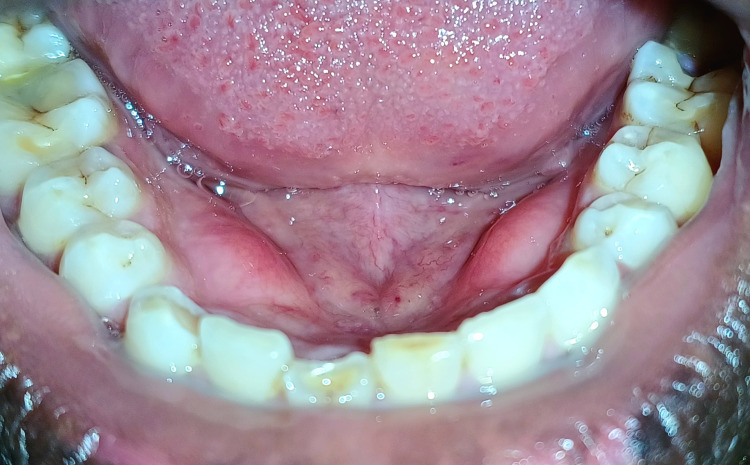 Figure 1
Peer Reviews
No public reviews on file for this paper yet. If you reviewed it on a platform where reviews are public (OpenReview, ICLR, NeurIPS, ICML), you can paste yours below so the community can read it here.
Videos
No videos yet. Explain this paper in a talk, walkthrough, or lecture? Add one.
Taxonomy
TopicsOropharyngeal Anatomy and Pathologies · Oral and Maxillofacial Pathology · Oral and gingival health research