Detection and counting of Leishmania intracellular parasites in microscopy images
Lariza María de la Caridad Portuondo-Mallet, Niurka Mollineda-Diogo, Rubén Orozco-Morales, Juan Valentín Lorenzo-Ginori

TL;DR
This paper presents a computational system for automatically detecting and counting Leishmania parasites in microscope images, improving efficiency and accuracy in drug testing.
Contribution
A novel image processing system for automatic detection and counting of Leishmania amastigotes in microscopy images is developed.
Findings
The system uses multilevel Otsu segmentation and watershed transform for object separation.
Classifier algorithms were evaluated for accurate amastigote detection.
The proposed method achieved high effectiveness with favorable sensitivity and precision metrics.
Abstract
Leishmaniasis is a disease caused by protozoan parasites of the genus Leishmania and has a high prevalence and impact on global health. Currently, the available drugs for its treatment have drawbacks, such as high toxicity, resistance of the parasite, and high cost. Therefore, the search for new, more effective, and safe drugs is a priority. The effectiveness of an anti-leishmanial drug is analyzed through in vitro studies in which a technician manually counts the intracellular form of the parasite (amastigote) within macrophages, which is slow, laborious, and prone to errors. To develop a computational system that facilitates the detection and counting of amastigotes in microscopy images obtained from in vitro studies using image processing techniques. Segmentation of objects in the microscope image that might be Leishmania amastigotes was performed using the multilevel Otsu method…
Genes, proteins, chemicals, diseases, species, mutations and cell lines named across the full text — each resolved to its canonical identifier and authoritative record.
Click any figure to enlarge with its caption.
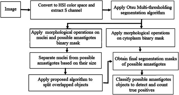 Figure 1
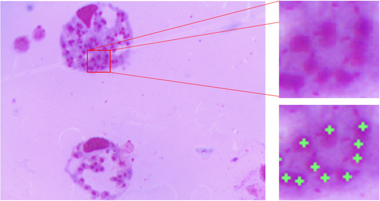 Figure 2
 Figure 3
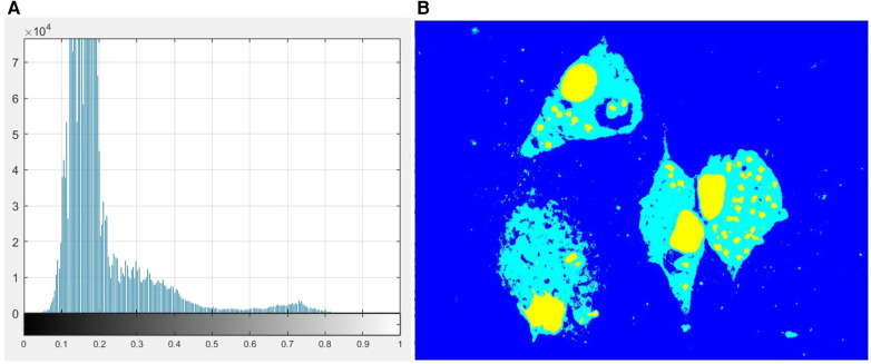 Figure 4
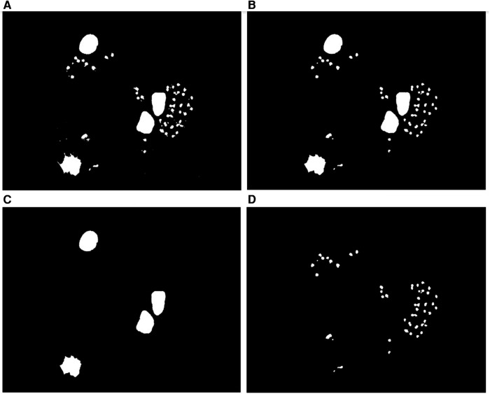 Figure 5
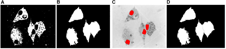 Figure 6
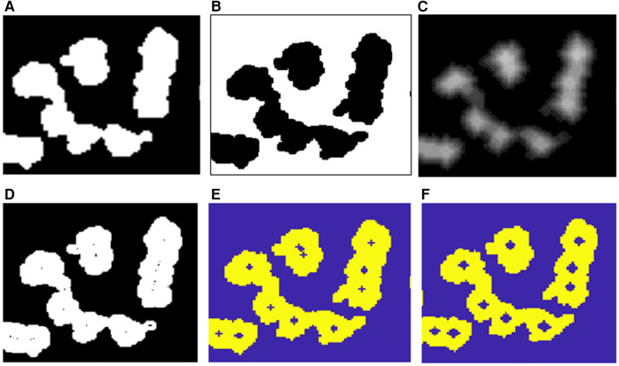 Figure 7
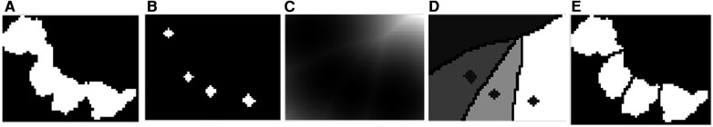 Figure 8
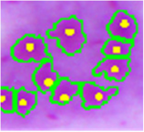 Figure 9
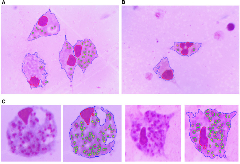 Figure 10
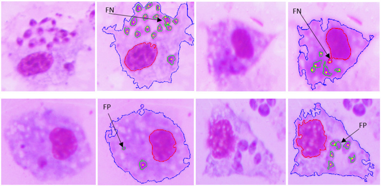 Figure 11
Peer Reviews
No public reviews on file for this paper yet. If you reviewed it on a platform where reviews are public (OpenReview, ICLR, NeurIPS, ICML), you can paste yours below so the community can read it here.
Videos
No videos yet. Explain this paper in a talk, walkthrough, or lecture? Add one.
Taxonomy
TopicsCell Image Analysis Techniques · Medical Image Segmentation Techniques · Image Processing Techniques and Applications