Histomorphometric Study of Non-critical Bone Defect Repair after Implantation of Magnesium-substituted Hydroxyapatite Microspheres
Jacqueline de Azerêdo Silva, George Gonçalves dos Santos, Iorrana Índira dos Anjos Ribeiro, Ana Maria Guerreiro Braga da Silva, Isabela Cerqueira Barreto, Marcos Almeida Matos, Maurício Andrade Barreto, Fúlvio Borges Miguel

TL;DR
This study examines how magnesium-substituted hydroxyapatite microspheres affect bone repair in rats, finding that both types of microspheres support healing but magnesium doesn't enhance it further.
Contribution
The study introduces a histomorphometric comparison of magnesium-substituted versus non-substituted hydroxyapatite in bone defect repair.
Findings
Biomaterials filled the defect and formed new osteoid matrix after 15 days.
At 45 days, all groups showed significant bone formation, but magnesium substitution did not improve results over standard hydroxyapatite.
Abstract
Objective The present study aims to analyze histomorphometrically the repair of a non-critical bone defect after implantation of hydroxyapatite (HA) microspheres substituted by magnesium (Mg). Methods Thirty rats were distributed into 3 experimental groups, evaluated at 15 and 45 days postoperatively: HAG (bone defect filled with HA microspheres); HAMgG (bone defect filled with HA microspheres replaced with 1 mol% Mg), and CG (bone defect without implantation of biomaterials). Results After 15 days, the biomaterials filled the entire defect extent, forming a new osteoid matrix between the microspheres. In the CG, this neoformation was restricted to the edges with the deposition of loose connective tissue with reduced thickness. At 45 days, new bone formation filled almost the entire extension of the bone defect in the 3 groups, with statistically significant osteoid deposition in…
Genes, proteins, chemicals, diseases, species, mutations and cell lines named across the full text — each resolved to its canonical identifier and authoritative record.
Click any figure to enlarge with its caption.
 Figure 1
 Figure 2
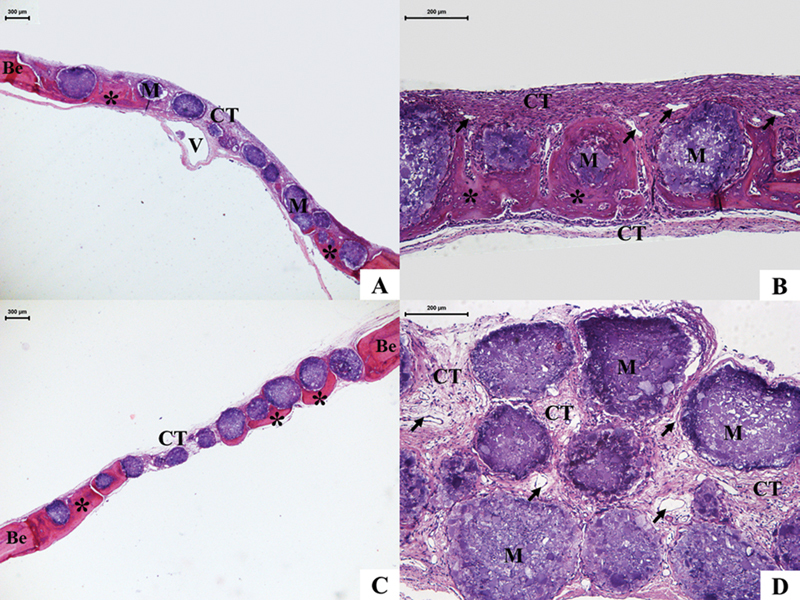 Figure 3
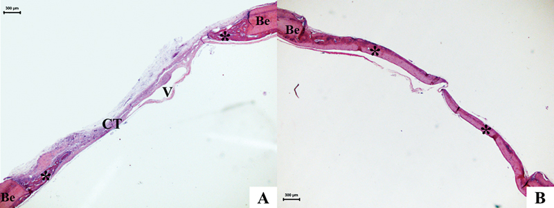 Figure 4
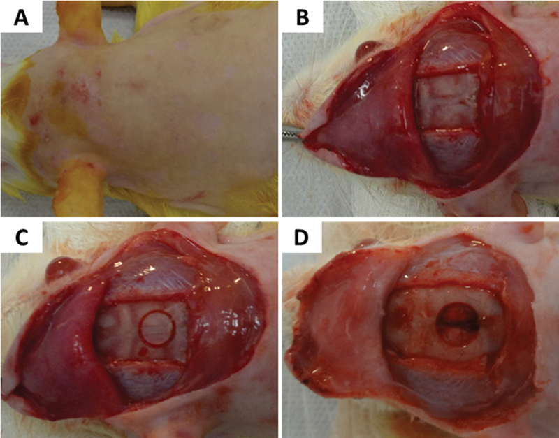 Figure 5
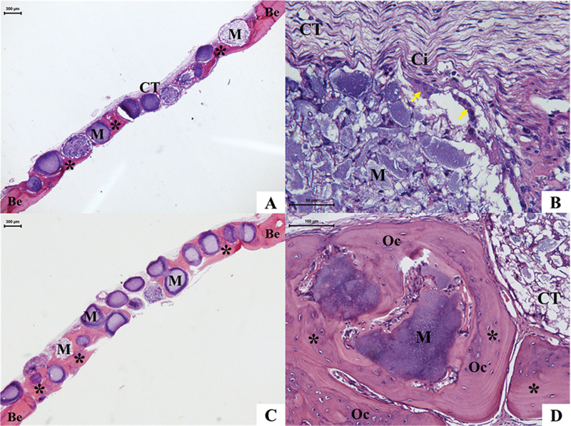 Figure 6
 Figure 7
 Figure 8
Peer Reviews
No public reviews on file for this paper yet. If you reviewed it on a platform where reviews are public (OpenReview, ICLR, NeurIPS, ICML), you can paste yours below so the community can read it here.
Videos
No videos yet. Explain this paper in a talk, walkthrough, or lecture? Add one.
Taxonomy
TopicsBone Tissue Engineering Materials · Orthopaedic implants and arthroplasty · Titanium Alloys Microstructure and Properties