Boundary-based registration improves sensitivity for detecting hypoperfusion in sporadic frontotemporal lobar degeneration
Sylvia Mihailescu, Quinn Hlava, Philip A. Cook, Maria Luisa Mandelli, Suzee E. Lee, Bradley F. Boeve, Bradford C. Dickerson, Maria Luisa Gorno-Tempini, Emily Rogalski, Murray Grossman, James Gee, Corey T. McMillan, Christopher A. Olm

TL;DR
Using boundary-based registration improves the detection of reduced blood flow in brain regions affected by frontotemporal lobar degeneration compared to traditional methods.
Contribution
Boundary-based registration (BBR) is shown to enhance sensitivity in detecting hypoperfusion in sporadic FTLD.
Findings
All registration methods showed significant hypoperfusion in frontal and temporal regions in patients compared to controls.
BBR methods performed similarly to manual registration in detecting hypoperfusion differences between patient groups.
Decreased perfusion in specific brain regions was associated with higher disease severity in sFTLD-TDP patients.
Abstract
Frontotemporal lobar degeneration (FTLD) is associated with FTLD due to tau (FTLD-tau) or TDP (FTLD-TDP) inclusions found at autopsy. Arterial Spin Labeling (ASL) MRI is often acquired in the same session as a structural T1-weighted image (T1w), enabling detection of regional changes in cerebral blood flow (CBF). We hypothesize that ASL-T1w registration with more degrees of freedom using boundary-based registration (BBR) will better align ASL and T1w images and show increased sensitivity to regional hypoperfusion differences compared to manual registration in patient participants. We hypothesize that hypoperfusion will be associated with a clinical measure of disease severity, the FTLD-modified clinical dementia rating scale sum-of-boxes (FTLD-CDR). Patients with sporadic likely FTLD-tau (sFTLD-tau; N = 21), with sporadic likely FTLD-TDP (sFTLD-TDP; N = 14), and controls (N = 50) were…
Genes, proteins, chemicals, diseases, species, mutations and cell lines named across the full text — each resolved to its canonical identifier and authoritative record.
Click any figure to enlarge with its caption.
 Figure 1
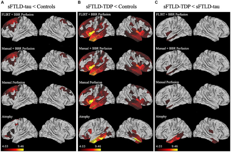 Figure 2
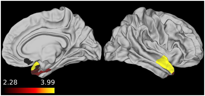 Figure 3
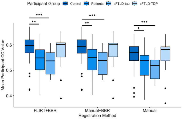 Figure 4
Peer Reviews
No public reviews on file for this paper yet. If you reviewed it on a platform where reviews are public (OpenReview, ICLR, NeurIPS, ICML), you can paste yours below so the community can read it here.
Videos
No videos yet. Explain this paper in a talk, walkthrough, or lecture? Add one.
Taxonomy
TopicsAdvanced Neuroimaging Techniques and Applications · Functional Brain Connectivity Studies · Advanced MRI Techniques and Applications