Color-coded circulation for visualizing swirling flow with a 3-dimensional helical stent in the superficial femoral artery
Mitsunari Maruyama, Aso Hiroya, Hisatoshi Araki, Rika Yoshida, Shinji Ando, Megumi Nakamura, Takeshi Yoshizako, Yasushi Kaji

TL;DR
This paper introduces a new imaging technique to visualize swirling blood flow after placing a 3D helical stent in a leg artery.
Contribution
The novelty is using color-coded circulation to visualize swirling flow from a 3D helical stent, which has not been reported before.
Findings
Color-coded circulation effectively visualizes swirling flow and blood vessel paths.
The technique may detect contrast agent distribution and blood flow missed by traditional angiography.
3D helical stents create swirling flow that increases wall shear stress and may prevent intimal hyperplasia.
Abstract
Color-coded circulation is a display method that generates dynamic color-coded images based on the time of arrival of contrast agents using parametric imaging to create video displays. By cyclically displaying information in color according to the arrival time of the contrast agent at each pixel, anatomical blood vessel paths and blood flow information can be simultaneously visualized. Three-dimensional (3D) helical stents increase wall shear stress due to swirling flow and prevent intimal hyperplasia. To the best of our knowledge, there are no reports on the visualization of this swirling flow using color-coded circulation. Here, we report the use of color-coded circulation to visualize the swirling flow following the placement of a 3D helical stent in the left superficial femoral artery. Color-coded circulation may facilitate the evaluation of contrast agent distribution and blood…
Genes, proteins, chemicals, diseases, species, mutations and cell lines named across the full text — each resolved to its canonical identifier and authoritative record.
Click any figure to enlarge with its caption.
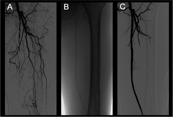 Figure 1
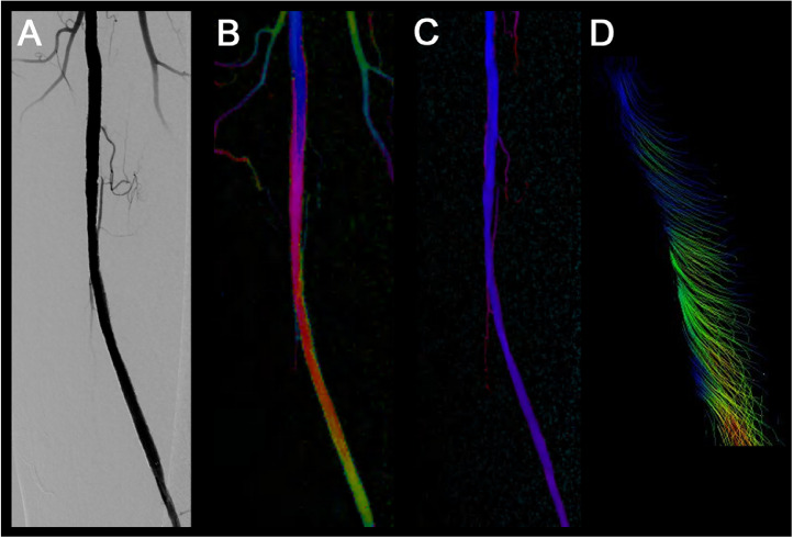 Figure 2
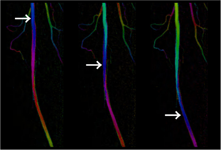 Figure 3
Peer Reviews
No public reviews on file for this paper yet. If you reviewed it on a platform where reviews are public (OpenReview, ICLR, NeurIPS, ICML), you can paste yours below so the community can read it here.
Videos
No videos yet. Explain this paper in a talk, walkthrough, or lecture? Add one.
Taxonomy
TopicsCoronary Interventions and Diagnostics · Peripheral Artery Disease Management · Cardiac and Coronary Surgery Techniques
Introduction
Parametric imaging produces a color-coded static map based on the time-density curve of digital subtraction angiography (DSA) contrast media density values. Color-coded circulation is a new imaging modality based on parametric imaging, which generates videos using stepwise shifts in the color scale [1]. This color-coded circulation allows repeated visualization of the flow of contrast agents.
There is a case report indicating that color-coded circulation was useful for evaluating the vessels that were mismatched with 2-dimensional angiosome in foot perfusion during endovascular therapy [1]. However, no other reports have reported on the usefulness of color-coded circulation.
The femoropopliteal artery is long and relatively straight under resting conditions, making it difficult for swirling flow to occur and resulting in low wall shear stress [2]. This creates an environment conducive to the development of atherocslerosis. Wall shear stress exceeding 1.5 Pa inhibits the formation of atheroma, while low wall shear stress less than 0.5 Pa is associated with the development of atherosclerosis and restenosis [3].
The 3-dimensional (3D) helical stent has a spiral shape. A spiral shape increases wall shear stress owing to swirling flow and reduces intimal hyperplasia [4,5]. In addition, the 3D helical shape minimizes the risk of vascular injury and stent fracture, which are common problems with straight stents in the superficial and popliteal arteries [6].
Herein, we report the use of color-coded circulation to visualize swirling flow following the deployment of a 3D helical stent in left superficial femoral artery (SFA) occlusion.
Case report
Three-D helical stent placement
A 90-year-old man presented with intermittent claudication of the left lower extremity. Computed tomography angiography revealed left SFA occlusion. Endovascular therapy was performed percutaneously through antegrade puncture of the left common femoral artery. The DSA image showed left SFA occlusion (Fig. 1A). Vessel preparation was performed by the use of a balloon (ULTRASCORE™ Scoring PTA balloon catheter), and stent deployment (Fig. 1B) was carried out using a 3D helical stent. A stent-in-stent technique was employed and stent placement was successful (Fig. 1C), with the left ankle-brachial index increasing from 0.54 to 0.78.Fig. 1. Three-dimensional (3D) helical stent placement. (A) Digital subtraction angiography (DSA) image before endovascular therapy. (B) 3D helical stent. (C) DSA image after stent placement. The stent was deployed using a 3D helical stent for left superficial femoral artery occlusion.Fig 1
Color-coded circulation and 4-dimensional (4D) flow magnetic resonance imaging (MRI)
Endovascular therapy was performed using an X-ray angiography system (Alphenix Sky). Color-coded circulation is a display method that shows static color-coded images based on the time of arrival of contrast agents through parametric imaging and presents them as videos on a workstation (Alphenix WorkStation Pro). By cyclically displaying information in color according to the arrival time of the contrast agent at each pixel, anatomical blood vessel paths and blood flow information can be simultaneously depicted. The DSA images were displayed using color-coded circulations when appropriate.
A streamlined image was obtained via 4D flow MRI using a 3-T MRI scanner (Ingenia Elition X) and an image analysis system (SYNAPSE VINCENT). Four-D flow MRI of the femur was performed using an electrocardiogram-gated, time-resolved, 3D phase-contrast MR sequence with 3-directional velocity encoding. The parameters were as follows: repetition time/echo time/flip angle of 6.4 ms/4.6 ms/12°, k-space segmentation/temporal resolution of 9.3 ms/51.5 ms, field of view of 380 × 380 mm, slice thickness of 3.0 mm, and voxel size of 1.83 × 3.26 × 3.00 mm³. The acquisition took approximately 3.08 min, producing 11 timeframes per R-R interval.
It was difficult to confirm the swirling flow in the DSA image (Fig. 2A) after stent placement. Layered colors were observed at the stent site (Fig. 2B, supplementary material: video displays); these were not present before stent placement (Fig. 2C). When 4D flow MRI of the stent exit site was performed later, swirling flow was observed on the streamlined image (Fig. 2D). Focusing on a single color, such as blue, revealed a spiral flow pattern (Fig. 3, white arrow, supplementary material: video displays).Fig. 2. Color-coded circulation image before and after stent placement. (A) Digital subtraction angiography (DSA) image after stent placement. (B) Color-coded circulation image after stent placement. (C) Color-coded circulation image before stent placement. (D) Streamline image by 4-dimensional flow magnetic resonance imaging at the stent exit site. It was difficult to confirm the presence of swirling flow using DSA (A). Layered colors are observed at the stent site (B, supplementary material: video displays) but are not present before stent placement (C). Swirling flow is visible in the streamlined image (D).Fig 2. Fig. 3Color-coded circulation image after stent placement. Focusing on a single color, such as blue, reveals a spiral flow pattern (white arrow, supplementary material: video displays).Fig 3
Discussion
The swirling flow in the BioMimics 3D vascular stent was visualized using color-coded circulation. Focusing on a single color revealed a spiral flow pattern. The layered colors in the images highlight the swirling flow within the stent. This color-coding technique facilitates the observation of contrast agents and blood flow, which are not easily detectable with DSA, by displaying them in distinct colors.
The BioMimics 3D stent is a 3D spiral-shaped device that creates a swirling flow within the stent. This design uses wall shear stress to promote long-term patency. When wall shear stress is applied, the vascular endothelial cells are not activated and do not secrete substances that promote arteriosclerosis, thereby suppressing intimal thickening and restenosis [5]. MIMICS-3D is a real-world European registry. After propensity score matching, clinically driven target lesion revascularization and primary patency with a 3D helical stent might not be influenced by lesion background, including chronic total occlusion, chronic limb-threatening ischemia, or highly calcified lesions [7]. The MIMICS 3D registry also demonstrated satisfactory results, comparable to drug-eluting stents, in patients with 3D helical stents [8].
Conclusion
We report the use of color-coded circulation to visualize the swirling flow following the placement of a 3D helical stent in the left SFA. Color-coded circulation may facilitate the evaluation of contrast agent distribution and blood flow, which may otherwise go undetected with DSA.
Patient consent
Informed consent was obtained for the publication of this case report.
Author contributions
All authors have made significant contributions to the manuscript and approved the final version for publication.
The reference list from the paper itself. Each links out to its DOI / PubMed record.
- 1Kawarada O.Color coded circulation in the field of infrapopliteal intervention Cardiovasc Interv Ther 361202113113310.1007/s 12928-020-00652-832077009 · doi ↗ · pubmed ↗
- 2Wood NB Zhao SZ Zambanini A Jackson M Gedroyc W Thom SA Curvature and tortuosity of the superficial femoral artery: a possible risk factor for peripheral arterial disease J Appl Physiol (1985)101520061412141810.1152/japplphysiol.00051.200616825527 · doi ↗ · pubmed ↗
- 3Malek AM Alper SL Izumo S.Hemodynamic shear stress and its role in atherosclerosis JAMA 2822119992035204210.1001/jama.282.21.203510591386 · doi ↗ · pubmed ↗
- 4Caro CG Seneviratne A Heraty KB Monaco C Burke MG Krams R Intimal hyperplasia following implantation of helical-centreline and straight-centreline stents in common carotid arteries in healthy pigs: influence of intraluminal flow J R. Soc Interface.108920132013057810.1098/rsif.2013.0578 PMC 380854524132200 · doi ↗ · pubmed ↗
- 5Sullivan TM Zeller T Nakamura M Caro CG Lichtenberg M.Swirling flow and wall shear: evaluating the Bio Mimics 3D helical centerline stent for the femoropopliteal segment Int. J Vasc. Med.20182018979517410.1155/2018/9795174 PMC 584635729682350 · doi ↗ · pubmed ↗
- 6Rits Jvan Herwaarden JA Jahrome AK Krievins D Moll FL.The incidence of arterial stent fractures with exclusion of coronary, aortic, and non-arterial settings Eur J Vasc. Endovasc Surg.363200833934510.1016/j.ejvs.2008.05.00518602847 · doi ↗ · pubmed ↗
- 7Rammos C Zeller T Piorkowski M Deloose K Hertting K Sesselmann V The Bio Mimics 3D helical centreline nitinol stent in chronic limb threatening ischaemia and complex lesions: three year outcomes of the MIMICS-3D Registry Eur J Vasc Endovasc Surg 676202492393210.1016/j.ejvs.2024.02.04338447693 · doi ↗ · pubmed ↗
- 8Piorkowski M Zeller T Rammos C Deloose K Hertting K Sesselmann V Bio Mimics 3D stent in femoropopliteal lesions: 3-year outcomes with propensity matching for drug-coated balloons J Cardiovasc Dev Dis 103202312610.3390/jcdd 1003012636975890 PMC 10051351 · doi ↗ · pubmed ↗