A Morphometric Study of the Retromolar Fossa and the Incidence of the Retromolar Foramen in Dry Human Mandibles
Dwiz Dwivedi, Noor Us Saba, Pratibha Shakya, Heena Singh, Navneet Chauhan

TL;DR
This study measures the retromolar fossa in human mandibles and finds a 60% incidence of retromolar foramina, important for avoiding surgical complications.
Contribution
The study provides new morphometric data and incidence rates of retromolar foramina in dry human mandibles.
Findings
The medial border of the retromolar trigone showed statistically significant differences between right and left sides (P=0.02).
Retromolar foramina were found in 60% of mandibles, with six unilateral and 12 bilateral cases.
Foramina were closer to the anterior border of the retromolar trigone than the ramus or lingula.
Abstract
Introduction: The mandible is the largest and strongest facial bone which plays a crucial role for various surgeries and diagnostic imaging. The retromolar fossa, located behind the third molar socket on each side, was observed for morphometry of anterior, medial, and posterior borders. The present study aimed to assess the retromolar fossa and the presence of retromolar foramen. Methods: This cross-sectional study was conducted on 30 dry mandibles of adult humans of unknown sex; morphometry of retromolar foramen was done using three parameters: anteriorly, laterally, and posteroinferiorly. Dimensions of the retromolar trigone were seen with notable differences between the right and left sides. Results: Dimensions of the anterior border had a mean of 12.34±1.175 mm on the right side and 12.56±1.46 mm on the left side. The mean of the medial border of the trigone on the right side was…
Genes, proteins, chemicals, diseases, species, mutations and cell lines named across the full text — each resolved to its canonical identifier and authoritative record.
Click any figure to enlarge with its caption.
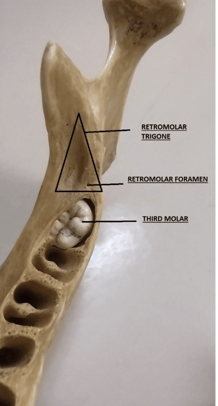 Figure 1
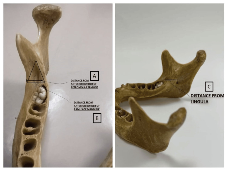 Figure 2
| Left Side | Right Side | |||||
| Mandible | Anterior border | Medial border | Lateral border | Anterior border | Medial border | Lateral border |
| 1 | 13.03 | 21.1 | 21.05 | 13.05 | 18.85 | 13.4 |
| 2 | 12.4 | 17.15 | 16.45 | 12.2 | 19.4 | 15.3 |
| 3 | 11.05 | 23.15 | 26.05 | 11.1 | 20.35 | 19.15 |
| 4 | 11.1 | 26.45 | 24.2 | 11.2 | 23.2 | 27.5 |
| 5 | 10.1 | 18.2 | 18.5 | 10.1 | 10.3 | 19.8 |
| 6 | 12.3 | 21.1 | 19.1 | 13.3 | 25.15 | 22.7 |
| 7 | 14.5 | 25.1 | 21.5 | 11.1 | 20.5 | 18.75 |
| 8 | 14.7 | 20.9 | 21.8 | 12.8 | 20.1 | 18.3 |
| 9 | 11.1 | 17.35 | 11.1 | 12.8 | 18.1 | 16.1 |
| 10 | 14.8 | 22.95 | 22.2 | 12.2 | 18.9 | 17.4 |
| 11 | 11.6 | 19.1 | 16.75 | 11.7 | 19.45 | 11.05 |
| 12 | 13.35 | 21.4 | 18.05 | 13.25 | 20.05 | 17.2 |
| 13 | 13.05 | 19.05 | 21.1 | 12.7 | 19.1 | 15.9 |
| 14 | 11.4 | 19.05 | 18.55 | 11.65 | 19.35 | 16.1 |
| 15 | 14.4 | 24.1 | 22.45 | 13.25 | 22.15 | 21.2 |
| 16 | 14.3 | 23.4 | 24.05 | 15.05 | 24.05 | 25.2 |
| 17 | 9.55 | 20.35 | 21.05 | 11.05 | 20.65 | 18.1 |
| 18 | 14.1 | 24.5 | 21.75 | 14.65 | 23.6 | 19.6 |
| 19 | 14.6 | 20.8 | 19.7 | 13.8 | 19.55 | 17.05 |
| 20 | 12.2 | 21.85 | 20.75 | 11.7 | 20.4 | 18.05 |
| 21 | 13.6 | 22.05 | 22.8 | 11.45 | 22.4 | 21.05 |
| 22 | 14.2 | 22.85 | 21.7 | 13.45 | 20.55 | 20.25 |
| 23 | 13.65 | 24.8 | 23.9 | 14.65 | 26.1 | 23.1 |
| 24 | 12.2 | 17.45 | 21.5 | 12.3 | 21.23 | 26.3 |
| 25 | 11.3 | 26.15 | 18.15 | 11.4 | 18.4 | 16.7 |
| 26 | 11.1 | 19.3 | 24.3 | 11.5 | 18.5 | 18.5 |
| 27 | 13.2 | 20.15 | 13.2 | 12.5 | 23.2 | 16.8 |
| 28 | 10.8 | 21.4 | 14.5 | 11.1 | 22.75 | 16.3 |
| 29 | 12.6 | 24.35 | 11.25 | 12.2 | 17.2 | 15.5 |
| 30 | 13.3 | 23.45 | 12.4 | 12.8 | 20.4 | 17.6 |
| Left Side | Right Side | ||||||
| S. no. of bone | Distance from the anterior border | Distance from the anterior border of the ramus | Distance from lingula | S. no. of bone | Distance from the anterior border | Distance from the anterior border of the ramus | Distance from lingula |
| 1 | Just at anterior border | 9.7 | 13.65 | 1 | 1.35 | 9.15 | 13.2 |
| 3 | 7.6 | 10.7 | 15.5 | 3 | Just behind anterior border | 10.1 | 21.5 |
| 4 | 7.05 | 10.1 | 12.5 | Just behind anterior border | 11.1 | 20.1 | |
| 5.1 | 9.35 | 12.2 | 4 | 7.6 | 10.1 | 13.5 | |
| 5.1 | 7.35 | 13.3 | 5 | 2.05 | 9.8 | 12.05 | |
| 7 | 9.8 | 15.8 | 9.4 | 7 | 10.2 | 8.85 | 13.7 |
| 8 | Just behind anterior border | 12.25 | 15.3 | 11 | 1.2 | 8.1 | 14.55 |
| Just at anterior border | 12.05 | 14.65 | 13 | 1.9 | 8.75 | 13.05 | |
| 11 | 1.05 | 8.45 | 13.1 | 6.1 | 5.1 | 11.05 | |
| 12 | Just at anterior border | 11.1 | 13.8 | 15 | Just at anterior border | 10.5 | 14.9 |
| 0.5 | 6.65 | 13.5 | 17 | Just behind anterior border | 9.1 | 12.3 | |
| 13 | Just at anterior border | 10.05 | 13.5 | 18 | Just at anterior border | 12.05 | 18.5 |
| 5.4 | 5.05 | 11.75 | 23 | 2.25 | 10.8 | 13.05 | |
| 17 | Just behind anterior border | 8.25 | 13.05 | 25 | 8.65 | 6.8 | 15.7 |
| 22 | 4.45 | 8.75 | 14.35 | 27 | 3 | 11 | 17.75 |
| 6.5 | 9.2 | 14.15 | 29 | 2.3 | 12.5 | 12.4 | |
| 23 | 4.05 | 10.05 | 15.6 | 30 | 8.5 | 9.35 | 13.3 |
| 9.85 | 7.05 | 15.5 | - | - | - | - | |
| 25 | 3.25 | 10.1 | 12.5 | - | - | - | - |
| 27 | 2.75 | 8.9 | 14.35 | - | - | - | - |
| 29 | 4.5 | 7.1 | 13.35 | - | - | - | - |
| 30 | 6.75 | 11.1 | 11.5 | - | - | - | - |
| Right Side (Mean) | Right Side (SD) | Left Side (Mean) | Left Side (SD) | P-Value (Single Side) | |
| Anterior border | 12.34526 | 1.175798 | 12.56568 | 1.462238 | 0.114703 |
| Medial border | 20.2298 | 2.845902 | 21.47896 | 2.570257 | 0.022268 |
| Lateral border | 18.33479 | 3.564504 | 19.20686 | 3.928224 | 0.074621 |
| Left Side (Mean) | Left Side (SD) | Right Side (Mean) | Right Side (SD) | |
| Distance from anterior border | 5.23125 | 3.287853153 | 4.591666667 | 3.517111532 |
| Distance from anterior border of ramus | 9.504545455 | 2.28545656 | 9.597058824 | 1.829486829 |
| Distance from lingula | 13.47727273 | 1.494890287 | 14.74117647 | 2.996520654 |
| Study | Country | Sample Size | Presence of Retromolar Foramen |
| Present study | India | 30 | 18 (60%) |
| Schejtman et al. (1967) [ | Argentina | 18 | 13 (72%) |
| Sawyer et al. (1991) [ | USA | 234 | 18 (7.69%) |
| Priya et al. (2005) [ | India | 157 | 20 (12.74%) |
| Kawai et al. (2012) [ | Japan | 46 | 24 (52.17%) |
| Galdámes et al. (2008) [ | Brazil | 294 | 38 (12.92%) |
| Athavale et al. (2013) [ | India | 71 bones and 10 cadavers | 10 (14.08%) |
| Potu et al. (2014) [ | India | 94 | 11 (11.70%) |
| Ren et al. (2023) [ | China | 123 | 39 (31.70%) |
Peer Reviews
No public reviews on file for this paper yet. If you reviewed it on a platform where reviews are public (OpenReview, ICLR, NeurIPS, ICML), you can paste yours below so the community can read it here.
Videos
No videos yet. Explain this paper in a talk, walkthrough, or lecture? Add one.
Taxonomy
TopicsDental Radiography and Imaging · Bone Tumor Diagnosis and Treatments · Facial Trauma and Fracture Management
Introduction
The mandible is the largest and strongest bone of the face. It consists of two ramus and a body. The upper border of the body has a "U" shaped alveolar arch to embed the teeth of the lower jaw in their sockets [1]. Retromolar fossae are two triangular areas present behind the socket of the third molar on each side. These are small depressions, having a temporal crest medially, a continuation of the anterior border of the ramus of the mandible laterally, and the posterior margin of the third molar anteriorly [2].
Retromolar foramina and their continuation into the retromolar canal can also be seen in these fossae. The presence of foramina and canals in retromolar trigone has clinically important neurovascular passages [3,4]. Knowledge of the retromolar fossa and the presence of retromolar foramen is important for surgeons in orthognathic surgeries, extraction of the third molar, periodontal wedge procedures, sagittal split osteotomy as well as for radiologists. It is a potential space to accommodate various neoplasms and oral cancers [5,6]. The present study aimed to assess the retromolar fossa and the presence of retromolar foramen.
Materials and methods
This cross-sectional study was conducted on 30 dry mandibles of adult humans of unknown sex in the Department of Anatomy, King George’s Medical University, Lucknow, India. This study was approved by the Institutional Ethics Committee with ref. code: XIX-PGTSC-IIB-IMR-S/P1 dated 05/08/2023. Mandibles with sockets of third molar teeth were selected, whereas distorted and damaged bones were excluded.
Morphometry of the retromolar fossa was done bilaterally on the mandible with the help of the vernier caliper. The anterior, medial, and lateral borders of the retromolar fossa were measured in millimetres (mm) to make a trigone just behind the third molar tooth (Figure 1).
Retromolar trigone showing its medial border, lateral border, and anterior border.
The location of the retromolar foramen was measured in a distance of millimetres (mm) from the anterior border, from the ramus of the mandible laterally, and from the lingula posteroinferiorly (Figure 2).
Measurements taken for the location of the retromolar foramen: (A) from the anterior border of the retromolar trigone; (B) from the anterior border of the ramus of the mandible; (C) from the lingula of the mandible on the same side as the retromolar foramen.
The study was analysed by using single tail-paired t-test in an Excel file (Microsoft® Corp., Redmond, WA, USA). Mean, standard deviation (SD) and P-value were calculated.
Results
Measurements of retromolar trigone (Table 1) and retromolar foramen were taken and tabulated for both the right and left sides of the mandibles (Table 2).
Borders of retromolar trigone
The length of the anterior border of retromolar trigone varies from 10.1 mm to 15.05 mm on the right side (mean=12.34±1.175), and 9.55 mm to 14.80 mm on the left side (mean=12.56±1.46) respectively. Similarly, the length of the medial border of the trigone on the right side was between 10.3 mm and 25.15 mm (mean=20.23±2.84), and 17.15 mm to 26.45 mm (mean=21.48±2.57) on the left side. Lastly, the length of the lateral border varies from 11.05 mm to 27.5 mm on the right side (mean=18.33±3.56), and 11.1 mm to 26.05 mm on the left side (mean=19.21±3.93). The P-value of the medial border was found to be statistically significant (P=0.02) (Table 3).
Retromolar foramen
The occurrence of retromolar foramen was seen in 18 out of 30 mandibles i.e., 60%. They were unilateral in six mandibles and bilateral in 12 mandibles. Unilateral double retromolar foramen was present on the left side in three mandibles and unilateral single retromolar foramen was seen on the right side in the other three mandibles. Three retromolar foramen on the left side were also observed in one of the mandibles having foramen bilaterally. These foramina were generally present near the anterior border of the retromolar trigone i.e., near the third molar socket. They were closer to the anterior border of the ramus in comparison to the lingula (Table 2).
The average distance of the foramen from the anterior border of the trigone was 4.59±3.52 mm on the right side, and 5.23±3.29 mm on the left side. The distance from the anterior border of the ramus was 9.59±1.83 mm on the right side, and 9.50±2.29 mm on the left side. This distance of foramen from the lingula was 14.74±2.99 mm on the right side, and 13.48±1.50 mm on the left side (Table 4).
Discussion
Various studies in India [7-9] and around the globe as Japan [10], China [11], Brazil [12], and the USA [13] have been carried out to observe the retromolar trigone and find the incidence of retromolar foramen. A wide range (8-72%) of variation was observed in comparing the findings of different studies. The probable cause of these differences can be related to the development of the mandible, and having many nerve canals in the initial stages of the embryonic period. These multiple nerve channels may or may not disappear in a later stage, leaving a range of presentations from a single canal in the mandible to a combination of numerous small passages taking origin from the chief mandibular canal [5]. Other causes might include the differences in populations in terms of their ethnicity, region, and climatic conditions.
In our study, the size of the trigone was observed in parallel with various other studies. Dimensions of the anterior border were similar to the observations of Potu et al. [9], whereas dimensions of the medial and lateral walls were quite less in the present study. This difference is attributed to the sample difference and regional differences in their study. The P-value in the present study was significant for the dimensions of the medial border of the trigone (Table 3).
The frequency of observation of retromolar foramina was 60% in the present study which was in line with the findings of Kawai et al. [10] and Schejtman et al. [14]. Incidence was very low in the studies of Priya et al., Athavale et al., Potu et al., Galdámes et al. and Sawyer et al. [7-9,12,13] as compared to the current study (Table 5). The retromolar foramen is an anatomical variation consisting of neurovascular supply through the retromandibular canal [15]. Any damage to the supply during surgeries can cause various complications as reported by Singh [16]. Singh reported complete paraesthesia of the buccal mucosa due to injury in the retromolar region during the third molar extraction. Branching of the nerves and vessels from the mandibular canal to exit from the retromandibular foramen might be responsible for incomplete anaesthesia of the retromolar region. The presence of variations in the vasculature in the retromandibular foramen and retromandibular canal may cause incomplete anaesthesia in this region during surgeries [3]. It is important to consider the presence of retromandibular foramen while performing various oral surgeries specifically during third molar extraction and sagittal split osteotomy. Also, the neoplasms of the retromolar region tend to metastasise through the vasculature into the surrounding tissues [4,17].
Conclusions
Awareness of the anatomical variations in the dimensions of the retromolar fossa and the incidence of retromolar foramen helps surgeons and anaesthetists to do various procedures in the oral cavity. The cancer of the trigone as well as the extraction of the third molar requires information regarding its dimensions and the presence of retromolar foramen within the trigone.
The reference list from the paper itself. Each links out to its DOI / PubMed record.
- 1Gray’s Anatomy - The Anatomical Basis of Clinical Practice 42nd Edition Standring S 667669 Amsterdam Elsevier 2020 https://www.amazon.in/Grays-Anatomy-Anatomical-Clinical-Practice/dp/0702077054
- 2Mandibular retromolar foramen and canal - a systematic review and meta-analysis Ann Maxillofac Surg Shah SP Mehta D 4444491020203370859310.4103/ams.ams_19_20PMC 7944007 · doi ↗ · pubmed ↗
- 3Clinical Anatomy and significance of the retromolar foramina and their canals: a literature review Cureus Truong MK He P Adeeb N Oskouian RJ Tubbs RS Iwanaga J 09201710.7759/cureus.1781 PMC 573201029255660 · doi ↗ · pubmed ↗
- 4Diagnostic approach to retromolar trigone cancer by multiplanar computed tomography reconstructions Can Assoc Radiol J Mazziotti S Pandolfo I D'Angelo T 3353446520142526737610.1016/j.carj.2014.04.001 · doi ↗ · pubmed ↗
- 5Incidence and anatomical properties of retromolar canal in an Iranian population: a cone beam computed tomography study Int J Dent Nikkerdar N Golshah A Norouzi M Falah-Kooshki S 9178973202020203221104810.1155/2020/9178973 PMC 7085402 · doi ↗ · pubmed ↗
- 6Cone beam CT findings of retromolar canals: report of cases and literature review Imaging Sci Dent Han SS Park CS 3093124320132438007210.5624/isd.2013.43.4.309PMC 3873321 · doi ↗ · pubmed ↗
- 7Retromolar foramen Indian J Dent Res Priya R Manjunath KY Balasubramanyam Balasubramanyam 1516162005 https://pubmed.ncbi.nlm.nih.gov/16375231/16375231 · pubmed ↗
- 8Bony and cadaveric study of retromolar region People's J Sci Res Athavale SA Vijaywargia M Deopujari R Kotgirwar S 141862013 https://pjsr.org/July 13_Dr.%20S.A.%20Athavale.pdf