DNA mismatch repair system expression in salivary gland tumors: A Systematic Review
Geórgya Mayara Travasso Torres Alves, Raisa Jordana Geraldine Severino-Lazo, Gleyson Kleber do Amaral-Silva, Fabiana Moura da Motta Silveira, Marianne de Vasconcelos Carvalho

TL;DR
This review examines how DNA mismatch repair proteins are expressed in salivary gland tumors and finds reduced levels linked to tumor development.
Contribution
The study systematically reviews MMR protein expression in salivary gland tumors, highlighting its potential role in oncogenesis.
Findings
Reduced expression of MMR proteins like hMSH2, hMLH1, hMSH3, and hMSH6 was observed in salivary gland tumors.
The reduction in MMR proteins correlates with both benign and malignant tumor development.
Further research is needed to clarify the role of MMR protein expression as a biomarker.
Abstract
The DNA mismatch repair (MMR) system serves as a sophisticated guardian of the precise functioning of the human genome. Dysregulation within this system is linked to the oncogenesis process. Reduced expression of MMR system proteins identified in salivary gland tumors (SGTs) suggests an increased risk of tumoral occurrence. This study aims to analyze the expression of MMR proteins in SGTs and discuss the relevance of this association to the development of these neoplasms. This review was conducted following the PRISMA guidelines and was registered in PROSPERO (CRD42023465590). A comprehensive search of the PubMed/MEDLINE, Web of Science, Scopus, Embase, and ProQuest (non-peer reviewed platform) was performed to answer the question “Do DNA MMR system proteins exhibit expression in SGTs?”. The methodological quality of the selected studies was assessed using the JBI’s Critical Appraisal…
Genes, proteins, chemicals, diseases, species, mutations and cell lines named across the full text — each resolved to its canonical identifier and authoritative record.
Click any figure to enlarge with its caption.
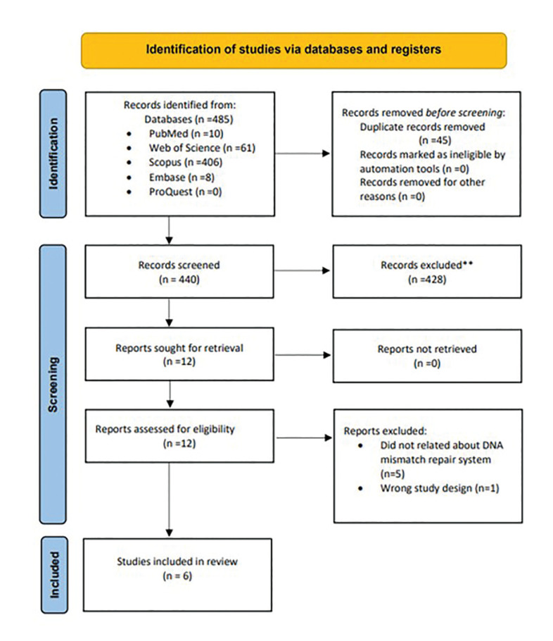 Figure 1
Peer Reviews
No public reviews on file for this paper yet. If you reviewed it on a platform where reviews are public (OpenReview, ICLR, NeurIPS, ICML), you can paste yours below so the community can read it here.
Videos
No videos yet. Explain this paper in a talk, walkthrough, or lecture? Add one.
Taxonomy
TopicsReligious and Theological Studies · Theology and Canon Law Studies
Introduction
Salivary gland tumors (SGTs) constitute a heterogeneous group of lesions characterized by morphological diversity and inherent biological behaviors, representing approximately 3% to 10% of neoplasms within the head and neck region (1,2). According to the World Health Organization’s (WHO) Classification of Head and Neck Tumors, both major and minor salivary glands exhibit a remarkable diversity of alterations in differentiation patterns and architectural changes. This results in an overall annual incidence, considering all SGTs, ranging from 0.4 to 13.5 cases per 100,000 inhabitants (3). Due to their complex clinicopathological features, accurate diagnosis can be challenging.
Understanding the molecular biology of SGTs is significant for differential diagnosis and appropriate clinical management (4,5). In the fifth edition of the WHO classification, molecular data and biomarker studies have become widely referenced for both malignant and benign salivary neoplasms. These studies elucidate tumor-specific genetic rearrangements and have proved to be an important direction in comprehending these variable tumors (3-5). In this context, the evaluation of various proteins expressed in these tumors may provide insights into oncogenesis, pathogenesis, and open new pathways for the development of target-specific therapies.
Precise DNA replication is essential for maintaining genomic integrity and transmitting genetic information accurately. Dysfunctions in the repair mechanisms, which safeguard cells against potential mutation burden, are associated with an increased risk of developing various tumors. The DNA mismatch repair (MMR) system acts as a sophisticated protector of the human genome, encoding a set of error-correcting proteins involved in the replication of genetic material, thus preventing mutations (6-7). This system comprises a cluster of genes whose main protein subunits work together: MutSα (hMSH2-hMSH6), MutSβ (hMSH2-hMSH3) and MutLα (hMLH1-hPMS2). These complexes operate at the initiation of the MMR pathway in the cell cycle by preferentially detecting base-pair mismatches, and major insertion/deletion mispairs loops, and subsequently executing the excision of these errors (6,8,9). Consequently, the role of this system demonstrates relevance and cannot be underestimated.
Despite the diagnostic diversity, the oncogenesis of SGTs remains poorly understood. However, the reduced expression of proteins encoded by the DNA MMR system has been identified in SGTs and is believed by some authors to be linked with the development of lesions. In contrast, conflicting data regarding the response of this system require further investigation. Hence, the aim of this systematic review of published studies is to assess the involvement of the DNA MMR system protein expression in SGTs and discuss the relevance of this association to the development of these neoplasms. Thereby, the insights acquired from the study of the expression of DNA MMR system proteins in SGTs assume particular significance.
Material and Methods
- Protocol and Registration
The present article followed the Preferred Reporting Items for Systematic Reviews and Meta-Analyses (PRISMA) checklist (10) and was registered in the International Prospective Register of Systematic Reviews (PROSPERO) under the registration number CRD42023465590.
- Eligibility Criteria
This systematic review followed a question formulated based on the “population, exposure, comparison, outcome, and study design of studies” (PECOS) criteria. The research question was: “Do DNA MMR system proteins exhibit expression in SGTs?”. The inclusion criteria addressed observational studies (cohort or cross-sectional) that evaluated the expression of DNA MMR system in patients with benign and/or malignant SGTs, describing the methods employed to detect the protein subunits comprising the system. Exclusion criteria included case reports, reviews, theses, dissertations, book chapters, studies reported in animals, studies with an uncertain diagnosis of SGTs, and those that did not elucidate the detection method for the MMR system.
- Search Strategy
A comprehensive search in electronic databases including PubMed/MEDLINE, Web of Science, Scopus, Embase, and ProQuest platform (non-peer-reviewed literature) was performed. No restrictions were imposed based on language or date. The search strategy is detailed in Table 1. Additionally, hand searching was performed in the reference list of included studies and in specific international journals in the field. To select included studies, the titles and abstracts of the articles were reviewed. Duplicates were removed using the (Rayyan Management software). Any discrepancies in the selection process between the investigators were resolved by a third researcher through discussion to reach a consensus.
- Data Collection Process
One researcher (GMTTA) collected data from the included articles, while a second researcher (RJGSL) checked all extracted data. The collected variables included the author, year, country, sample size, gender, mean age, location of tumors, pathological diagnosis of SGTs, type of tumors, the DNA MMR biomarkers, and the amount of the expression of DNA MMR system proteins.
Results
- Screening and Selection of the Papers
The comprehensive search, detailed in Fig. 1, initially identified a total of 485 relevant studies across various databases: 10 in PubMed/MEDLINE, 61 in Web of Science, 406 in Scopus, 8 in Embase, and 0 in ProQuest (non-peer-reviewed platform). After eliminating duplicates, 12 articles were selected for full-text analysis. Six studies met the inclusion criteria and underwent data extraction. Inter-rater agreement, assessed by Cohen’s Kappa coefficient during the article selection phase, demonstrated an “almost perfect agreement” between reviewers (kappa = 0.99). The methodological quality and risk of bias was assessed using the JBI Critical Appraisal Checklist for Analytical Cross-Sectional Studies (11). The JBI analysis is described in Table 2.
Figure 1PRISMA flow diagram showing the study identification and selection process.
- Description of the Studies
A detailed overview of the included studies is presented in Table 3. This systematic review analyzed six cross-sectional studies published between 2001 and 2022. All the included studies evaluated the expression of the DNA MMR system in benign and malignant SGTs (12-17). The total sample comprised 236 tissue specimens from patients, with 50 males and 92 females. The mean age ranged from 36.29 to 60 years. The majority of tumors were located in the major salivary glands, with pleomorphic adenoma being the most frequently diagnosed benign pathological type in 104 cases (44.06%). Adenoid cystic carcinoma was the most frequently diagnosed malignant pathological type, representing 32 cases (13.52%). Benign lesions constituted the majority with 142 cases (60.15%), as opposed to malignant lesions evaluated in 84 cases (39.85%).
- DNA Mismatch Repair Proteins Expression
Regarding the percentage of cases, variable expression of the DNA MMR system was observed, ranging from 0% to 100% between benign and malignant SGTs. The range indicated a relevant heterogeneity between SGTs. However, considering the percentage of marked cells in SGTs, the expression of DNA MMR biomarkers showed noTable features. Tobón-Arroyave et al. (15) focusing only on benign SGTs, reported a underexpression of marked cells for biomarkers, ranging from 4.15 ± 3.05% to 7.27 ± 2.50% which aligned with the total mean. Castrilli et al. (12) showed important reduction levels of marked cell expression in benign STGs, varying from 14.0 ± 12.6% to 31.1 ± 22.6% which also showed a decreased expression reported by the total mean. Statistical analysis revealed a significant difference in hHMS2 protein expression among the benign lesion groups used in both studies (Mann-Whitney test, p=0.003). Similarly, significant difference was observed concerning hMLH1 protein expression (p=0.000). Benign SGTs showed reduced expression of DNA MMR system proteins. Soares et al. (14) focusing only on malignant SGTs, reported an expression ranging from 56.5% to 62.5%. However, they showed a reduction in the total mean expression in the labeled cells, from 29.25% to 45.25%. Castrilli et al. (12) comprising a wide range of malignant SGTs, showed a similar balanced reduction in the marked cells as emphasized by the total mean expression, from 56.1 ± 31.6% to 27.9 ± 26.0%. Significant differences were observed in hHMS2 and hMLH1 proteins comparing malignant lesions in both studies (p=0.002) and (p=0.000), respectively. Malignant SGTs also showed reduced expression of DNA MMR system proteins. Nevertheless, Amaral-Silva et al. (13) showed a decreased levels of marked cells expression in both malignant and benign SGTs, ranging from 4.27 ± 5.35% to 46.47 ± 18.06% and 1.73 ± 1.20% to 46.37 ± 22.35%, respectively. The total mean for hMSH3 (6.25 ± 6.95%) and hMSH6 (10.81 ± 6.45%) also reported a lower proportion of malignant SGTs compared to hMSH2 and hMLH1. The available data on DNA MMR system expression is summarized in Table 4. Detailed results of total mean expression in marked cells are presented in the Supplementary Material.
Discussion
The DNA MMR system is widely recognized as an essential mechanism for genome stability, playing a pivotal role in repairing base-base or insertion-deletion errors during DNA replication (6,7). This process has a significant impact on mutagenic suppression in dividing cells (8,12-14,18,19). Overall, we found a reduced expression of proteins encoded by the DNA MMR system in the percentage of marked cells. This result suggests an interesting association with the development of these tumors since the uncorrected spontaneous mutation by the system induces an increase in cell proliferation and tumor invasion. Furthermore, increased risk through MMR deficient expression has been observed in important syndromic manifestations such as Lynch Syndrome, Muir-Torre Syndrome, and Turcot Syndrome (7,8,9,20).
Immunohistochemistry (IHC) has been a potent tool for scrutinizing the deficient protein expression of DNA MMR subunits in diverse tumors for more than two decades (20-22). All studies in this systematic review used this method to evaluate the DNA MMR proteins expression in SGTs (12-17). This review showed that the system expression of SGTs data was typically categorized into the positive percentage of marked cells and the positive percentage of cases. When, considering the percentage of cases without accounting for the percentage of cells marked by each subunit expressing this repair, implies the creation of a gap in the understanding of this phenomenon. Conversely, considering the percentage of marked cells may offer a more nuanced elucidation of the results by acknowledging through measurement of the individual contributions of MMR proteins in tumor cells (12-17).
Regarding the hHMS2 protein, a balanced reduced nuclear expression in cells within SGTs was identified for our analysis. This protein subunit is linked to the function of recognizing DNA damage (6,8,9). Interestingly, the analysis showed that the decreased expression of this protein is more noTable in benign SGTs. Castrilli et al. (12) showed that compared to malignant tumors, benign tumors exhibited a heightened loss of total mean expression of this protein, 31.1± 22.6%. In accordance, Tobón-Arroyave et al. (15) reported a substantial underexpression of marked cells for this biomarker in pleomorphic adenoma, 7.27 ± 2.50%. This could probably be explained that, since the DNA MMR system is involved in a wide range of activities linked to primary cell stability, reduced expression means equivalent loss of cell checkpoint control. The literature associates deficient expression of this protein with a higher risk of extracolonic tumors (18). However, dysfunctions in the expression of DNA MMR system proteins have been linked to Lynch Syndrome. This condition is characterized by an increased risk of several cancers affecting multiple anatomical regions, encompassing the head and neck. These related neoplasms include colorectal cancer, endometrial cancer, ovarian cancer, stomach cancer, small intestine cancer, urinary tract cancer, biliary tract cancer, brain tumors (typically glioblastoma/Turcot Syndrome), sebaceous adenomas, sebaceous adenocarcinomas (Muir-Torre Syndrome), keratoacanthomas, pancreatic cancer, and prostate cancer (20). This comprehensive scope underscores how deficiencies in DNA MMR protein expression can impact a variety of organs and tissues.
Similarly, examinations of the hMLH1 protein in SGTs exhibited relevant findings. In accordance with the available data, malignant and benign SGTs demonstrated expression reduction of marked cells for this biomarker. Tobón-Arroyave et al. (15) showed that underexpressed percentages in labeled cells for this biomarker (4.15 ±3.05%). Underlining this association with benign SGTs, Castrilli et al. (12) also demonstrated a decrease in total mean expression in benign SGTs (14.0 ± 12.6%). Interestingly, a noteworthy underexpression in hMLH1 and hHMS2 was directly reported in sebaceous adenocarcinoma associated with MTS, ranging from 28% to 2% respectively, highlighting this type of malignant SGTs development (14). This has already been mentioned in the WHO Classification of Head and Neck Tumors (3). The reduced expression of this protein correlates with the progression from preneoplastic lesions to oral squamous cell carcinoma and behavior of oral invasive malignancies (22). In addition, deficient MLH1 expression is associated with the development of colorectal cancer at a younger age and occurrence of metastasis in breast cancer cases (18).
Intriguingly, contradictory findings were observed for both hHMS2 and hMLH1 in warthin tumors, the second most common benign neoplasm of the salivary glands (1,2,23). Castrilli et al.'s (12) results showed total case negativity for the system aligned with their cell markings (0% expression), while Hunt (16) reported total positivity of cases (100% expression) in this tumor type. However, considering the system's working mechanism, these disparities could probably be explained by the fact that mutation's behavior can cause generalized or only areas of zonal defects in neoplastic tissues. Most notably, Amaral- Silva et al. (13) reported that marked cells of warthin tumors showed an interesting underexpression for the hMSH6 biomarker (1.73 ± 1.20%), ranging from 0.1 - 4.6%. Meanwhile, dysfunctions in both hMSH2 and hMLH1 expression is also correlated with higher levels of bone invasion, as well as the presence of metachronous neoplasms (22).
Recent investigations introduced a new dimension to the discussion by analyzing two previously unstudied DNA MMR system subunits, hMSH3 and hMSH6. Remarkably, malignant and benign SGTs presented a lower percentage of marked cells expressed for hMSH3, with their lowest expression represented by 4.27 ± 5.35% and 7.30 ± 3.41%, respectively (13). Amaral-Silva et al. (11) reported that malignant tumors showed an underexpression with a total mean of 6.25 ± 6.95% in cells marked for the activity of hMSH3 biomarker. Malignant SGTs exhibited a lower total mean than benign SGTs, suggesting a higher lack of hMSH3 expression. Deficient expression of hMSH3 protein is commonly observed in esophageal carcinoma - present in 91% of tumors compared to 76% in adjacent normal esophageal tissue (13). Similarly, the hMSH6 biomarker also demonstrated a significant underexpression in labeled cells in malignant (10.81 ± 6.45%) and benign SGTs (5.45 ± 5.05%). In addition, deficiency of hMSH6 expression is associated with colorectal cancer and genetic alterations in breast cancer, potentially impacting the response to immunotherapies (18,20).
In summary, this systematic review underscores the intricacy of the DNA MMR system protein expression in SGTs. However, it is essential to acknowledge some limitations of the studies. The scarcity of primary studies on this subject and the absence of studies with larger samples due to the unusual lesions hindered further statistical analysis, thereby limiting the scope of our results. Moreover, further investigations are warranted to better elucidate the precision of measuring protein expression in the DNA MMR system for SGTs and explore its implications. The protein expression presents the potential to contribute to a more robust understanding of the role of the DNA MMR system in salivary gland tumorigenesis, thereby providing valuable insights for clinical decision-making and potential target therapeutic interventions in the future, ultimately leading to better outcomes for patients.
Conclusions
The analysis conducted in this systematic review suggests an interesting association between reduced expression of the DNA MMR system proteins and the development of malignant and benign SGTs.
The reference list from the paper itself. Each links out to its DOI / PubMed record.
- 1Alsanie I Rajab S Cottom H Adegun O Agarwal R Jay A Distribution and Frequency of Salivary Gland Tumours: An International Multicenter Study Head Neck Pathol 2022161043543562229610.1007/s 12105-022-01459-0PMC 9729635 · doi ↗ · pubmed ↗
- 2Fonseca FP Carvalho Mde Vde Almeida OP Rangel AL Takizawa MC Bueno AG Clinicopathologic analysis of 493 cases of salivary gland tumors in a Southern Brazilian population Oral Surg Oral Med Oral Pathol Oral Radiol 201211423092276940910.1016/j.oooo.2012.04.008 · doi ↗ · pubmed ↗
- 3SkálováA Hyrcza MD Leivo I Update from the 5th Edition of the World Health Organization Classification of Head and Neck Tumors: Salivary Glands Head Neck Pathol 20221640533531298010.1007/s 12105-022-01420-1PMC 9018948 · doi ↗ · pubmed ↗
- 4Schvartsman G Pinto NA Bell D Ferrarotto R Salivary gland tumors: Molecular characterization and therapeutic advances for metastatic disease Head Neck 201941239473055284810.1002/hed.25468 · doi ↗ · pubmed ↗
- 5Fonseca FP Sena Filho M Altemani A Speight PM Vargas PA Molecular signature of salivar gland tumors: potential use as diagnostic and prognostic marker J Oral Pathol Med 201645101102599036910.1111/jop.12329 · doi ↗ · pubmed ↗
- 6Li GM Mechanisms and functions of DNA mismatch repair Cell Res 20081885981815715710.1038/cr.2007.115 · doi ↗ · pubmed ↗
- 7Olave MC Graham RP Mismatch repair deficiency: The what, how and why it is important Genes Chromosomes Cancer 202261314213483726810.1002/gcc.23015 · doi ↗ · pubmed ↗
- 8Ijsselsteijn R Jansen J Gde Wind NDNA mismatch repair-dependent DNA damage responses and cancer DNA Repair (Amst)2020931029233308726410.1016/j.dnarep.2020.102923 · doi ↗ · pubmed ↗