Albright’s Hereditary Osteodystrophy: A Rare Genetic Disorder Diagnosed on Standard Radiography
Catherine Dessard, Jacques Malghem, Lokmane Taihi

TL;DR
This paper discusses how Albright’s Hereditary Osteodystrophy, a rare genetic disorder, can be diagnosed using standard radiography.
Contribution
The novelty lies in highlighting the use of standard radiography for diagnosing a rare genetic condition.
Findings
Albright’s Hereditary Osteodystrophy can be diagnosed using characteristic radiological features.
Standard radiography is sufficient for identifying the disorder in some cases.
Abstract
Teaching point: Some genetic syndromes have characteristic features that allow for their diagnosis to be made based on radiological findings.
Genes, proteins, chemicals, diseases, species, mutations and cell lines named across the full text — each resolved to its canonical identifier and authoritative record.
Click any figure to enlarge with its caption.
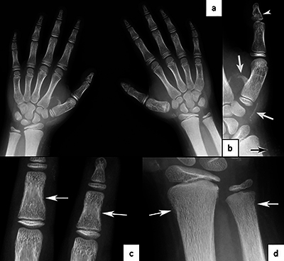 Figure 1
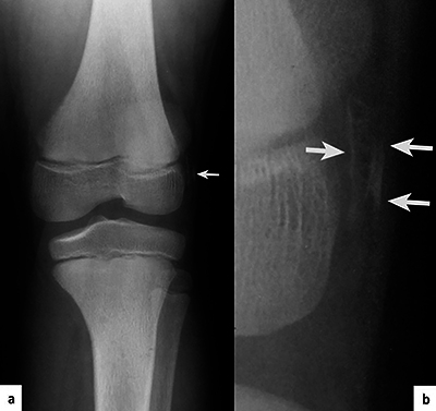 Figure 2
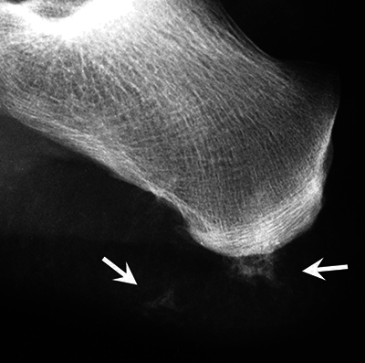 Figure 3
Peer Reviews
No public reviews on file for this paper yet. If you reviewed it on a platform where reviews are public (OpenReview, ICLR, NeurIPS, ICML), you can paste yours below so the community can read it here.
Videos
No videos yet. Explain this paper in a talk, walkthrough, or lecture? Add one.
Taxonomy
TopicsDermatological and Skeletal Disorders · Hypertrophic osteoarthropathy and related conditions · Medical and Biological Sciences
Case History
A nine-year-old child was referred for a hands X-ray examination for short stature investigation (Figure 1). A previous knee X-ray was available (Figure 2), and a foot X-ray was performed (Figure 2).
a) Hands radiography for bone age assessment. Slight brachymetacarpy of the 3rd, 4th and 5th metacarpals and brachyphalangy (P2R1/P2R5). b) Small distal phalanx of both thumbs due to premature fusion of growth plate (arrow head), pseudo-exostosis and linear ossification (arrows). c) and d) Resorption of subperiosteal bone (arrows).
Thin linear ossifications of the soft tissues (arrows) on the lateral side of the lateral condyle, parallel to the skin, visible on the knee X-ray (a) and the magnified view (b).
These X-ray images reveal several semiological elements:
Brachytelephalangy of the thumbs (Figure 1b, arrowhead).Brachymetacarpies and other brachyphalangies of different degrees (Figure 1a).Calcifications/ossifications in the soft tissues and pseudo-exostosis (Figure 1b, Figure 2, Figure 3, arrows).Some foci of subperiosteal resorption (Figure 1c, Figure 1d, arrows).
Calcifications/ossifications of the soft tissues next to the posterior tuberosity of the calcaneus.
These elements are indicative of Albright’s hereditary osteodystrophy (pseudohypoparathyroidism) associated with secondary hyperparathyroidism. The radiological diagnosis was confirmed by genetic analysis.
Comments
Albright’s hereditary osteodystrophy is a genetic disorder with its expression depending on the mode of inheritance and the type of guanine nucleotide binding protein alpha–stimulating activity polypeptide (GNAS) gene mutation. This mutation may induce resistance to parathyroid hormone.
It is associated with morphological anomalies such as short stature, round face, obesity, skeletal involvement (brachymetacarpy, brachymetatarsy, brachyphalangy, exostoses, …), calcifications/ossifications of the soft tissues, and calcifications of the basal ganglia of the brain. Signs of secondary hyperparathyroidism (subperiosteal resorption) may be observed in children.
There are two types, with similar clinical manifestation. Pseudohypoparathyroidism (PHP), or type 1, is associated with hypocalcemia and hyperphosphatemia and with the body’s not responding to parathyroid hormone. Pseudopseudohypoparathyroidism (PPHP), or type 2, is characterized by normocalcemia and a normal response to parathyroid hormone.
The range of skeletal anomalies is widely variable. However, the distal phalanx of the thumb is the most commonly shortened bone. This sign is highly suggestive of PHP/PPHP [1].
Brachymetacarpy of the 4th finger is most frequently observed. This non-specific sign can be found in other diseases such as Turner syndrome. However, when this sign is associated with brachytelephalangy of the thumb, it becomes a more specific sign [1].
The condition is often asymmetrical.
Conclusion
This rare condition can be diagnosed based on standard X-rays.
The reference list from the paper itself. Each links out to its DOI / PubMed record.
- 1Steinbach HL, Young DA. The roentgen appearance of pseudohypoparathyroidism (PH) and pseudo-pseudohypoparathyroidism (PPH). Differentiation from other syndromes associated with short metacarpals, metatarsals and phalanges. Am J Roentgenol Radium Ther Nucl Med. 1966;97(1):49–66. DOI: 10.2214/ajr.97.1.495938050 · doi ↗ · pubmed ↗