The genome sequence of the Ruby Tiger, Phragmatobia fuliginosa (Linnaeus, 1758)
Douglas Boyes, Owen T. Lewis, Lapo Ragionieri, Andrew Mongue, Scott Emrich

TL;DR
This paper presents the genome sequence of the Ruby Tiger moth, including its chromosomal structure and gene annotations.
Contribution
The study provides a high-quality genome assembly and gene annotation for Phragmatobia fuliginosa.
Findings
The genome assembly spans 629.4 megabases and includes 28 chromosomal pseudomolecules.
Gene annotation identified 13,338 protein-coding genes using Ensembl.
The mitochondrial genome is 15.4 kilobases in length and was also assembled.
Abstract
We present a genome assembly from an individual male Phragmatobia fuliginosa (the Ruby Tiger; Arthropoda; Insecta; Lepidoptera; Erebidae). The genome sequence is 629.4 megabases in span. Most of the assembly is scaffolded into 28 chromosomal pseudomolecules, including the assembled Z sex chromosome. The mitochondrial genome has also been assembled and is 15.4 kilobases in length. Gene annotation of this assembly on Ensembl identified 13,338 protein coding genes.
Genes, proteins, chemicals, diseases, species, mutations and cell lines named across the full text — each resolved to its canonical identifier and authoritative record.
Click any figure to enlarge with its caption.
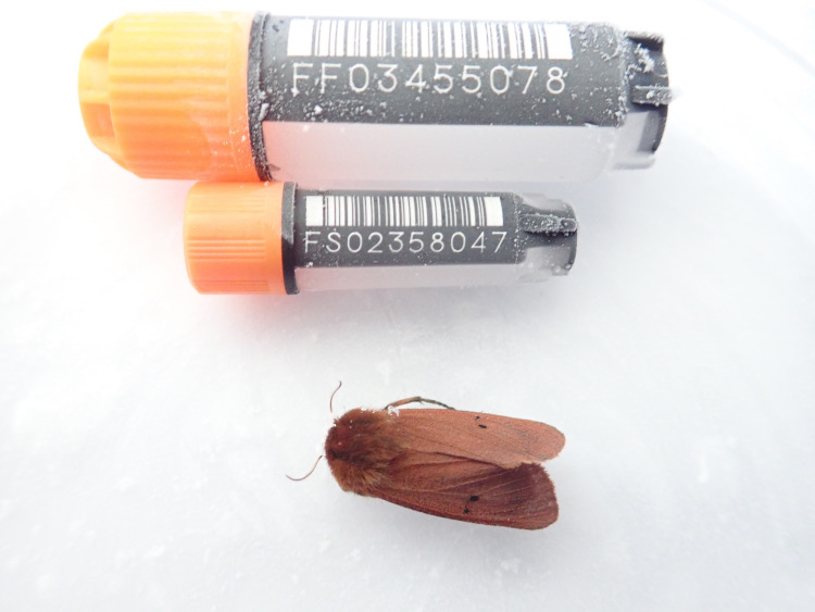 Figure 1
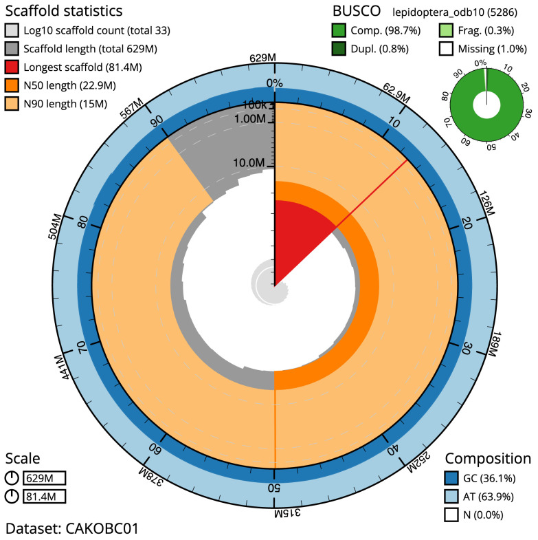 Figure 2
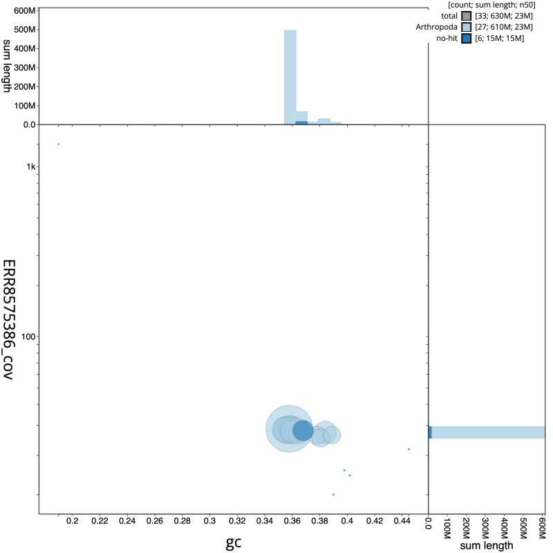 Figure 3
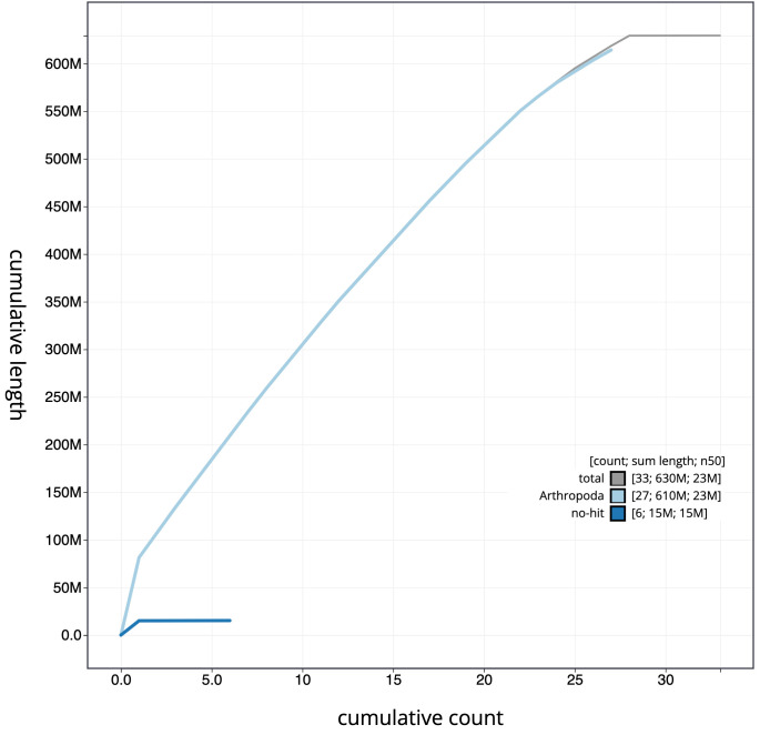 Figure 4
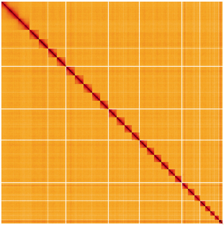 Figure 5
| Project accession data | ||
|---|---|---|
| Assembly identifier | ilPhrFuli1.1 | |
| Species |
| |
| Specimen | ilPhrFuli1 | |
| NCBI taxonomy ID | 214311 | |
| BioProject | PRJEB50747 | |
| BioSample ID | SAMEA7701498 | |
| Isolate information | ilPhrFuli1: male, abdomen (DNA sequencing); head and thorax
| |
| Assembly metrics
|
| |
| Consensus quality (QV) | 66.8 |
|
|
| 100% |
|
| BUSCO
| C:98.7%[S:97.9%,D:0.8%],
|
|
| Percentage of assembly mapped to
| 99.97% |
|
| Sex chromosomes | Z chromosomes |
|
| Organelles | Mitochondrial genome assembled. |
|
| Raw data accessions | ||
| PacificBiosciences SEQUEL II | ERR8575386 | |
| Hi-C Illumina | ERR8571673 | |
| Genome assembly | ||
| Assembly accession | GCA_932526445.1 | |
|
| GCA_932526455.1 | |
| Span (Mb) | 629.4 | |
| Number of contigs | 73 | |
| Contig N50 length (Mb) | 14.1 | |
| Number of scaffolds | 32 | |
| Scaffold N50 length (Mb) | 22.9 | |
| Longest scaffold (Mb) | 81.4 | |
| Genome annotation | ||
| Number of protein-coding genes | 13,338 | |
| Number of non-coding genes | 2,396 | |
| Number of gene transcripts | 22,406 | |
| INSDC accession | Chromosome | Size (Mb) | GC% |
|---|---|---|---|
| 1 | 26.74 | 35.9 | |
| 2 | 25.9 | 35.9 | |
| 3 | 25.37 | 35.6 | |
| 4 | 25.2 | 35.5 | |
| 5 | 25.01 | 35.8 | |
| 6 | 24.85 | 35.7 | |
| 7 | 24.31 | 36.1 | |
| 8 | 23.41 | 35.6 | |
| 9 | 23.17 | 35.8 | |
| 10 | 22.87 | 35.9 | |
| 11 | 22.68 | 35.7 | |
| 12 | 21.22 | 35.6 | |
| 13 | 21.2 | 35.5 | |
| 14 | 21.01 | 35.9 | |
| 15 | 20.88 | 36 | |
| 16 | 20.82 | 36 | |
| 17 | 20.03 | 36.1 | |
| 18 | 19.47 | 36.4 | |
| 19 | 18.72 | 36.3 | |
| 20 | 18.18 | 38.4 | |
| 21 | 18.09 | 36 | |
| 22 | 15.35 | 36.5 | |
| 23 | 15.04 | 36.8 | |
| 24 | 14.14 | 36.7 | |
| 25 | 11.92 | 37.8 | |
| 26 | 11.65 | 38.1 | |
| 27 | 10.68 | 38.9 | |
| Z | 81.38 | 35.8 | |
| MT | 0.02 | 19.2 |
| Software tool | Version | Source |
|---|---|---|
| BlobToolKit | 4.0.7 |
|
| Hifiasm | 0.16.1-r375 |
|
| HiGlass | 1.11.6 |
|
| MitoHiFi | 2 |
|
| PretextView | 0.2 |
|
| purge_dups | 1.2.3 |
|
| YaHS | yahs-1.1.91eebc2 |
|
- —Wellcome Trust
Peer Reviews
No public reviews on file for this paper yet. If you reviewed it on a platform where reviews are public (OpenReview, ICLR, NeurIPS, ICML), you can paste yours below so the community can read it here.
Videos
No videos yet. Explain this paper in a talk, walkthrough, or lecture? Add one.
Taxonomy
TopicsGenomics and Phylogenetic Studies · Genetic diversity and population structure · Chromosomal and Genetic Variations
Species taxonomy
Eukaryota; Metazoa; Ecdysozoa; Arthropoda; Hexapoda; Insecta; Pterygota; Neoptera; Endopterygota; Lepidoptera; Glossata; Ditrysia; Noctuoidea; Erebidae; Arctiinae; Phragmatobia; Phragmatobia fuliginosa (Linnaeus, 1758) (NCBI:txid214311).
Background
The ruby tiger Phragmatobia fuliginosa is a distinctive moth in the subfamily Arctiiinae, the only representative of its genus recorded in the UK. In southern Britain, adult moths have pinkish-red or pinkish-brown forewings and mostly bright pink hindwings that are usually hidden when the moth is settled. Moths from northern Britain are generally darker and have been placed in the subspecies borealis (Staudinger) ( Waring et al., 2017).
Phragmatobia fuliginosa has a range that extends across much of Europe and Asia, as well as parts of northern North America ( GBIF Secretariat, 2022). It has a wide distribution in Great Britain and Ireland, occurring mostly in in open habitats, and is absent only from Shetland. Adults are occasionally active during the day but are more likely to be recorded at light ( South, 1961). In northern Britain there is typically a single annual generation ( Waring et al., 2017), but in southern Britain there are usually two generations, with adult moths recorded in small numbers from April until June, and in much higher numbers during July and August ( Randle et al., 2019). The apparent high abundance of the second generation relative to the first may in part result from the late summer generation being more attracted to light traps ( Waring et al., 2017).
The spherical white eggs of P. fuliginosa are deposited in batches, and the larvae are polyphagous, consuming a wide variety of mostly herbaceous plants, with a particular fondness for ragworts ( Senecio spp.) ( Henwood et al., 2020). The hairy larvae overwinter fully-grown. South (1961) comments that “the vitality of caterpillars is extraordinary”, reporting an observation of a larva that was embedded in ice for at least 14 days without apparent harm. In the spring, the dark-coloured larvae bask in sunshine to raise their body temperature well above ambient, and the speedy larvae are often observed crossing roads and paths.
Male pheromones used in P. fuliginosa courtship are derived from pyrrolizidine alkaloids (PAs) obtained during larval feeding ( Krasnoff & Roelofs, 1990). A genome sequence for Phragmatobia fuliginosa will facilitate studies into molecular adaptations to polyphagy, the evolution of pheromone-based courtship, and contribute to a growing data set of resources for understanding lepidopteran biology more widely.
The genome of Phragmatobia fuliginosa was sequenced as part of the Darwin Tree of Life Project, a collaborative effort to sequence all named eukaryotic species in the Atlantic Archipelago of Britain and Ireland. Here we present a chromosomally complete genome sequence for Phragmatobia fuliginosa, based on one male specimen from Wytham Woods, Oxfordshire, UK.
Genome sequence report
The genome was sequenced from one male Phragmatobia fuliginosa specimen ( Figure 1) collected from Wytham Woods, Oxfordshire, UK (latitude 51.77, longitude –1.34). A total of 35-fold coverage in Pacific Biosciences single-molecule HiFi long was generated. Primary assembly contigs were scaffolded with chromosome conformation Hi-C data. Manual assembly curation corrected 29 missing joins or mis-joins and removed seven haplotypic duplications, reducing the assembly length by 2.82% and the scaffold number by 15.79%, and decreasing the scaffold N50 by 2.33%.
Photograph of the Phragmatobia fuliginosa (ilPhrFuli1) specimen used for genome sequencing.
The final assembly has a total length of 629.4 Mb in 32 sequence scaffolds with a scaffold N50 of 22.9 Mb ( Table 1). Most (99.97%) of the assembly sequence was assigned to 28 chromosomal-level scaffolds, representing 27 autosomes, and the Z sex chromosome. Chromosome-scale scaffolds confirmed by the Hi-C data are named in order of size ( Figure 2– Figure 5; Table 2). The assembly has a BUSCO v5.3.2 ( Manni et al., 2021) completeness of 98.7% (single 97.9%, duplicated 0.8%) using the lepidoptera_odb10 reference set. While not fully phased, the assembly deposited is of one haplotype. Contigs corresponding to the second haplotype have also been deposited.
Table 1.: Genome data for Phragmatobia fuliginosa, ilPhrFuli1.1.
Genome assembly of Phragmatobia fuliginosa, ilPhrFuli1.1: metrics.The BlobToolKit Snailplot shows N50 metrics and BUSCO gene completeness. The main plot is divided into 1,000 size-ordered bins around the circumference with each bin representing 0.1% of the 629,457,366 bp assembly. The distribution of scaffold lengths is shown in dark grey with the plot radius scaled to the longest scaffold present in the assembly (81,383,725 bp, shown in red). Orange and pale-orange arcs show the N50 and N90 scaffold lengths (22,865,098 and 15,039,256 bp), respectively. The pale grey spiral shows the cumulative scaffold count on a log scale with white scale lines showing successive orders of magnitude. The blue and pale-blue area around the outside of the plot shows the distribution of GC, AT and N percentages in the same bins as the inner plot. A summary of complete, fragmented, duplicated and missing BUSCO genes in the lepidoptera_odb10 set is shown in the top right. An interactive version of this figure is available at https://blobtoolkit.genomehubs.org/view/ilPhrFuli1.1/dataset/CAKOBC01/snail.
Genome assembly of Phragmatobia fuliginosa, ilPhrFuli1.1: GC coverage.BlobToolKit GC-coverage plot. Scaffolds are coloured by phylum. Circles are sized in proportion to scaffold length. Histograms show the distribution of scaffold length sum along each axis. An interactive version of this figure is available at https://blobtoolkit.genomehubs.org/view/ilPhrFuli1.1/dataset/CAKOBC01/blob.
Genome assembly of Phragmatobia fuliginosa, ilPhrFuli1.1: cumulative sequence.BlobToolKit cumulative sequence plot. The grey line shows cumulative length for all scaffolds. Coloured lines show cumulative lengths of scaffolds assigned to each phylum using the buscogenes taxrule. An interactive version of this figure is available at https://blobtoolkit.genomehubs.org/view/ilPhrFuli1.1/dataset/CAKOBC01/cumulative.
Genome assembly of Phragmatobia fuliginosa, ilPhrFuli1.1: Hi-C contact map.Hi-C contact map of the ilPhrFuli1.1 assembly, visualised using HiGlass. Chromosomes are shown in order of size from left to right and top to bottom. An interactive version of this figure may be viewed at https://genome-note-higlass.tol.sanger.ac.uk/l/?d=Cc6lMkieRfalxi0HQfR6Xw.
Table 2.: Chromosomal pseudomolecules in the genome assembly of Phragmatobia fuliginosa, ilPhrFuli1.
Genome annotation report
The P. fuliginosa genome assembly GCA_932526445.1 (ilPhrFuli1.1) was annotated using the Ensembl rapid annotation pipeline ( Table 1; Ensembl accession number GCA_932526445.1). The resulting annotation includes 22,406 transcribed mRNAs from 13,338 protein-coding and 2,396 non-coding genes.
Methods
Sample acquisition and nucleic acid extraction
A male P. fuliginosa specimen (ilPhrFuli1) was collected from Wytham Woods, Oxfordshire (biological vice-county: Berkshire) (latitude 51.77, longitude –1.34) on 13 June 2020. The specimen was taken from woodland habitat by Douglas Boyes (University of Oxford) using a light trap. The specimen was identified by Douglas Boyes using field ID and preserved on dry ice.
DNA was extracted at the Tree of Life laboratory, Wellcome Sanger Institute. The ilPhrFuli1 sample was weighed and dissected on dry ice with tissue set aside for Hi-C sequencing. Abdomen tissue was cryogenically disrupted to a fine powder using a Covaris cryoPREP Automated Dry Pulveriser, receiving multiple impacts. High molecular weight (HMW) DNA was extracted using the Qiagen MagAttract HMW DNA extraction kit. HMW DNA was sheared into an average fragment size of 12–20 kb in a Megaruptor 3 system with speed setting 30. Sheared DNA was purified by solid-phase reversible immobilisation using AMPure PB beads with a 1.8X ratio of beads to sample to remove the shorter fragments and concentrate the DNA sample. The concentration of the sheared and purified DNA was assessed using a Nanodrop spectrophotometer and Qubit Fluorometer and Qubit dsDNA High Sensitivity Assay kit. Fragment size distribution was evaluated by running the sample on the FemtoPulse system.
Sequencing
Pacific Biosciences HiFi circular consensus DNA sequencing libraries were constructed according to the manufacturers’ instructions. DNA sequencing was performed by the Scientific Operations core at the WSI on Pacific Biosciences SEQUEL II (HiFi) instrument. Hi-C data were also generated from head and thorax tissue of ilPhrFuli1 using the Arima v2 kit and sequenced on the Illumina NovaSeq 6000 instrument.
Genome assembly
Assembly was carried out with Hifiasm ( Cheng et al., 2021) and haplotypic duplication was identified and removed with purge_dups ( Guan et al., 2020). The assembly was then scaffolded with Hi-C data ( Rao et al., 2014) using YaHS ( Zhou et al., 2023). The assembly was checked for contamination and corrected as described previously ( Howe et al., 2021). Manual curation was performed using HiGlass ( Kerpedjiev et al., 2018) and Pretext ( Harry, 2022). The mitochondrial genome was assembled using MitoHiFi ( Uliano-Silva et al., 2022), which performed annotation using MitoFinder ( Allio et al., 2020). The genome was analysed, and BUSCO scores were generated within the BlobToolKit environment ( Challis et al., 2020). Table 3 contains a list of all software tool versions used, where appropriate.
Genome annotation
The Ensembl gene annotation system ( Aken et al., 2016) was used to generate annotation for the Phragmatobia fuliginosa assembly (GCA_932526445.1). Annotation was created primarily through alignment of transcriptomic data to the genome, with gap filling via protein-to-genome alignments of a select set of proteins from UniProt ( UniProt Consortium, 2019).
Ethics and compliance issues
The materials that have contributed to this genome note have been supplied by a Darwin Tree of Life Partner. The submission of materials by a Darwin Tree of Life Partner is subject to the Darwin Tree of Life Project Sampling Code of Practice. By agreeing with and signing up to the Sampling Code of Practice, the Darwin Tree of Life Partner agrees they will meet the legal and ethical requirements and standards set out within this document in respect of all samples acquired for, and supplied to, the Darwin Tree of Life Project. All efforts are undertaken to minimise the suffering of animals used for sequencing. Each transfer of samples is further undertaken according to a Research Collaboration Agreement or Material Transfer Agreement entered into by the Darwin Tree of Life Partner, Genome Research Limited (operating as the Wellcome Sanger Institute), and in some circumstances other Darwin Tree of Life collaborators.
The reference list from the paper itself. Each links out to its DOI / PubMed record.
- 1Aken BL Ayling S Barrell D : The Ensembl gene annotation system. Database (Oxford). 2016;2016:baw 093. 10.1093/database/baw 093 27337980 PMC 4919035 · doi ↗ · pubmed ↗
- 2Allio R Schomaker-Bastos A Romiguier J : Mito Finder: Efficient automated large-scale extraction of mitogenomic data in target enrichment phylogenomics. Mol Ecol Resour. 2020;20(4):892–905. 10.1111/1755-0998.13160 32243090 PMC 7497042 · doi ↗ · pubmed ↗
- 3Challis R Richards E Rajan J : Blob Tool Kit - Interactive Quality Assessment of Genome Assemblies. G 3 (Bethesda). 2020;10(4):1361–1374. 10.1534/g 3.119.400908 32071071 PMC 7144090 · doi ↗ · pubmed ↗
- 4Cheng H Concepcion GT Feng X : Haplotype-resolved de novo assembly using phased assembly graphs with hifiasm. Nat Methods. 2021;18(2):170–175. 10.1038/s 41592-020-01056-5 33526886 PMC 7961889 · doi ↗ · pubmed ↗
- 5GBIF Secretariat: Phragmatobia fuliginosa (Linnaeus, 1758). GBIF Backbone Taxonomy. 2022; (Accessed: 27 February 2023). Reference Source
- 6Guan D Mc Carthy SA Wood J : Identifying and removing haplotypic duplication in primary genome assemblies. Bioinformatics. 2020;36(9):2896–2898. 10.1093/bioinformatics/btaa 025 31971576 PMC 7203741 · doi ↗ · pubmed ↗
- 7Harry E : Pretext View (Paired R Ead TEX Ture Viewer): A desktop application for viewing pretext contact maps. 2022; (Accessed: 19 October 2022). Reference Source
- 8Henwood B Sterling P Lewington R : Field Guide to the Caterpillars of Great Britain and Ireland.London: Bloomsbury. 2020. Reference Source