Feature-Based Classification of Mild Cognitive Impairment and Alzheimer’s Disease Based on Optical Coherence Tomographic Angiographic Image
Sarinporn Visitsattapongse, Areerat Maneerat, Adisak Trinavarat, Chatchawan Rattanabannakit, Ekkaphop Morkphrom, Vorapun Senanarong, Varalak Srinonprasert, Dittapong Songsaeng, La-ongsri Atchaneeyasakul, Chuchart Pintavirooj

TL;DR
This paper introduces a machine learning tool that uses eye scans to detect early signs of Alzheimer's disease and mild cognitive impairment.
Contribution
The novelty lies in using OCT-A images with novel histogram-based features for classifying Alzheimer’s and mild cognitive impairment.
Findings
A diagnostic model achieved 92.17% accuracy in classifying Alzheimer’s and mild cognitive impairment.
Features like vessel density and retinal thickness were effective in distinguishing disease stages.
The study used a large local database to ensure model effectiveness across diverse populations.
Abstract
Alzheimer’s disease is a type of neurodegenerative disorder that is characterized by the progressive degeneration of brain cells, leading to cognitive decline and memory loss. It is the most common cause of dementia and affects millions of people worldwide. While there is currently no cure for Alzheimer’s disease, early detection and treatment can help to slow the progression of symptoms and improve quality of life. This research presents a diagnostic tool for classifying mild cognitive impairment and Alzheimer’s diseases using feature-based machine learning applied to optical coherence tomographic angiography images (OCT-A). Several features are extracted from the OCT-A image, including vessel density in five sectors, the area of the foveal avascular zone, retinal thickness, and novel features based on the histogram of the range-filtered OCT-A image. To ensure effectiveness for a…
Genes, proteins, chemicals, diseases, species, mutations and cell lines named across the full text — each resolved to its canonical identifier and authoritative record.
Click any figure to enlarge with its caption.
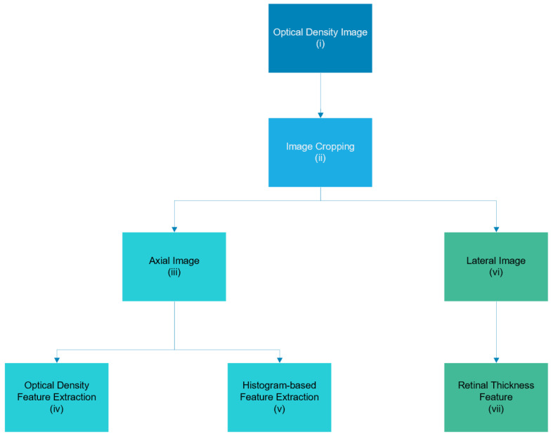 Figure 1
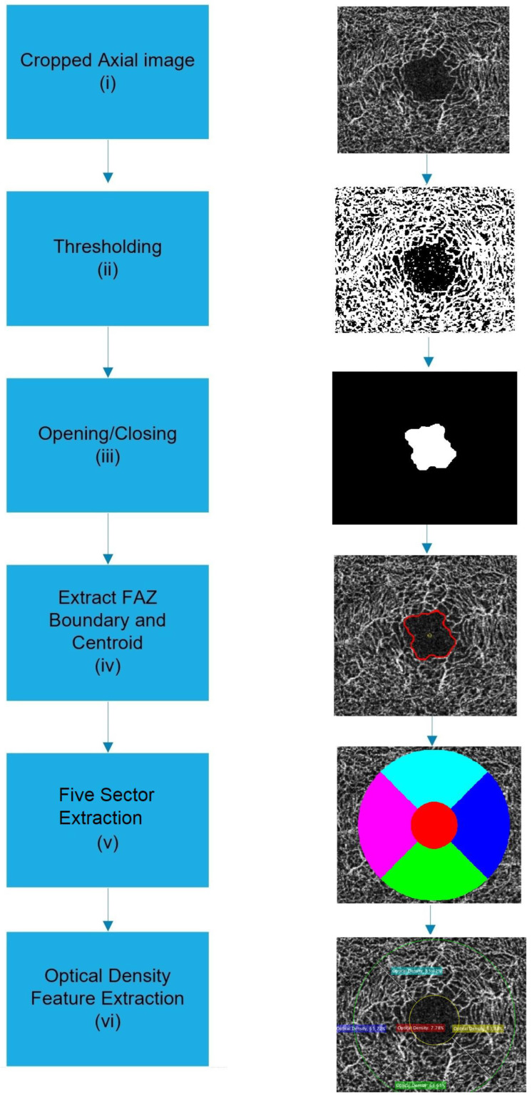 Figure 2
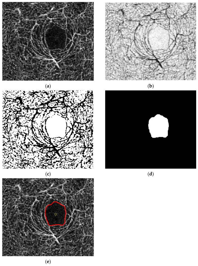 Figure 3
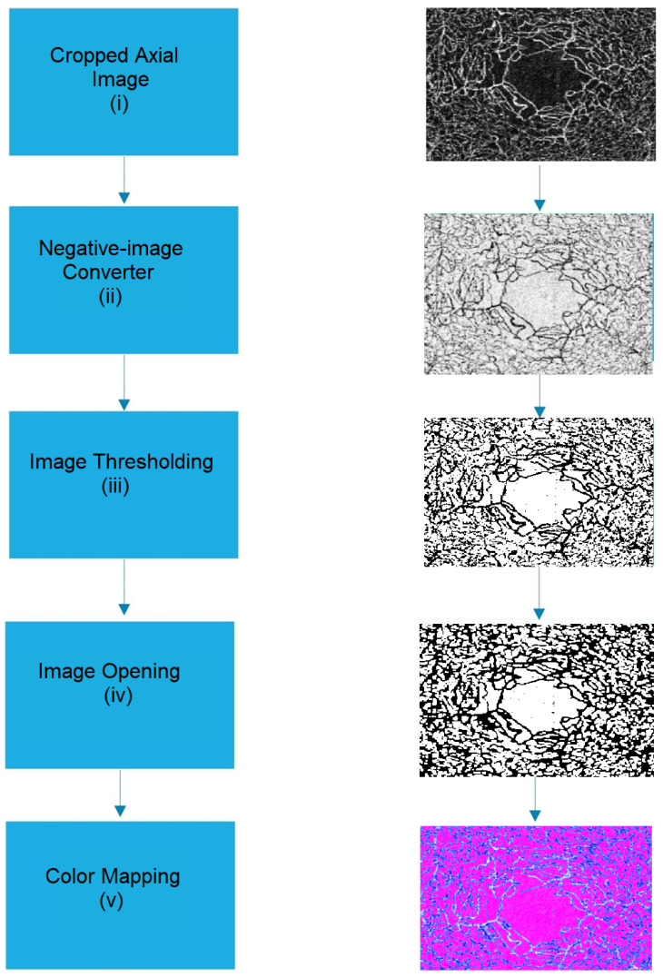 Figure 4
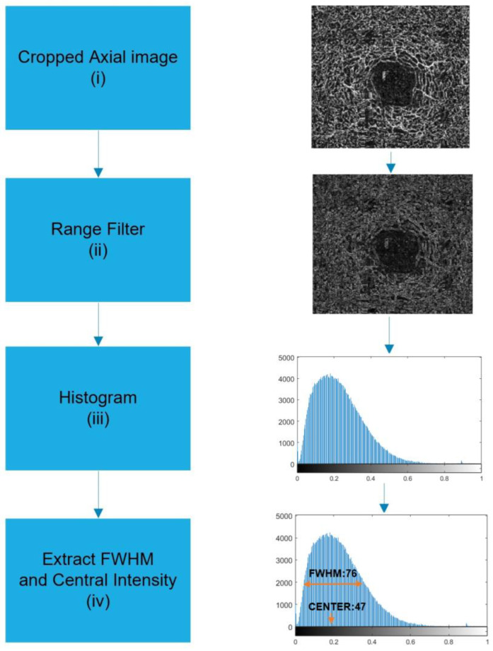 Figure 5
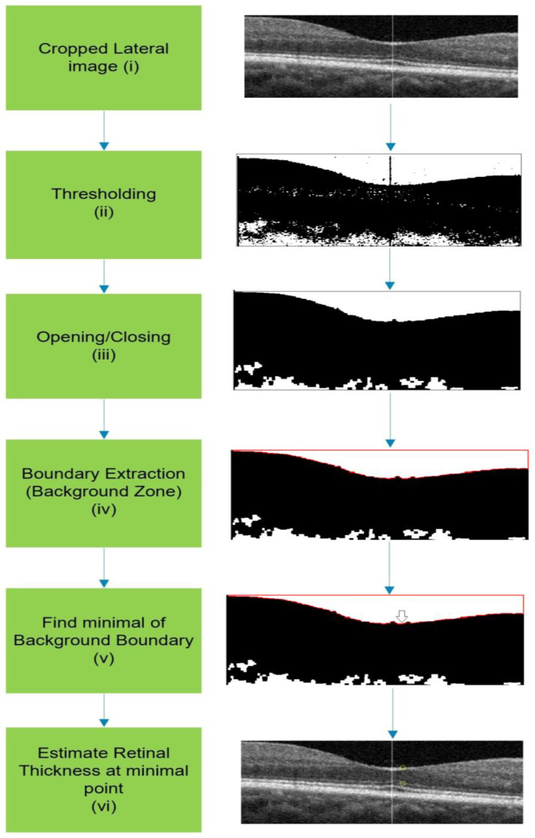 Figure 6
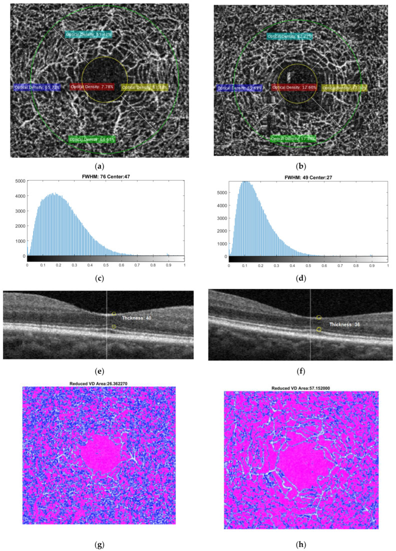 Figure 7
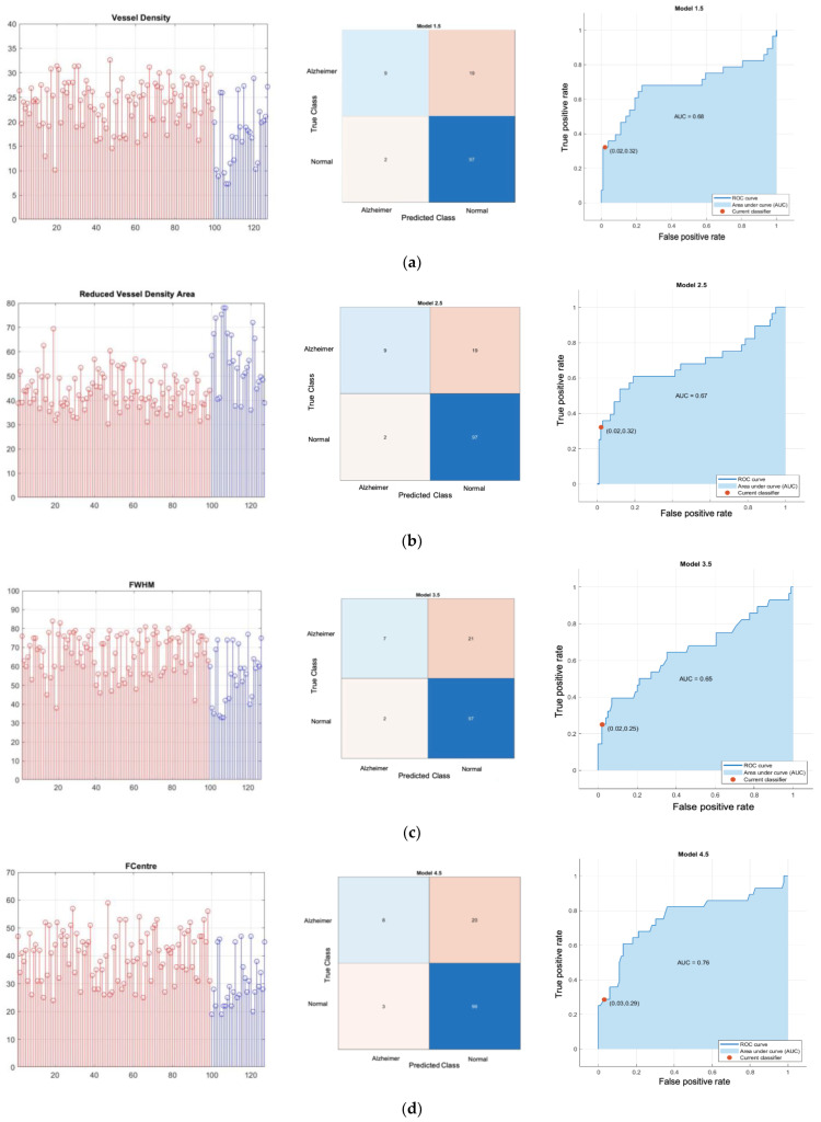 Figure 8
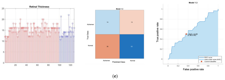 Figure 9
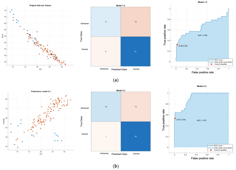 Figure 10
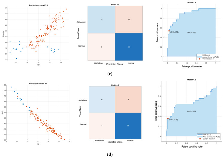 Figure 11
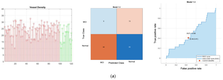 Figure 12
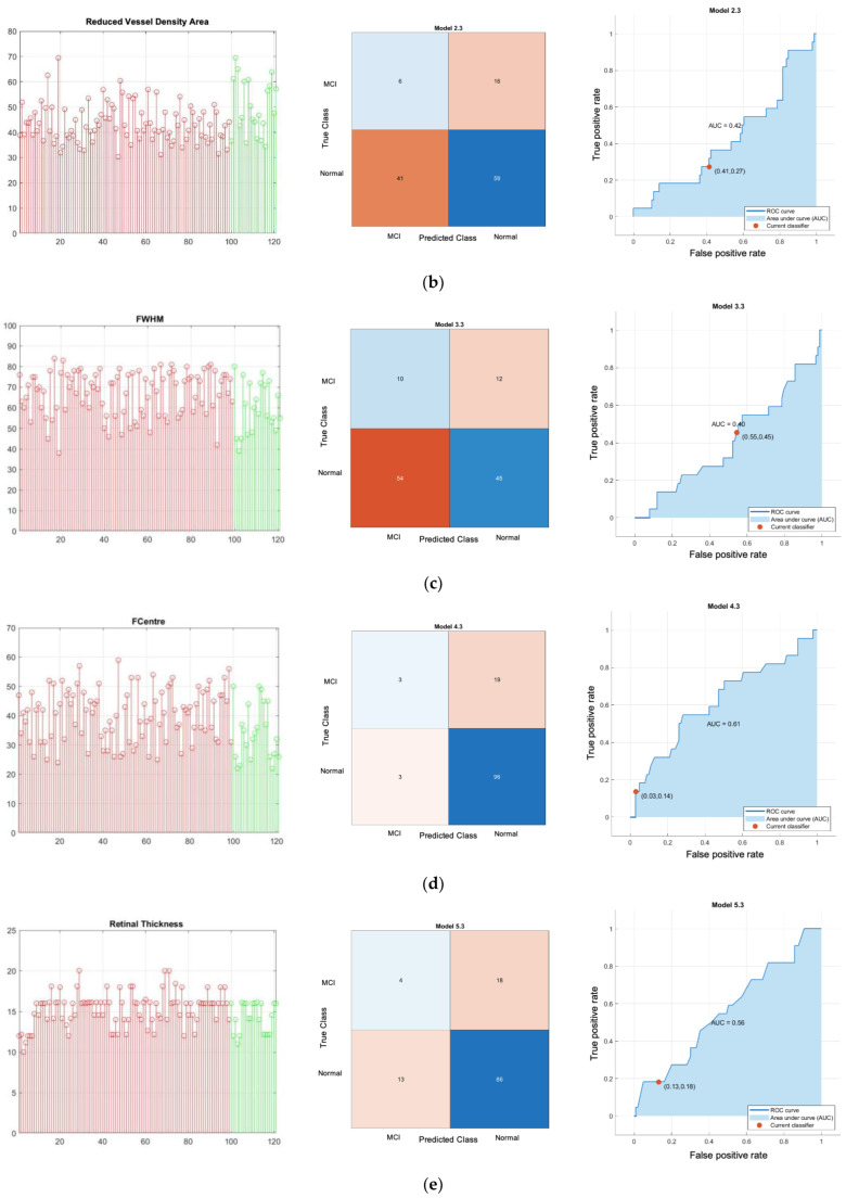 Figure 13
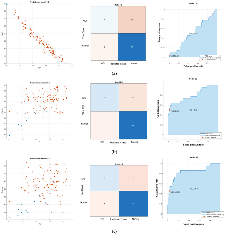 Figure 14
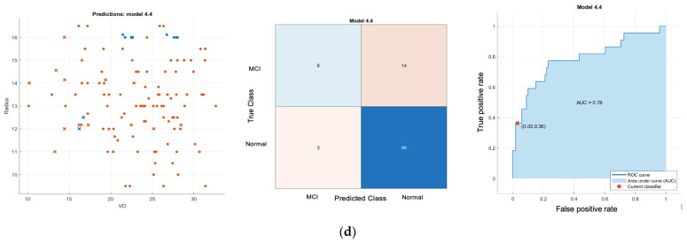 Figure 15
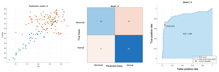 Figure 16
Peer Reviews
No public reviews on file for this paper yet. If you reviewed it on a platform where reviews are public (OpenReview, ICLR, NeurIPS, ICML), you can paste yours below so the community can read it here.
Videos
No videos yet. Explain this paper in a talk, walkthrough, or lecture? Add one.
Taxonomy
TopicsImmune cells in cancer · Erythrocyte Function and Pathophysiology · Acute Myeloid Leukemia Research