Transvaginal ultrasound diagnosis of a rare entity: Premenopausal ovarian hyperthecosis
Benedetta Cornelli, Wouter Froyman, Giulia Garofalo

TL;DR
This paper discusses the use of transvaginal ultrasound to diagnose a rare condition called premenopausal ovarian hyperthecosis.
Contribution
The paper introduces ultrasound features that can help identify ovarian hyperthecosis in premenopausal women.
Findings
Ovarian hyperthecosis can present as a heterogeneous ovary with central vascularization and peripheral follicles on ultrasound.
The presence of these ultrasound features, with or without hyperandrogenism, should prompt consideration of OH in expert hands.
Abstract
Ovarian hyperthecosis (OH) is a benign pathology, less common in premenopause. Literature is poor on its ultrasound (US) characteristics. We suggest that a heterogeneous ovary at US, with a central vascularisation and follicles to the periphery, with or without hyperandrogenism, should lead to consider OH in the hands of experts.
Genes, proteins, chemicals, diseases, species, mutations and cell lines named across the full text — each resolved to its canonical identifier and authoritative record.
Click any figure to enlarge with its caption.
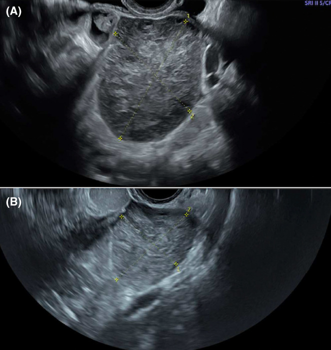 Figure 1
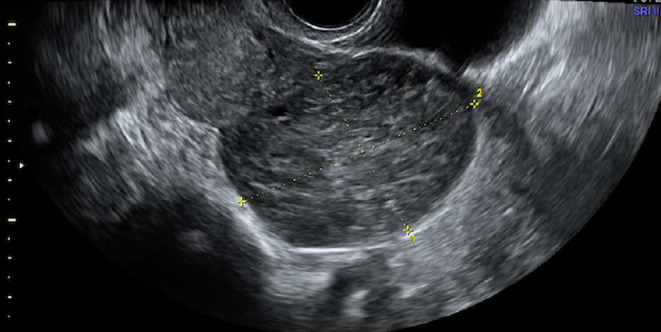 Figure 2
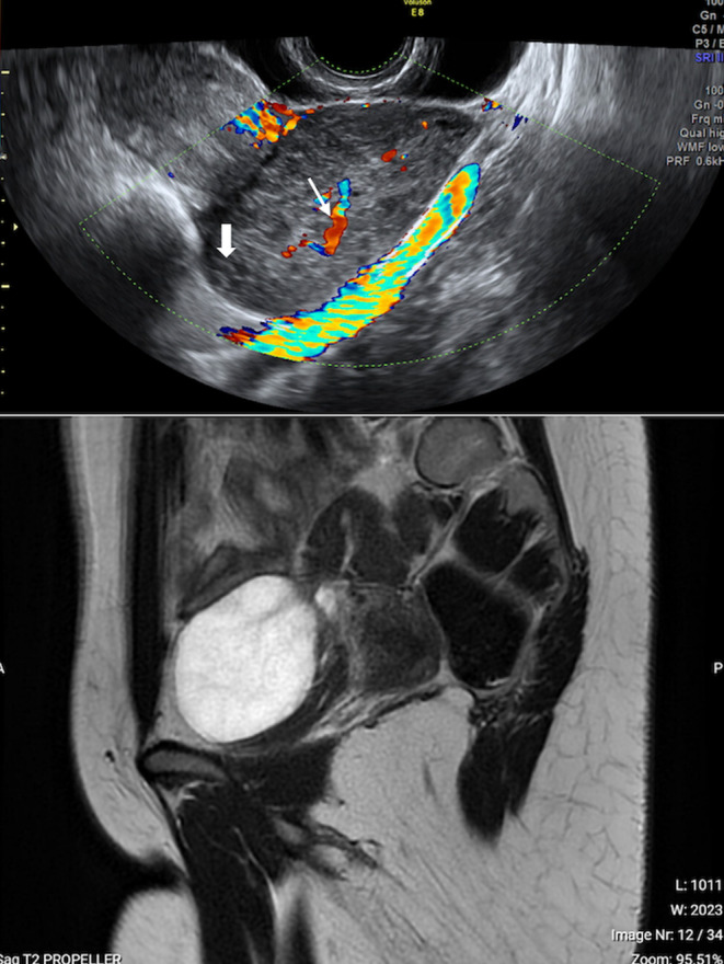 Figure 3
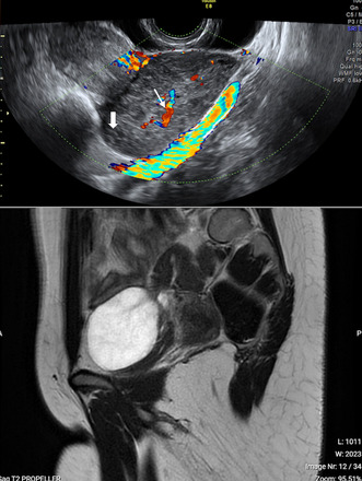 Figure 4
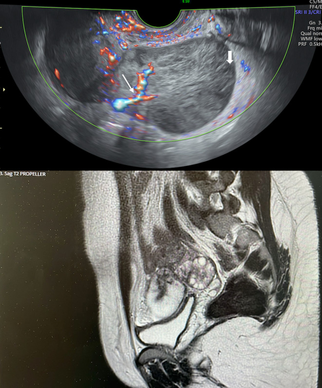 Figure 5
 Figure 6
Peer Reviews
No public reviews on file for this paper yet. If you reviewed it on a platform where reviews are public (OpenReview, ICLR, NeurIPS, ICML), you can paste yours below so the community can read it here.
Videos
No videos yet. Explain this paper in a talk, walkthrough, or lecture? Add one.
Taxonomy
TopicsGerman History and Society