Human Primary Lens Epithelial Cultures on Basal Laminas Studied by Synchrotron-Based FTIR Microspectroscopy for Understanding Posterior Capsular Opacification
Sofija Andjelic, Marko Hawlina

TL;DR
This study uses advanced infrared spectroscopy to compare the molecular profiles of cultured and postoperative lens epithelial cells on basal laminas to better understand posterior capsular opacification.
Contribution
The study introduces synchrotron-based FTIR microspectroscopy to differentiate molecular profiles of cultured and postoperative lens epithelial cells on basal laminas.
Findings
Cultured LECs on bls showed higher collagen contribution based on specific infrared spectral features.
Postoperative LECs on bls exhibited a higher cell contribution, indicated by distinct spectral peak ratios.
SR-FTIR revealed greater LEC contribution in postoperative lens epithelia compared to cultured LECs.
Abstract
Human primary lens epithelial cultures serve as an in vitro model for posterior capsular opacification (PCO) formation. PCO occurs when residual lens epithelial cells (LECs) migrate and proliferate after cataract surgery, differentiating into fibroblastic and lens fiber-like cells. This study aims to show and compare the bio-macromolecular profiles of primary LEC cultures and postoperative lens epithelia LECs on basal laminas (bls), while also analyzing bls and cultured LECs separately. Using synchrotron radiation-based Fourier transform infrared (SR-FTIR) (Bruker, Karlsruhe, Germany) microspectroscopy at the Spanish synchrotron light source ALBA, we observed that the SR-FTIR measurements were predominantly influenced by the strong collagen absorbance of the bls. Cultured LECs on bls showed a higher collagen contribution, indicated by higher vas CH3, CH2 and CH3 wagging and deformation,…
Genes, proteins, chemicals, diseases, species, mutations and cell lines named across the full text — each resolved to its canonical identifier and authoritative record.
Click any figure to enlarge with its caption.
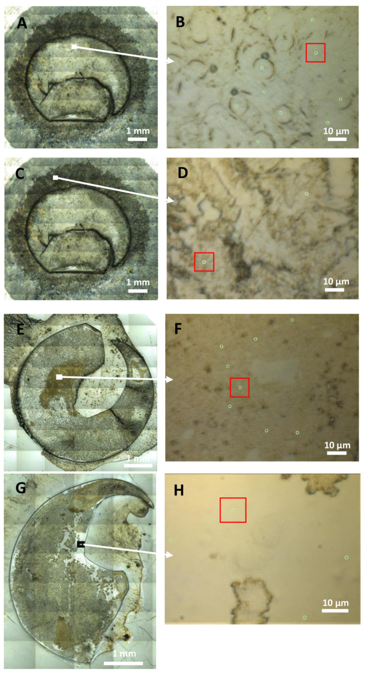 Figure 1
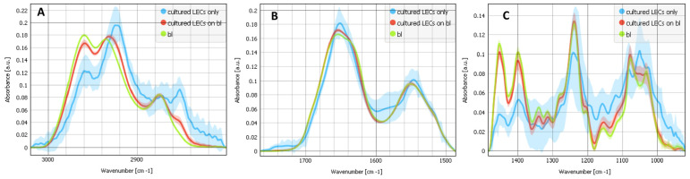 Figure 2
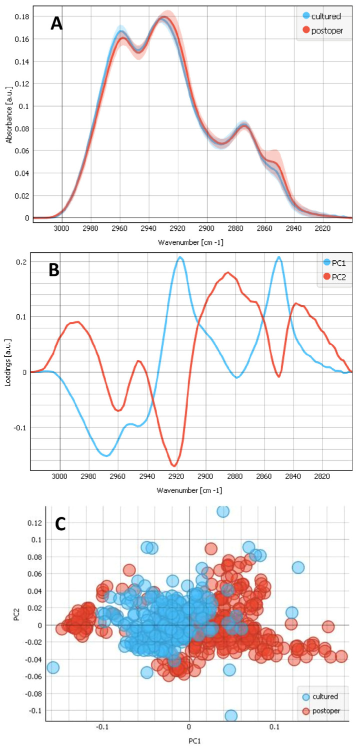 Figure 3
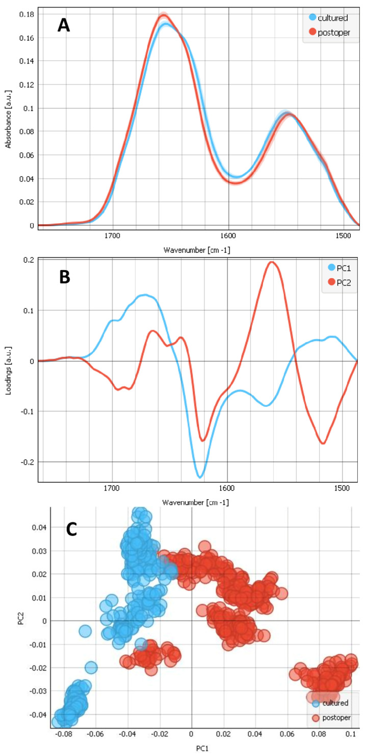 Figure 4
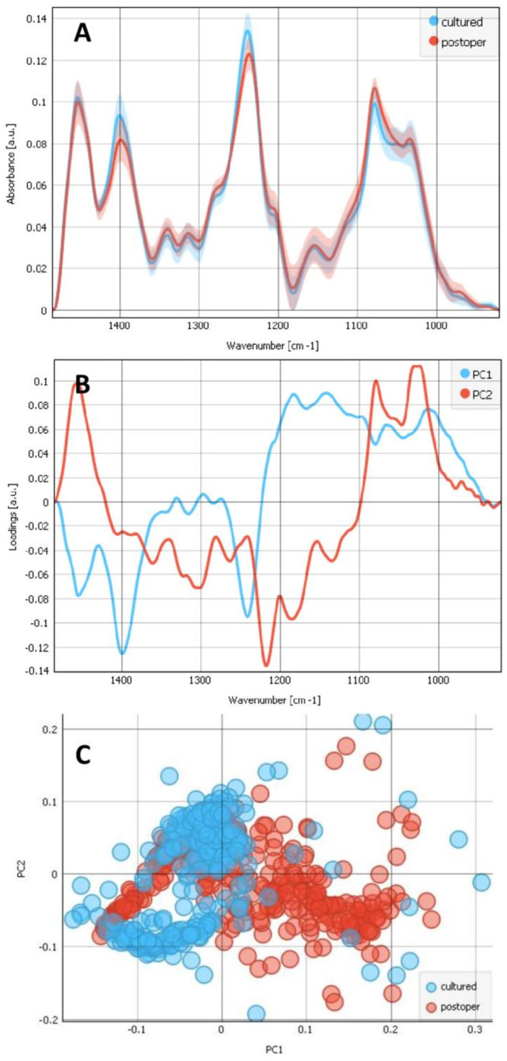 Figure 5
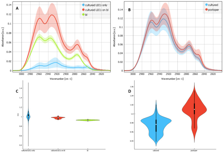 Figure 6
Peer Reviews
No public reviews on file for this paper yet. If you reviewed it on a platform where reviews are public (OpenReview, ICLR, NeurIPS, ICML), you can paste yours below so the community can read it here.
Videos
No videos yet. Explain this paper in a talk, walkthrough, or lecture? Add one.
Taxonomy
TopicsIntraocular Surgery and Lenses · Connexins and lens biology · Corneal surgery and disorders