Posttraumatic Cutaneous Meningioma with a “Meningiolipoma” Pattern Presenting as a Nasal Bridge Mass
Dong Ren, Jerry Lou, Edward Kuan, Mari Perez-Rosendahl, William H. Yong

TL;DR
A rare case of a skin tumor resembling a meningioma, appearing on the nasal bridge after trauma, is reported and analyzed.
Contribution
Presentation of a unique posttraumatic cutaneous meningioma with a 'meningiolipoma' pattern in an uncommon nasal bridge location.
Findings
The tumor exhibited a 'meningiolipoma' pattern, a rare histological variant.
No prior cases of cutaneous meningioma in the nasal bridge were identified in the literature.
Diagnosis required a combination of imaging, histopathology, and immunohistochemistry.
Abstract
Meningiomas are tumors originating from arachnoid meningothelial cells. Occasionally, meningiomas are identified outside the central nervous system, and are referred to as extracranial meningiomas (EMs). The vast majority of EMs are an extension from an intracranial or intraspinal tumor. However, primary EMs may arise from extracranial sites with the most common sites being the skin and scalp subcutis, which are further categorized as cutaneous meningiomas (CMs). CMs are rare cutaneous tumors with similar ultrastructural and cytologic findings compared to those of intracranial meningiomas, but with a wide range of histologic differences. Therefore, an assessment using a panel of investigative tools, including imaging, histopathology, and immunohistochemistry, is required to determine the diagnosis of CMs. Here, we report the case of a 64-year-old gentleman presenting with a…
Genes, proteins, chemicals, diseases, species, mutations and cell lines named across the full text — each resolved to its canonical identifier and authoritative record.
Click any figure to enlarge with its caption.
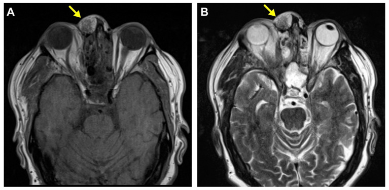 Figure 1
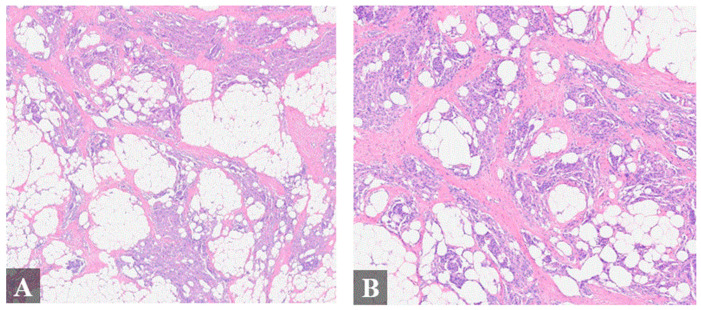 Figure 2
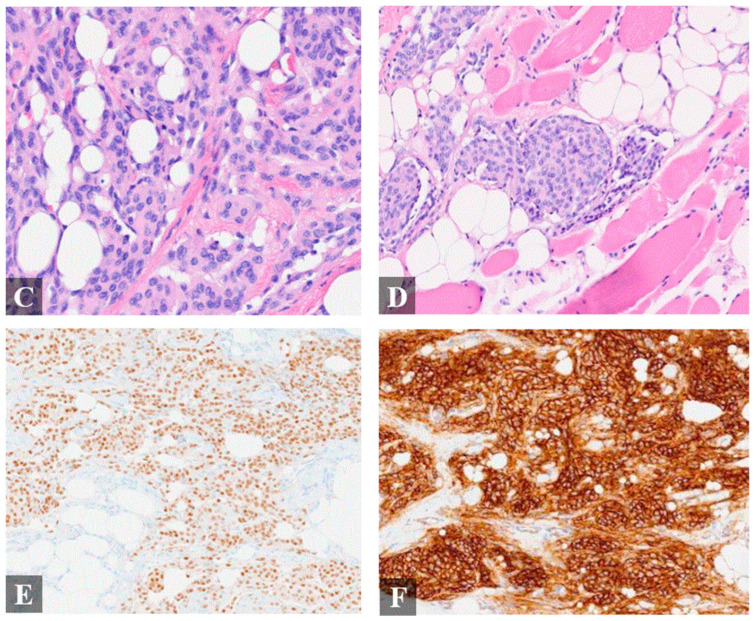 Figure 3
Peer Reviews
No public reviews on file for this paper yet. If you reviewed it on a platform where reviews are public (OpenReview, ICLR, NeurIPS, ICML), you can paste yours below so the community can read it here.
Videos
No videos yet. Explain this paper in a talk, walkthrough, or lecture? Add one.
Taxonomy
TopicsMeningioma and schwannoma management · Neurofibromatosis and Schwannoma Cases · Head and Neck Surgical Oncology
Meningiomas are the most frequently reported primary central nervous system (CNS) neoplasms in adults, accounting for over one-third of all CNS tumors [1]. They are classified as intracranial extra-axial neoplasms by the World Health Organization (WHO) due to their origin from arachnoid meningothelial cells. While most intracranial meningiomas are typically diagnosed via medical imaging and subsequent histopathologic examination, extracranial meningiomas are easily misdiagnosed due to their rarity and a broad spectrum of differential diagnoses with benign nerve sheath tumors and skin tumors. The diagnosis is typically made only after histopathologic examination in most cases [2,3]. Complete surgical excision is the first-line treatment for the majority of symptomatic and enlarging extracranial meningiomas, and patients exhibit favorable prognosis and a low recurrence rate [4].
Extracranial meningiomas can be divided into primary and secondary extracranial meningiomas (PEMs and SEMs) based on the tumor origin, where SEMs present at a secondary location away from an existing primary intracranial tumor, whereas PEMs refer to the meningiomas that arise from extracranial sites without a known primary intracranial tumor [5]. The common sites for PEMs are the head and neck region, including the skin and the scalp subcutis; sinonasal PEMs are less common [6]. Although extracranial meningiomas are rare, extension and metastasis from intracranial or intraspinal tumors comprise the vast majority of extracranial meningiomas. Therefore, the exclusion of a primary intracranial meningioma is required for the diagnosis of a PEM.
Primary cutaneous meningiomas are usually present at birth, with a high occurrence on the scalp, forehead, and paravertebral regions [7]; these are designated as type I lesions thought to derive from ectopic arachnoid cells [8]. Type II lesions usually present in adulthood and are derived from meningothelial cells located in the cutis. Type II lesions have been speculated to arise from ectopic meningothelial cells, potentially displaced from the arachnid during head trauma. Type III lesions are defined by direct extension from intracranial or intraspinal tumors [8]. Herein, we report an unusual case of a Type II cutaneous meningioma involving the nasal bridge that may be associated with remote head trauma (Figure 1 and Figure 2).
The reference list from the paper itself. Each links out to its DOI / PubMed record.
- 1Ostrom Q.T. Gittleman H. Farah P. Ondracek A. Chen Y. Wolinsky Y. Stroup N.E. Kruchko C. Barnholtz-Sloan J.S. CBTRUS statistical report: Primary brain and central nervous system tumors diagnosed in the United States in 2006–2010 Neuro-oncology 201315(Suppl. S 2)ii 1ii 5610.1093/neuonc/not 15124137015 PMC 3798196 · doi ↗ · pubmed ↗
- 2Agaimy A. Buslei R. Coras R. Rubin B.P. Mentzel T. Comparative study of soft tissue perineurioma and meningioma using a five-marker immunohistochemical panel Histopathology 201465607010.1111/his.1236624393170 · doi ↗ · pubmed ↗
- 3Koutlas I.G. Scheithauer B.W. Folpe A.L. Intraoral perineurioma, soft tissue type: Report of five cases, including 3 intraosseous examples, and review of the literature Head Neck Pathol.2010411312010.1007/s 12105-010-0177-320401642 PMC 2878625 · doi ↗ · pubmed ↗
- 4Liu H. Qian H. Li X. Zuo F. Meng X. Liu S. Wan J. Clinial Features, Individualized Treatment and Long-Term Surgical Outcomes of Skull Base Meningiomas with Extracranial Extensions Front. Oncol.202010105410.3389/fonc.2020.0105432714869 PMC 7340145 · doi ↗ · pubmed ↗
- 5Simpson M.T. Sneddon K.J. Extracranial meningioma of the oral cavity Br. J. Oral Maxillofac. Surg.19872552052510.1016/0266-4356(87)90146-X 3480004 · doi ↗ · pubmed ↗
- 6Miedema J.R. Zedek D. Cutaneous meningioma Arch. Pathol. Lab. Med.201213620821110.5858/arpa.2010-0505-RS 22288971 · doi ↗ · pubmed ↗
- 7Zeikus P. Robinson-Bostom L. Stopa E. Primary cutaneous meningioma in association with a sinus pericranii J. Am. Acad. Dermatol.200654(Suppl. S 2)S 49S 5010.1016/j.jaad.2004.08.05816427994 · doi ↗ · pubmed ↗
- 8Arnold M. Cleaver D. Cleaver N. Type II Cutaneous Meningioma Dermatol. Surg.2020461449145110.1097/DSS.000000000000212031453903 · doi ↗ · pubmed ↗