Molecular Detection of Anaplasma phagocytophilum in Cats in Europe and Associated Risk Factors
Vera Geisen, Nikola Pantchev, Yury Zablotski, Olga Kim, Majda Globokar Vrhovec, Katrin Hartmann, Michéle Bergmann

TL;DR
This study found that Anaplasma phagocytophilum infections in cats are more common than previously thought, especially in Northern Europe and during summer, highlighting the need for tick prevention.
Contribution
The study provides new evidence that A. phagocytophilum infections in cats are underreported and identifies geographic and seasonal risk factors.
Findings
Anaplasma phagocytophilum DNA was detected in 7.5% of 1015 European cats.
Infections were significantly more common in Northern Europe compared to Central and Southern Europe.
Infections occurred more frequently during summer than in winter.
Abstract
Although Anaplasma (A.) phagocytophilum infection in cats is considered to be less frequent compared with dogs, there is evidence indicating that the risk of the infection in cats might be underestimated. The study aimed to find out if infections in cats are underestimated and to discover which factors increase the risk of infection. Blood samples of 1015 cats across Europe were tested for A. phagocytophilum DNA from 2017 to 2022. The number of samples sent for testing increased over the 6 years. Anaplasma phagocytophilum DNA was found in 76 out of 1015 cats (7.5%). Infections were more common in Northern Europe than in Central or Southern Europe. During summer, the number of positive samples was significantly higher compared with winter (p = 0.047). The risk for A. phagocytophilum infection in cats should not be underestimated, especially in Northern Europe. Preventing tick bites is…
Genes, proteins, chemicals, diseases, species, mutations and cell lines named across the full text — each resolved to its canonical identifier and authoritative record.
Click any figure to enlarge with its caption.
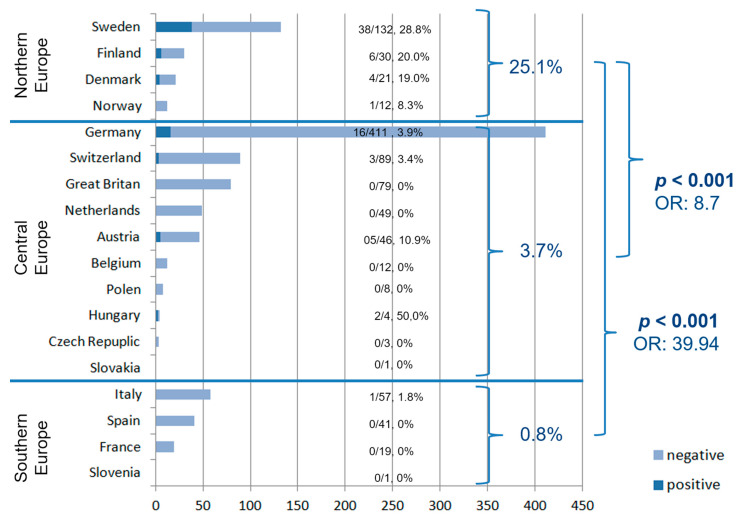 Figure 1
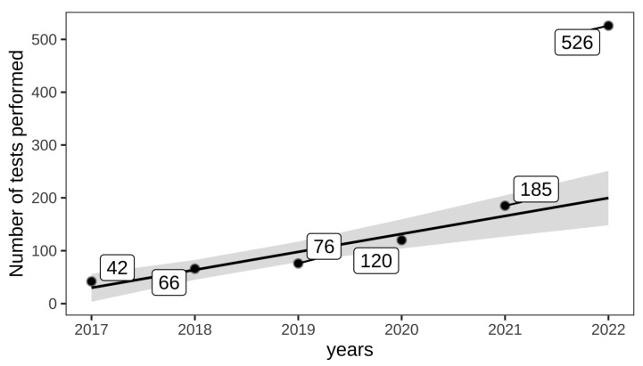 Figure 2
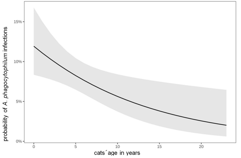 Figure 3
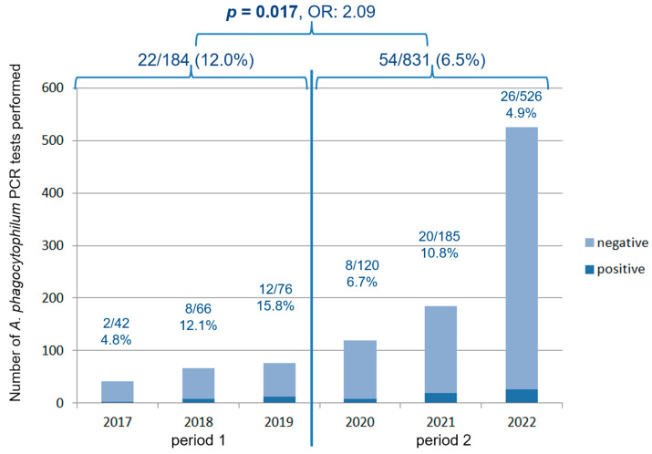 Figure 4
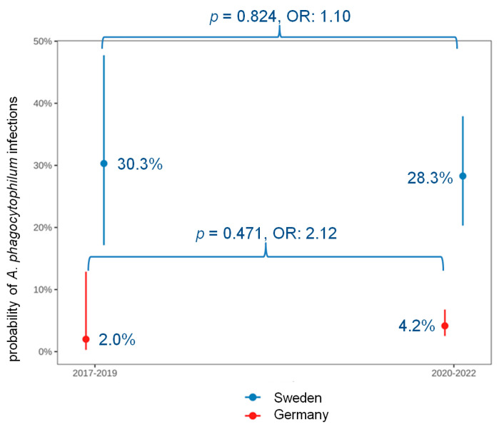 Figure 5
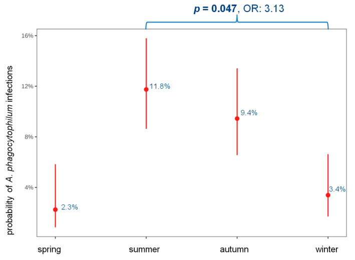 Figure 6
Peer Reviews
No public reviews on file for this paper yet. If you reviewed it on a platform where reviews are public (OpenReview, ICLR, NeurIPS, ICML), you can paste yours below so the community can read it here.
Videos
No videos yet. Explain this paper in a talk, walkthrough, or lecture? Add one.
Taxonomy
TopicsVector-borne infectious diseases · Dermatological diseases and infestations · Viral Infections and Vectors
1. Introduction
Anaplasma (A.) phagocytophilum is an emerging pathogen that affects humans and various other species, including dogs and cats [1,2,3,4]. Anaplasma phagocytophilum is a Gram-negative, obligate intracellular bacterium [5]. Its main vector in Europe is Ixodes (I.) ricinus [6]; however, other Ixodes ticks (e.g., I. trianguliceps, I. hexagonus, and I. ventalloi) also have been documented as vectors [7,8,9,10]. The exact role of A. phagocytophilum as a pathogen in cats is not fully clear. Some cats are asymptomatically infected, and a few cats develop clinical signs, which are primarily unspecific, including lethargy, fever, and anorexia [11,12,13,14]. Main laboratory changes in cats (if present) are thrombocytopenia, leukopenia, leukocytosis, lymphopenia, and anemia [11,12,15]. Since anaplasmosis is an acute disease with a short incubation period, accurate diagnosis relies on direct detection of the pathogen (via PCR or the identification of morulae in neutrophilic granulocytes) in combination with compatible clinical signs and/or laboratory changes [16].
Although A. phagocytophilum infection in cats is considered to be less frequent compared with dogs [16,17,18], there is evidence indicating that infection risk in cats might be underestimated [12,13,15,19].
As observed in dogs, the prevalence of antibodies against A. phagocytophilum in cats is notably high. The antibodies’ prevalence in cats was determined to be 4.5–33.3% in Italy [20,21], 30% in Austria [12,22], 24% in Switzerland [12], 9.1–23% in Germany [12,23,24], 22.1% in Sweden [25], and 1.8–8.4% in Spain [26,27,28,29]. However, considering that anaplasmosis is an acute, self-limiting disease, the presence of antibodies is not indicative of a clinical manifestation compared with direct detection of the pathogen [30,31].
Direct detection of the pathogen via PCR yielded (less often) positive results. In cats in Italy, prevalence ranged from 0–23.1% [20,21,32]; in Portugal, from 0–0.6% [33,34]; in Germany, from 0.3–3% [12,17,23,24]; in the United Kingdom (UK), 1.7% [35], and in Spain, from 0–1% [26,29,36]. However, the majority of cited studies are not up to date, and a comparative study of A. phagocytophilum infections in cats across European countries has not been conducted.
In the meantime, several clinical case reports and series on clinical manifestations of feline anaplasmosis have been published, including a recent study involving 27 A. phagocytophilum-positive cats from Germany (molecular prevalence: 3%), Switzerland (molecular prevalence: 10%), and Austria (molecular prevalence: 8%) [12]. The aims of the present study were to investigate whether the risk of A. phagocytophilum bacteremia in cats in Europe is underestimated and to compare the results across Europe. Furthermore, the influence of cats’ gender and age, and the years of the samples’ submission and seasonality (spring, summer, autumn, winter) on A. phagocytophilum infection was evaluated. Additionally, it was investigated wether a pattern in PCR submissions could be observed over time.
2. Materials and Methods
Blood samples of 1015 cats were tested for A. phagocytophilum DNA. Blood samples derived from submissions by veterinarians from different European countries within 6 years (2017–2022) to a commercial laboratory (IDEXX Laboratories, Kornwestheim, Germany) (Table 1). A single A. phagocytophilum PCR was requested for 651 samples, while 26 samples were submitted for a tick panel, 242 samples for an anemia panel (available since 2022), and 96 samples for a fever panel (available since 2022), all including a PCR for A. phagocytophilum. Further information about the reasons for submission was not available due to the nature of the study (a retrospective evaluation of laboratory submissions), but it can be assumed that a clinical suspicion was raised by the submitting veterinarian in the majority of the cases. All samples were included in which information on the date of sample’s submission, country, postal code, and city of the submitting veterinarian, age and sex of the sampled cats; and the result of the A. phagocytophilum PCR could be obtained from the laboratory’s computer system. Samples from outside Europe were excluded.
A. phagocytophilum DNA was amplified by real-time PCR from whole blood according to Dyachenko and colleagues (2012) at IDEXX Laboratories Inc. Kornwestheim, Germany [37]. The extraction of total DNA from whole blood was carried out utilizing the QIAamp DNA Blood BioRobot MDx kit (QIAGEN, Hilden, Germany), adhering to the guidelines provided by the manufacturer. Real-time PCR was carried out utilizing the LightCycler 480 system (Roche, Mannheim, Germany), using custom-designed forward and reverse primers in addition to hydrolysis probes. The genes selected as the target genes for detection of the pathogen via real-time PCR was msp2 of A. phagocytophilum (accession number DQ519570). The real-time PCR assays were shown to have a reproducible average analytical sensitivity of 10 DNA molecules per reaction.
Statistical analysis was performed using R Version 4.3.1. Robust linear regression was used to determine whether there was a trend of either an increase or decrease in PCR requests submitted by veterinarians over time. To determine associations between the cats’ origin (country and geographic distribution) and A. phagocytophilum infection, univariable logistic regression was performed. For these analyses, Europe was divided into three geographical regions: Northern Europe (Sweden, Finland, Denmark, and Norway), Central Europe (Netherlands, UK, Germany, Poland, Switzerland, Austria, Hungary, Belgium, Czech Republic, and Slovakia), and Southern Europe (France, Spain, Italy, and Slovenia). Associations of the cats’ sex and age, the years of sample’ submission (years 2017–2019 versus 2020–2022), and seasonality with A. phagocytophilum infection were evaluated by multivariable logistic regression. Since all four predictors were considered clinically relevant, no backward selection of the variables was performed. To exclude multicollinearity among the predictors, the generalized variance-inflation factors (GVIFs) were calculated for the logistic regression. Due to the lower number of samples in some years, data were consolidated into 2 periods of equal length: period 1: 2017–2019 and period 2: 2020–2022. In countries with more than 100 samples submitted overall, an association between the period of the samples’ submission (2017–2019 versus 2020–2022) and A. phagocytophilum infection was evaluated using bivariable logistic regression. For all analyses, a p-value of <0.05 was considered significant.
3. Results
3.1. Population
Of the 1015 sampled cats, 302/864 (35.0%) were female and 562/864 (65.0%) were male; sex was unknown in 151 cats. The age was known in 781 cats and ranged from 0.17–23 years (median: 5 years); the age of 234 cats was unknown. Of the 1015 cats, 411 originated from Germany (40.5%), 132 from Sweden (13.0%), 89 from Switzerland (8.8%), 79 from the UK (7.8%), 57 from Italy (5.6%), 49 from the Netherlands (4.8%), 46 from Austria (4.5%), 41 from Spain (4.0%), 30 from Finland (3.0%), 21 from Denmark (2.1%), 19 from France (1.9%), 12 from Belgium, 12 from Norway (1.2%), 8 from Poland (0.8%), 4 from Hungary (0.4%), 3 from Czech Republic (0.3%), and 1 each from Slovenia and Slovakia (0.1%) (Figure 1).
3.2. Submissions to the Laboratory
PCR orders submitted to the laboratory increased significantly over the past 6 years (p = 0.002) (Figure 2) with a mean increase of 34 tests per year. Since the numbers exhibited an outlier-like explosion in 2022, robust linear regression was used to determine the pattern of submission, in which the outliers had less weight.
3.3. Anaplasma Phagocytophilum-Infected Cats
Anaplasma phagocytophilum DNA was detected in 76/1015 of cats (7.5%, 95% CI: 6.0–9.3%). Of 302 female cats, 21 (6.9%) were infected, and of 562 male cats, 48 (8.5%) were infected. There was no significant difference in sex between A. phagocytophilum-negative and -positive cats (multivariable logistic regression: p = 0.600) (Table 1).
The median age of A. phagocytophilum-infected cats was 3 years (range: 0.25–16 years); that of A. phagocytophilum-noninfected cats was 5 years (range: 0.17–23 years). The risk of A. phagocytophilum infection significantly decreased with the cats’ age (p = 0.013, odds ratio (OR): 0.92) (Table 1, Figure 3).
The highest percentage of A. phagocytophilum infections was detected in Sweden (38/132; 28.8%) and Finland (6/30; 20%). This was only outnumbered by Hungary, where, however, only 4 samples were tested (2 out of 4 were positive) (Figure 1). Figure 4 shows the proportion of A. phagocytophilum-infected cats out of the number of tested cats, and the percentage for each year.
Anaplasma phagocytophilum infection was significantly more common in Northern Europe when compared with Central (p < 0.001, OR: 8.70) and Southern Europe (p < 0.001, OR: 39.94) (Figure 1). While throughout Europe, the percentage of A. phagocytophilum infections significantly decreased over the past 3 years (2020–2022) compared with the previous 3 years (2017–2019) (p = 0.017, OR: 2.09) (Figure 4), it did not change significantly in Germany (p = 0.471) and Sweden (p = 0.824) (Figure 5).
Multivariable logistic regression revealed a significantly higher likelihood for A. phagocytophilum infection during summer compared with winter (p = 0.047, OR: 3.13) (Figure 6, Table 1). Other significant seasonal differences were not observed.
4. Discussion
PCR requests for A. phagocytophilum submitted to the laboratory significantly increased over the past 6 years, while the percentage of infection significantly decreased. This trend is likely attributed to the growing awareness among veterinarians regarding A. phagocytophilum infection and its integration in laboratory panels.
In total, A. phagocytophilum DNA was detected in 76 out of 1015 (7.5%) cats. The notably high percentage of A. phagocytophilum-positive PCR results was unexpected. The percentage was high, particularly in Northern Europe (25.1% (8.3–28.8%)), which can be explained by the northward expansion of the vectors. Several decades ago, Ixodes ticks were scarce in Northern Europe [38,39], but their range has expanded northward over time. This was accompanied by a significant increase in the population density of ticks, as seen, for example, in Sweden [40,41,42], the country with the highest number of A. phagocytophilum infections in the present study. Steadily rising temperatures are (at least partially) associated with the northward migration of the ticks’ hosts, such as deer and moose [40]. Consequently, this migration facilitates the broader distribution of Ixodes ticks, which require temperatures of more than 5–7 °C for their activity [43,44]. On the other hand, surprisingly, a recent study discovered that I. ricinus in Sweden exhibited activity even at temperatures as low as −5 °C [45]. Furthermore, one study revealed that A. phagocytophilum can induce the expression of an antifreeze glycoprotein gene in Ixodes ticks, enhancing the ticks’ cold resistance [46]. A further explanation for the high prevalence of Ixodes ticks in Northern Europe is the explosive proliferation of the ticks’ main host, the red deer. In the early 1990s, red deer experienced a rapid increase in population due to an outbreak of scabies in red foxes, leading to a reduction in the deer’s natural predators [47]. Another important fact is the high prevalence of A. phagocytophilum in both the vector and reservoir hosts. The prevalence of A. phagocytophilum in I. ricinus by PCR was reported to be 0.7–15.0% in Sweden [48,49], 3.5–9.6% in Finland [50,51], and 40.5% in Denmark [52], in comparison with only 3.1–6.5% in Germany [53,54,55,56]. A molecular prevalence of 26.3% was observed in Swedish moose [25], 23.9% in Swedish cattle [57], and even 42.6% in Danish roe deer [58]. A further point to consider regarding the high number of *A. phagocytophilum-*positive cats in Northern Europe is their lifestyle. Many cats in this region have outdoor access, making them more susceptible to tick infestations and thus infection [59]. Additionally, many studies have shown that I. ricinus prefers wooded areas, which are very common in Northern European countries and thus predispose this area to a high(er) tick prevalence [60,61,62]. In this context, the afforestation of open vegetation types, as are currently occurring through some climate protection projects in Scandinavia, have likely also led to increased tick density [62].
In Central Europe, the rate of A. phagocytophilum infections was relatively high (3.9%, 16/411) paralleling the results of a recent study that documented a comparable rate of 3% (18/619) [12]. This contrasts starkly with earlier research, where the molecular prevalence ranged from only 0.3–0.4% [12,17,23,24]. Also, the prevalence of A. phagocytophilum in Ixodes ticks in Germany exhibited a notable increase, escalating from 1% in 2004 [63] to as high as 6.5% in studies conducted between 2010 and 2021 [53,54,55,56]. However, tests have improved over the years, which has potentially impacted the increase in positive results as well.
In the present study, a notably low percentage of infected cats was observed in Southern Europe, with only 1 out of 118 positive samples (0.8%). This finding contrasts with an older study conducted in Northern Italy, which reported a notably higher prevalence of 23.1% [20]. However, all cats sampled in that study in 2014 were stray cats, which were certainly not protected against ectoparasites. Owned cats, in comparison, are more likely to receive regular ectoparasite prophylaxis, especially in regions endemic for chronic persistent vector-borne diseases in Southern Europe; this would also explain the controversially low number of A. phagocytophilum infections in the current study and in multiple previous studies (≤1%) [21,26,28,32,33,34,36,64]. Another contributing factor for the low rate in Southern Europe is the lower prevalence of Ixodes ticks in Mediterranean regions, since this ticks require higher humidity levels to thrive [43].
So far, relatively little information is available about the association of the age of cats with A. phagocytophilum bacteremia. In the present study, A. phagocytophilum-infected cats were relatively young (median age: 3 years), and there was a significant decrease in positive rates with increasing age (Figure 2). The median age in dogs is 7–8 years [2,65,66]. The low median age in cats could indicate an age-related resistance in older cats, similar to what is observed with Bartonella spp. infection in cats [67]. This could potentially be attributed to a differential function of the cat’s immune system as opposed to that of dogs [68].
Anaplasma phagocytophilum infection was significantly more likely in summer compared with winter. This observation aligns with the reported tick activity [69] and is similar to findings of other studies examining at the seasonal presence of A. phagocytophilum [66,70]. However, it is noteworthy that some studies reported a bimodal distribution of A. phagocytophilum infection, with peaks occurring in spring and autumn [65].
The limitation of the study is the preselection of samples, as only those submitted to the laboratory were examined, thereby deviating from a purely prevalence-based investigation.
5. Conclusions
In conclusion, the present study highlights that infections with A. phagocytophilum confirmed by PCR in cats are more common than expected. Anaplasma phagocytophilum infection should be considered as important infections, particularly in Northern Europe. Therefore, implementing effective tick prevention measures is crucial for the management of feline health and are as important in non-Mediterranean regions as in Mediterranean ones.
The reference list from the paper itself. Each links out to its DOI / PubMed record.
- 1Dziegiel B. Adaszek L. Kalinowski M. Winiarczyk S. Equine granulocytic anaplasmosis Res. Vet. Sci.20139531632010.1016/j.rvsc.2013.05.01023790982 · doi ↗ · pubmed ↗
- 2Chirek A. Silaghi C. Pfister K. Kohn B. Granulocytic anaplasmosis in 63 dogs: Clinical signs, laboratory results, therapy and course of disease J. Small Anim. Pr.20185911212010.1111/jsap.1278729171663 · doi ↗ · pubmed ↗
- 3Dahlgren F.S. Mandel E.J. Krebs J.W. Massung R.F. Mc Quiston J.H. Increasing incidence of Ehrlichia chaffeensis and Anaplasma phagocytophilum in the United States, 2000–2007 Am. J. Trop. Med. Hyg.20118512413110.4269/ajtmh.2011.10-061321734137 PMC 3122356 · doi ↗ · pubmed ↗
- 4De Arcangeli S. Balboni A. Serafini F. Battilani M. Dondi F. Anaplasma phagocytophilum infection in thrombocytopenic dogs Vet. Ital.201854737810.12834/Vet It.1070.5796.229631317 · doi ↗ · pubmed ↗
- 5Dumler J.S. Barbet A.F. Bekker C.P. Dasch G.A. Palmer G.H. Ray S.C. Rikihisa Y. Rurangirwa F.R. Reorganization of genera in the families Rickettsiaceae and Anaplasmataceae in the order Rickettsiales: Unification of some species of Ehrlichia with Anaplasma, Cowdria with Ehrlichia and Ehrlichia with Neorickettsia, descriptions of six new species combinations and designation of Ehrlichia equi and ‘HGE agent’ as subjective synonyms of Ehrlichia phagocytophila Int. J. Syst. Evol. Microbiol.2001512145216510.10 · doi ↗ · pubmed ↗
- 6Shaw S.E. Birtles R.J. Day M.J. Arthropod-transmitted infectious diseases of cats J. Feline Med. Surg.2001319320910.1053/jfms.2001.014911795958 PMC 10822301 · doi ↗ · pubmed ↗
- 7Nijhof A.M. Bodaan C. Postigo M. Nieuwenhuijs H. Opsteegh M. Franssen L. Jebbink F. Jongejan F. Ticks and associated pathogens collected from domestic animals in the Netherlands Vector Borne Zoonotic Dis.2007758559510.1089/vbz.2007.013017979540 · doi ↗ · pubmed ↗
- 8Bown K.J. Lambin X. Telford G.R. Ogden N.H. Telfer S. Woldehiwet Z. Birtles R.J. Relative importance of Ixodes ricinus and Ixodes trianguliceps as vectors for Anaplasma phagocytophilum and Babesia microti in field vole (Microtus agrestis) populations Appl. Env. Microbiol.2008747118712510.1128/AEM.00625-0818820068 PMC 2592922 · doi ↗ · pubmed ↗