Cutaneous Ulcers and Response to Treatment in a Child with Anti-MDA5 Dermatomyositis
Mamadapur Mahabaleshwar, Ramaswamy Subramanian

Abstract
Genes, proteins, chemicals, diseases, species, mutations and cell lines named across the full text — each resolved to its canonical identifier and authoritative record.
Click any figure to enlarge with its caption.
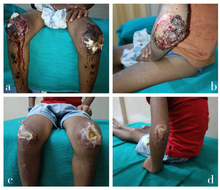 Figure 1
Peer Reviews
No public reviews on file for this paper yet. If you reviewed it on a platform where reviews are public (OpenReview, ICLR, NeurIPS, ICML), you can paste yours below so the community can read it here.
Videos
No videos yet. Explain this paper in a talk, walkthrough, or lecture? Add one.
Taxonomy
TopicsInflammatory Myopathies and Dermatomyositis · Eosinophilic Disorders and Syndromes · Immunodeficiency and Autoimmune Disorders
CLINICAL IMAGE
A 6-year-old female presented with painful skin ulcers with discharge over both elbows and knees for 10 days. There was no fever, muscle weakness, oral ulcers, joint pain, or cardiorespiratory complaints. On examination, she had ulcers over both elbows, knees, and lateral malleoli with crusting. Gottron papules were present (Figure 1A–B). Muscle power was 5/5. Investigation revealed CRP of 48.61 mg/L, Hb of 9.4gm/dl. CPK was 25U/L. Serum LDH was 251 IU/L.LFT was normal. ANA was negative. Her extended myositis profile was positive for Anti-MDA5 antibody. CT chest was normal. She was managed with IV immunoglobulin 2gm/kg over 5 days, and IV methylprednisolone 15mg/kg for 3 days. She was discharged with Prednisolone and Tacrolimus. One month later the skin ulcers had healed (Figure 1C–D).
(A–B) Ulcers over both knees and elbow with crusting. (C–D) Healed ulcers at follow-up following immuno-suppression.
DISCUSSION
Juvenile Dermatomyositis is an autoimmune disease characterised by inflammation, autoimmunity, and vasculopathy^1^ with protean manifestations. Juvenile MDA 5 myositis prevalence may vary from 10–40% of JDM cases.^2^ Antimelanoma differentiation-associated gene 5 (MDA5) antibody-positive dermatomyositis usually presents with minimal or no muscle weakness,^3,4^ cutaneous ulcers which may be deep and punched out in the extensor aspects, arthritis, and ILD. Treatment recommendations vary in the management of anti-MDA5 JDM. The triple combination therapy (high-dose glucocorticoids, calcineurin inhibitor, and intravenous cyclophosphamide) has been widely used. Evidence for the role of JAK inhibitors, rituximab, plasma exchange, and polymyxin B perfusion is limited and is used in refractory cases.^5^
Early diagnosis and initiation of aggressive combined immunosuppression help reduce damage and improve survival.
The reference list from the paper itself. Each links out to its DOI / PubMed record.
- 1Mehta P Machado PM Gupta L. Understanding and managing anti-MDA 5 dermatomyositis, including potential COVID-19 mimicry. Rheumatol Int 2021 Jun;41(6):1021–36.33774723 10.1007/s 00296-021-04819-1PMC 8000693 · doi ↗ · pubmed ↗
- 2Mamyrova G Kishi T Shi M Targoff IN Huber AM Curiel RV Anti-MDA 5 autoantibodies associated with juvenile dermatomyositis constitute a distinct phenotype in North America. Rheumatology 2021 Apr 1;60(4):1839–49.33140079 10.1093/rheumatology/keaa 429PMC 8023991 · doi ↗ · pubmed ↗
- 3Mariampillai K Granger B Amelin D Guiguet M Hachulla E Maurier F Development of a New Classification System for Idiopathic Inflammatory Myopathies Based on Clinical Manifestations and Myositis-Specific Autoantibodies. JAMA Neurol 2018 Dec;75(12):1528–37.30208379 10.1001/jamaneurol.2018.2598 PMC 6583199 · doi ↗ · pubmed ↗
- 4Yasin SA Schutz PW Deakin CT Sag E Varsani H Simou S Histological heterogeneity in a large clinical cohort of juvenile idiopathic inflammatory myopathy: analysis by myositis autoantibody and pathological features. Neuropathol Appl Neurobiol 2019;45(5):495–512.30378704 10.1111/nan.12528 PMC 6767402 · doi ↗ · pubmed ↗
- 5Shunsuke Furuta MY. New therapies in anti-MDA 5 antibody-positive dermatomyositis. Curr Opin Rheumatol 2023 Sep 8;35:1–8.37682061 10.1097/BOR.0000000000000979 · doi ↗ · pubmed ↗