Dermoscopic features of papillated Bowen’s disease
Jonathan Stevens, Claudia Schroder, Magdalena Delgado

Abstract
Genes, proteins, chemicals, diseases, species, mutations and cell lines named across the full text — each resolved to its canonical identifier and authoritative record.
Click any figure to enlarge with its caption.
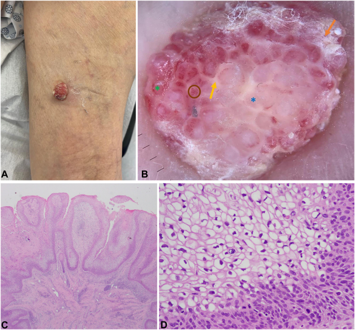 Figure 1
Peer Reviews
No public reviews on file for this paper yet. If you reviewed it on a platform where reviews are public (OpenReview, ICLR, NeurIPS, ICML), you can paste yours below so the community can read it here.
Videos
No videos yet. Explain this paper in a talk, walkthrough, or lecture? Add one.
Taxonomy
TopicsCancer and Skin Lesions · Nonmelanoma Skin Cancer Studies · Nail Diseases and Treatments
Clinical presentation
A 90-year-old female with controlled hypertension, presented with a 6-months history of an asymptomatic progressively growing pink lesion in the right thigh. There is no personal or family history of skin cancer. Clinical examination showed a 10 × 15 mm well-defined symmetrical erythematous tumor in the posterior region of the right thigh (Fig 1, A).Fig 1A, Macroscopic physical examination: a 10 × 15 mm well-defined symmetrical erythematous tumor in the posterior region of the right thigh. B, Dermatoscopic features: milky red honeycomb appearance with whitish septa (yellow arrow) separating whitish lacunae (blue asterisk) in the center and red lacunae on the periphery (green asterisk), some with glomerular vessels inside (brown circle), with small continuous whitish structureless areas on the periphery (orange arrow). C, Histopathological examination showing papillomatous exophytic proliferation, consisting of axes of fibroconnective tissue covered by squamous epithelium formed by atypical cells, in a significant proportion with clear cytoplasm (2× magnification). D, Atypical squamous intraepithelial proliferation with keratinocytes with clear cytoplasm and well-defined borders, nuclear pleomorphism, and mitosis (40× magnification).
Dermatoscopic appearance
Dermoscopy revealed a milky red honeycomb appearance with whitish septa separating whitish lacunae in the center and red lacunae on the periphery, some with glomerular vessels inside, with small continuous whitish structureless areas on the periphery (Fig 1, B). Complete surgical removal was performed.
Histologic diagnosis
Histopathology revealed intraepidermal squamous cell carcinoma, clear cell papillomatous/papillated subtype (Fig 1, C and D).Key messageBowen’s disease usually manifests as a slowly enlarging erythematous scaly patch or plaque.1 An uncommon variant of Bowen’s disease showing a verrucous appearance with a prominent clear cell change on histopathology was recently reported in 2017.1Given the rarity of this presentation, the differential diagnosis should include pagetoid Bowen's disease, extramammary Paget’s disease, clear cell acanthoma, superficial spreading melanoma, sebaceous carcinoma, and trichilemmal carcinoma.1Detailed descriptions of its dermoscopic features have been quite limited.2 There may be some difficulty recognizing the characteristic vascular structures and surface scales of conventional Bowen's disease.2 We propose a new dermoscopy feature called “milky red honeycomb”: whitish septa separating whitish lacunae in the center, red lacunae on the periphery with some glomerular vessels inside, and small continuous whitish structureless areas on the periphery.
Conflicts of interest
None disclosed.
The reference list from the paper itself. Each links out to its DOI / PubMed record.
- 1Lee D.Y.Choi K.H.Park S.H.Lee J.Y.Yoon T.Y.Lobulated Bowen's disease with a clear cell change Ann Dermatol 29420174874902876130010.5021/ad.2017.29.4.487PMC 5500717 · doi ↗ · pubmed ↗
- 2Namiki T.Ichiyama S.Funasaka Y.Dermoscopy of pigmented papillated Bowen disease: a report of two cases J Dermatol 4432017 e 23e 242720770110.1111/1346-8138.13467 · doi ↗ · pubmed ↗