Magnetic Resonance Imaging in the Assessment and Management of Post-injection-Site Morphea
Praveen K Sharma, Sharmeela S, Evangeline P Christina, Dhivya Gunasekaran

TL;DR
This paper presents a case where MRI was used to assess and manage morphea, a skin condition that can develop after an injection.
Contribution
The paper highlights the use of MRI in distinguishing between inflammation and fibrosis in post-injection morphea.
Findings
MRI revealed skin layer thickening and inflammation in a patient with morphea.
MRI helped differentiate between active inflammation and fibrosis in the affected area.
Abstract
Morphea, a form of localized scleroderma, can significantly affect individuals by causing skin tightening and discoloration. We describe the case of a 22-year-old woman who presented with progressive skin changes and discomfort in her right gluteal region following a history of an intramuscular injection in the right gluteal region. Clinical examination suggested morphea, prompting us to conduct an MRI to better understand the extent and nature of her condition. The MRI results revealed thickening of the skin layers and signs of inflammation, helping us differentiate between active inflammation and fibrosis. This case illustrates how MRI can provide crucial insights for managing morphea effectively.
Genes, proteins, chemicals, diseases, species, mutations and cell lines named across the full text — each resolved to its canonical identifier and authoritative record.
Click any figure to enlarge with its caption.
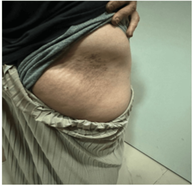 Figure 1
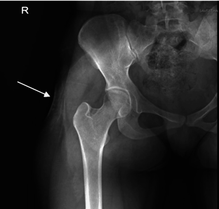 Figure 2
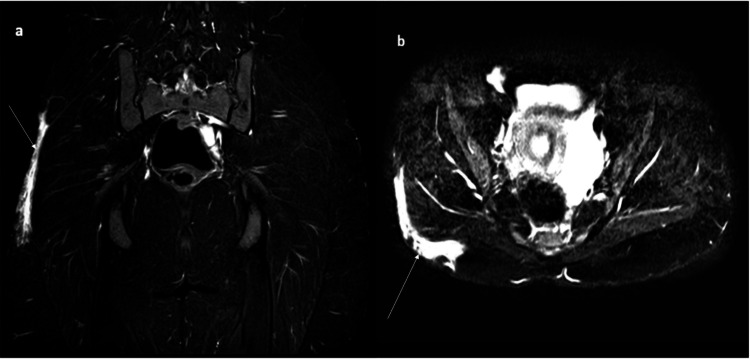 Figure 3
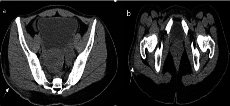 Figure 4
Peer Reviews
No public reviews on file for this paper yet. If you reviewed it on a platform where reviews are public (OpenReview, ICLR, NeurIPS, ICML), you can paste yours below so the community can read it here.
Videos
No videos yet. Explain this paper in a talk, walkthrough, or lecture? Add one.
Taxonomy
TopicsSystemic Sclerosis and Related Diseases · Body Contouring and Surgery · Dermatologic Treatments and Research