SET8 inhibition preserves PTEN to attenuate kidney cell apoptosis in cisplatin nephrotoxicity
Xu Yang, Yingjie Guan, George Bayliss, Ting C. Zhao, Shougang Zhuang

TL;DR
Inhibiting SET8 protects kidney cells from cisplatin damage by preserving PTEN, reducing cell death and DNA damage.
Contribution
This study reveals a novel protective mechanism of SET8 inhibition in cisplatin-induced kidney injury through PTEN preservation.
Findings
SET8 inhibition with UNC0379 reduces kidney cell apoptosis and DNA damage in cisplatin-induced AKI.
PTEN preservation by SET8 inhibition restores autophagy and reduces DDR in renal cells.
Blocking PTEN negates the protective effects of SET8 inhibition, confirming PTEN's role in the mechanism.
Abstract
The aberrant expression of SET8, a histone methyltransferase that mediates H4 lysine 20 mono-methylation (H4K20me1), is implicated in the pathogenesis of various tumors, however, its role in acute kidney injury (AKI) is unknown. Here we showed that SET8 and H4K20me1 were upregulated in the murine kidney with AKI induced by cisplatin, along with increased renal tubular cell injury and apoptosis and decreased expression of E-cadherin and Phosphatase and Tensin Homolog (PTEN). Suppression of SET8 by UNC0379 improved renal function, attenuated tubule damage, and restored expression of PTEN, but not E-cadherin. UNC0379 was also effective in lessening cisplatin-induced DNA damage response (DDR) as indicated by reduced expression of γ-H2AX, p53, p21, and alleviating cisplatin-impaired autophagy as shown by retained expression of Atg5, Beclin-1, and CHMP2A and enhanced levels of LC3-II in the…
Genes, proteins, chemicals, diseases, species, mutations and cell lines named across the full text — each resolved to its canonical identifier and authoritative record.
Click any figure to enlarge with its caption.
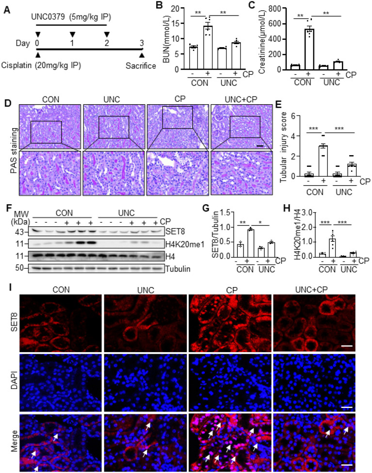 Figure 1
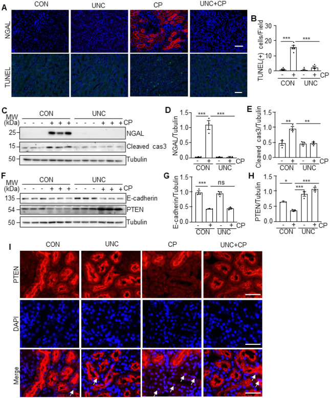 Figure 2
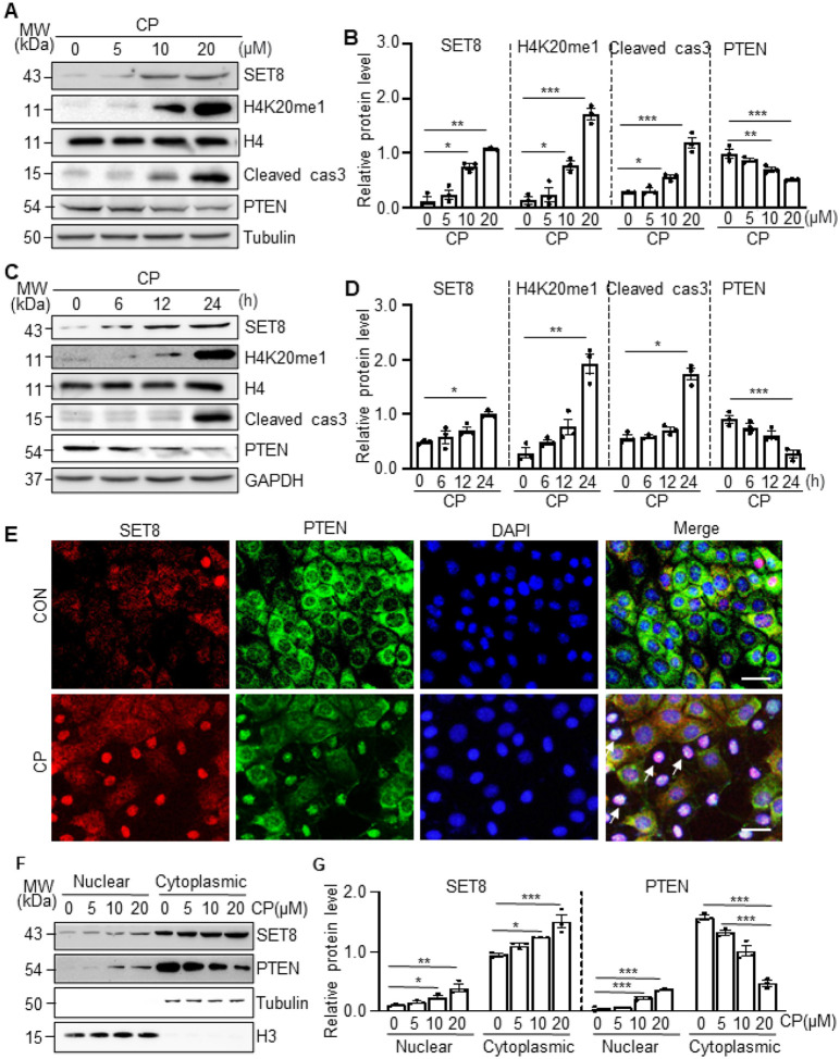 Figure 3
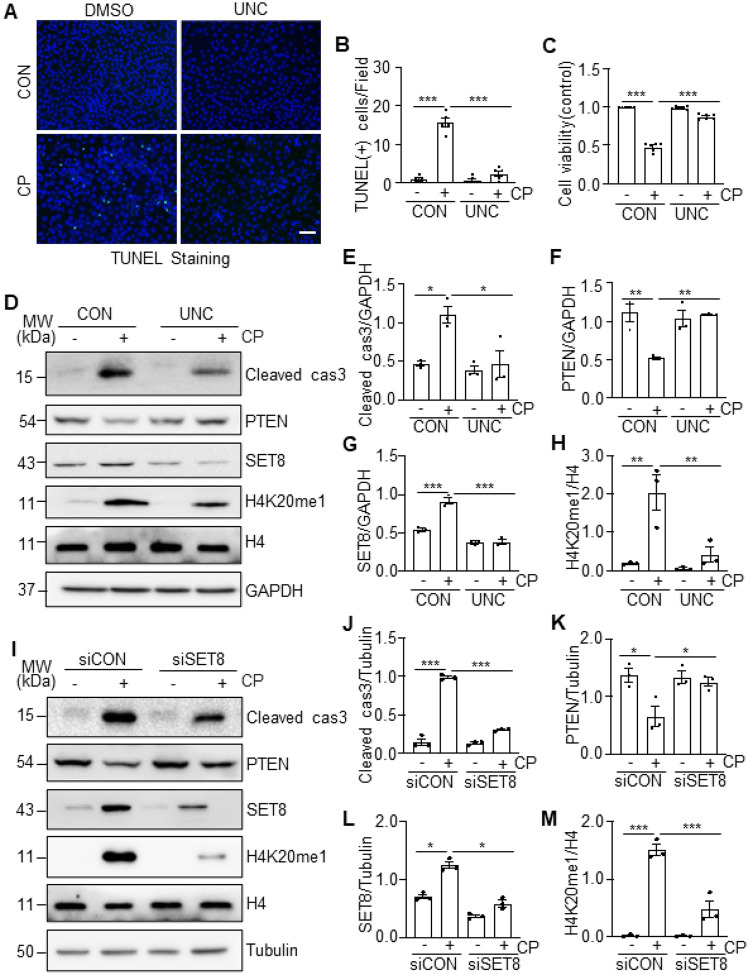 Figure 4
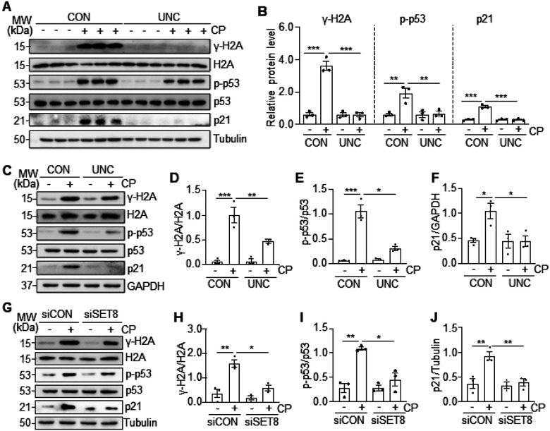 Figure 5
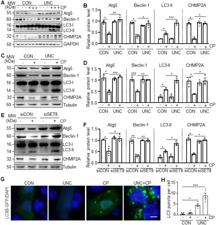 Figure 6
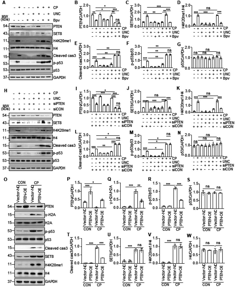 Figure 7
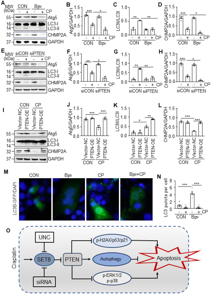 Figure 8
Peer Reviews
No public reviews on file for this paper yet. If you reviewed it on a platform where reviews are public (OpenReview, ICLR, NeurIPS, ICML), you can paste yours below so the community can read it here.
Videos
No videos yet. Explain this paper in a talk, walkthrough, or lecture? Add one.
Taxonomy
TopicsChemotherapy-induced organ toxicity mitigation · Renal and related cancers · Epigenetics and DNA Methylation
INTRODUCTION
Cisplatin is one of the most widely used chemotherapy drugs to treat various tumors (1). However, the uptake of cisplatin by renal proximal tubules results in cellular death and acute kidney injury (AKI) (2). AKI affects approximately 10%-20% of cancer patients who receive cisplatin treatment (3). To date, the pathogenesis of cisplatin-induced AKI remains incompletely understood, and effective renoprotective approaches are not available.
Accumulation of cisplatin in renal tubular cells induces various intracellular stress responses, including DNA damage response (DDR) and autophagy (4). DDR is a cellular event leading to DNA repair, cell cycle arrest, senescence, and/or apoptosis, which is orchestrated by various signaling cascades, including phosphorylation of the histone variant H2A (γ-H2AX) and p53 (5). γ-H2AX is an early step in the cellular response to DNA damage and is involved in the initiation of DDR, while phosphorylation of p53 is a critical downstream effector of DDR and responsible for triggering cell cycle arrest by upregulation of p21 and inducing apoptosis by activation of caspase 3 (5, 6). In cisplatin nephrotoxicity, autophagy is also rapidly activated in kidney tubular cells and required to protect the kidney from injury and apoptosis (7). However, the precise mechanisms that underlie DDR and autophagy induction by cisplatin in renal epithelial cells are still elusive.
Recent studies have suggested the importance of phosphatase and tensin homolog (PTEN) in regulating kidney cell apoptosis via p53 and autophagy in AKI. It has been reported that pharmacological inhibition of PTEN aggravates cisplatin-induced AKI and tubular cell apoptosis by activation of p53 signaling pathways (8), while activation of PTEN protects the kidney against AKI apoptosis by promoting autophagy (9). Moreover, PTEN overexpression promotes renal epithelium repair by restoring CHMP2A-mediated phagosome closure (10). Although PTEN expression and activation are indispensable for renoprotection, it is often downregulated in the kidney with AKI-induced diverse insults including cisplatin (8, 10, 11). Thus, identifying and targeting the mechanisms that lead to the loss of PTEN in the kidney may be a novel strategy to treat AKI.
PTEN stability and enzymatic activity are regulated by multiple mechanisms, including epigenetic silencing and post-transcriptional and posttranslational modifications (12). Among several types of posttranslational modifications, histone methylation has been shown to regulate cells’ sensitivity to DNA-damaging agents, including chemotherapeutics and radiotherapeutics. For example, NSD2, a methyltransferase, can induce PTEN methylation, and subsequently recruit PTEN to DNA damage sites to regulate DDR and cellular sensitivity to DNA damaging agents in tumor cells (13). SET8 (also known as PR-set7/9, SETD8, KMT5A), another member of the SET8 domain-containing methyltransferase family, specifically catalyzes mono-methylation of histone H4 lysine 20 (H4K20me1) (14) and has been implicated in multiple biological processes, including DNA replication, DNA damage repair, and gene transcription (15–17). However, the role of SET8 in AKI and renal epithelial cell death has not been investigated yet.
In this study, we explored the role and mechanism of SET8 in cisplatin nephrotoxicity. Our results showed that cisplatin treatment increased expression of SET8 and H4K20me1 with downregulation of PTEN in the kidneys of mice and cultured murine proximal tubular epithelial cells (TKPTs). Targeted inhibition of SET8 attenuates AKI and reduces renal tubular cell apoptosis via a mechanism associated with PTEN preservation and subsequent suppressing DNA damage and retaining autophagy.
RESULTS
Inhibition of SET8 by UNC0379 attenuates cisplatin-induced AKI in mice
To determine the role of SET8 in AKI, we examined the effect of SET8 inhibition on cisplatin-induced AKI in a mouse model using UNC0379, a highly selective inhibitor of SET8 (18–20). As depicted in Fig. 1A, UNC0379 was administered once daily for three consecutive days. Blood urea nitrogen (BUN) and serum creatinine (SCr) were measured to reflect the changes in renal function. The results in Fig. 1B, C demonstrated a significant elevation in BUN and SCr levels in the cisplatin group compared to the control group, which is consistent with progressive renal tubular injury as indicated by tubular distention, swelling and necrosis, and loss of brush border (Fig. 1D, E). Administration of UNC0379 significantly reduced the levels of BUN and SCr and attenuated renal tubular damage in mice following cisplatin treatment (Fig. 1B–E).
Western blot analysis of whole kidney lysates revealed that cisplatin treatment resulted in increased expression SET8 and its epigenetic activation marker, H4K20me1, in the injured kidney, compared with sham controls. Administration of UNC0379 reduced cisplatin-induced upregulation of SET8 and H4K20me1 without alteration of histone H4 levels (Fig. 1F–H). Immunofluorescence (IF) staining indicated that SET8 was predominantly expressed in renal tubular cells with most being distributed in the cytoplasm and a small portion in the nucleus in vehicle or UNC0379 treated kidneys. However, SET8 was also observed in the nucleus of renal tubular cells following cisplatin treatment, and UNC0379 largely inhibited this response (Fig. 1I).
Collectively, these data indicate that inhibition of SET8 with UNC0379 improves renal function and mitigates renal tubule damage in mice with cisplatin treatment, suggesting that SET8 activation contributes to the development of AKI.
UNC0379 inhibits renal tubular cell injury and apoptosis and preserves PTEN
expression in the murine kidney following cisplatin treatment
We further investigated the effect of UNC0379 on renal tubular cell injury and apoptosis in the murine model of cisplatin-induced AKI. As indicated by IF and immunoblot analysis, expression of neutrophil gelatinase-associated lipocalin (NGAL), a biomarker of kidney injury, was induced in the renal tubules of cisplatin-treated mice; UNC0379 administration totally blocked its expression (Fig. 2A, C–D). The TdT-mediated dUTP nick-end labeling (TUNEL) assay also displayed an increased number of TUNEL (+) cells in cisplatin-alone treated kidneys compared with vehicle-treated kidneys, and UNC0379 treatment largely reduced this population (Fig. 2A, B). In parallel, cisplatin enhanced the level of cleaved caspase 3 (Cleaved cas3), a major marker of apoptosis in the kidney; caspase 3 was reduced to the basal levels in the presence of UNC0379 (Fig. 2C, E). In contrast, cisplatin treatment led to downregulation of E-cadherin and PTEN; application of UNC0379 not only restored, but also enhanced expression of PTEN, but this treatment did not increase E-cadherin expression (Fig. 2F–H). IF staining illustrated that PTEN was extensively expressed in renal tubules of the vehicle or UNC0379 alone treated animals, cisplatin treatment remarkedly reduced PTEN expression, while UNC0379 restored its expression (Fig. 2I). Notably, PTEN was primarily located in the cytoplasm of renal tubules of the vehicle or UNC0379-alone treated animals. Cisplatin increased PTEN distribution in the nucleus, and UNC0379 diminished this response. These data suggest that SET8 activation is critically involved in renal tubular cell injury and apoptosis and is required for PTEN suppression and redistribution of PTEN in the nucleus of injured proximal tubular cells.
Cisplatin induces SET8 and H4K20me1 expression, promotes caspase 3 cleavage and
represses PTEN expression in cultured TKPTs
To confirm the role of SET8 in cisplatin-induced apoptosis of renal tubule cells in vitro, we first examined the expression of SET8 and H4K20me1, along with caspase 3 cleavage and PTEN expression, in cultured TKPTs following cisplatin exposure. As shown in Fig. 3A–B, exposure of TKPTs to cisplatin led to a dose-dependent increase of SET8 and H4K20me1 with a significant increase observed at a dose of 10μM and a further increase at 20μM. The time course study with 20 μM of cisplatin demonstrated a significant increase of SET8 and H4K20me1 at 24 hours (Fig. 3C–D). Coincident with the upregulation of SET8 and H4K20me1, Cleaved cas3 was increased, and the expression of PTEN was reduced in a dose-dependent and time-dependent manner, with significant change at a cisplatin dose of 20μM (Fig. 3A–B) and 24 hours (Fig. 3, C–D). IF staining indicated that SET8 and PTEN were mainly located in the cytoplasm of normally cultured TKPTs, and both were clearly observed in the nucleus of some cells following cisplatin treatment (Fig. 3E). Along with this, immunoblot analysis of the proteins extracted from the cytoplasm and nuclei showed a large amount of SET8 and PTEN in the cytoplasm and a small portion of them in the nucleus of TKPTs. Moreover, induction of the nuclear location of SET8 and PTEN was dependent on cisplatin dose. Significant increases of nuclear SET8 and PTEN were induced by 10–20μM. Of note, cisplatin treatment dose-dependently increased both the cytoplasmic and nuclear SET8 and nuclear PTEN, accompanied by reduced cytoplasmic PTEN (Fig. 3F–G). These data, together with in vivo observations, suggest that cisplatin induces activation of SET8, which is accompanied by downregulation and nuclear translocation of PTEN and activation of caspase 3.
inhibition of SET8 by UNC0379 or siRNA silencing reduces cisplatin-induced apoptosis
in cultured TKPTs
To investigate the role of SET8 in regulating renal tubular cell apoptosis and PTEN expression in culture, we first examined the effect of UNC0379 on cisplatin-induced cellular apoptosis, cleaved cas3, and expression of PTEN. As shown in Fig. 4A–C, treatment with UNC0379 mitigated cisplatin-induced apoptosis, as evidenced by reduced the number of TUNEL-positive cells and increased cell survival. UNC0379 also decreased expression levels of cleaved cas3 (Fig. 4D–E) and restored expression of PTEN (Fig. 4D, F). UNC0379 reduced cisplatin-induced expression of SET8 and H4K20me1 without altering the expression level of total histone 4 (Fig. 4D, G–H), indicating the effectiveness of this inhibitor. Next, we examined the effect of siRNA specifically designed for SET8 on the apoptosis of cisplatin-induced renal tubular cell apoptosis. Silencing SET8 also effectively suppressed expression levels of cleaved cas3 (Fig. 4I, J), reduced the number of TUNEL-positive cells (Supplemental Fig. 1A, B), and enhanced cell viability (Supplemental Fig. 1C) in TKPTs exposed to cisplatin. Moreover, transfection of SET8 siRNA largely reduced the expression level of SET8 and H4K20me1 and preserved the expression of PTEN (Fig. 4I, K–M). These results indicated that SET8 activation is critically involved in cisplatin-induced apoptosis and downregulation of PTEN in renal tubular cells.
SET8 mediates cisplatin-induced DDR in the kidney and cultured TKPTs.
DNA damage is the primary event of renal tubular cell apoptosis, and SET8 has been reported to be associated with DDR in tumor cells (21). To understand whether SET8-induced renal tubular cell apoptosis is associated with DDR following cisplatin treatment, we investigated the effect of SET8 inhibition on the phosphorylation of H2AX (γ-H2AX) and p53 (p-p53) and expression of p21 in vivo and in vitro (5). In the kidney of mice, cisplatin treatment led to increased expression of γ-H2AX, p-53, and p21, UNC0379 administration significantly reduced γ-H2AX and p21 expression and phosphorylated p53 level (Fig. 5A–B). Similarly, in the cultured TKPTs, exposure of cisplatin resulted in increases of all these protein levels, and UNC0379 treatment was also effective in reducing their expression and phosphorylation levels (Fig. 5C–F). Similar results were observed in TKPTs transfected with SET8 siRNA and exposed to cisplatin as well (Fig. 5G–J). Therefore, we suggest that SET8 activation is necessary for induction of DDR by cisplatin in the kidney and renal epithelial cells.
SET8 mediates the impairment of autophagy in the kidney and cultured TKPTs following
cisplatin treatment
In contrast to DNA damage, autophagy plays a protective role in AKI and renal tubular apoptosis induced by cisplatin (4). Here we further investigated the possible involvement of SET8 in autophagy by examining the expression of several molecules required for the formation of autophagosome, including Atg5, Beclin-1, CHMP2A, and LC3-I/II. As shown in Fig. 6A–B, Atg5, Beclin-1, and CHMP2A were abundantly expressed in control kidneys and dramatically reduced following cisplatin treatment; administration of UNC0379 restored their expression. An abundance of LC3-I, but not LC3-II (the autophagic form of LC3), was also detected in control kidneys. Cisplatin induced an obvious accumulation of LC3-II, which was further increased upon UNC0379 treatment, however, UNC0379 did not significantly increase LC3-II expression in control kidneys. Similarly, treatment with either UNC0379 or SET8 siRNA also preserved the expression of Atg5, Beclin-1, and CHMP2A and enhanced levels of LC3-II in TKPTs following cisplatin exposure (Fig. 6C–F).
Since redistribution of LC3 from the cytosol to a punctate autophagosome staining is an indication of autophagy, we also assessed the effect of UNC0379 on LC3 distribution in TKPTs. In the end, TKPTs were transfected with a green fluorescent protein (GFP)-LC3 fusion plasmid and then treated with cisplatin for 24h, followed by examination by fluorescence microscopy. In the control TKPTs, very few transfected (GFP-labeled) cells had punctate LC3 staining; cisplatin treatment increased the percentage of LC3 punctate cells to 40% (Fig. 6G–H). Given that autophagy induction is a defense response against apoptosis induced by cisplatin, these data suggest that SET8 activation may promote renal epithelial cell death by apoptosis, at least in part by abrogating the autophagic prosurvival response.
Cisplatin-induced PTEN downregulation is required for SET8 to induce DDR and
apoptosis in renal epithelial cells
To understand the relationship between PTEN expression and DNA damage and apoptosis, we further examined the effect of Bpv, an efficient PTEN-specific inhibitor (22) and a siRNA specifically silencing PTEN on cisplatin-induced apoptosis and phosphorylation of p53 and H2AX in cultured TKPTs. In agreement with the above observations, cisplatin reduced the expression of PTEN and increased the expression of SET8, H4K20me1, p-p53, and cleaved cas3; UNC0379 restored the expression of PTEN and reduced expression of p-p53 and cleaved cas3. Interestingly, treatment with either Bpv or PTEN siRNA in the absence of UNC0379 potentiated cisplatin-induced expression of p-p53 and cleaved cas3 but did not affect expression of SET8 and H4K20me1; inhibition of PTEN with Bpv or siRNA suppressed UNC0379-elicited restoration of PTEN and abolished the inhibitory effect of UNC0379 on the expression of p-p53 and cleaved cas3 in TKPTs exposed to cisplatin. As γ-H2AX is an early event in response to DNA damage and PTEN was reported to be able to dephosphorylate γ-H2AX(13), we also examined the effect of SET8 inhibition on the expression of γ-H2AX. As shown in supplemental Fig. 3A and B, inhibition of PTEN with Bpv or its siRNA increased cisplatin-induced expression of γ-H2AX compared with those treated with vehicle or control siRNA. These data suggest that PTEN preservation is vital for SET8 inhibition to antagonize DNA damage and apoptosis.
To verify the role of PTEN in cisplatin-induced DDR and apoptosis and in relation to SET8, we investigated the effect of PTEN overexpression on the phosphorylation of H2AX and p53 as well as cleavage of caspase 3. Figure 7O–W demonstrated an increase of PTEN expression in TKPTs transfected with PTEN plasma vector compared with those transfected with control vectors; cisplatin treatment remarkably reduced PTEN expression in control vector transfecting cells and reduced its expression in PTEN transfecting cells. PTEN overexpression did not affect expression of SET8 and H4K20me1 levels, but significantly reduced expression levels of γ-H2AX and p-53 and cleaved cas3.
In summary, these data support the notion that SET8 induces apoptosis through a mechanism associated with repression of PTEN and consequent induction of DDR.
Cisplatin-induced downregulation of PTEN contributes to the impairment of autophagy
in renal epithelial cells
We proceeded to investigate the role of PTEN in the regulation of autophagy in renal epithelial cells following cisplatin treatment by PTEN suppression and overexpression. As shown in Fig. 8A–I, suppression of PTEN with either Bpv or siPTEN further reduced expression of Atg5, LC3-II, and CHMP2A, while overexpression of PTEN restored expression of Atg5 and CHMP2A and enhanced the expression of LC3-II in TKPTs exposed to cisplatin. Figure 8M–N further demonstrated that blocking PTEN by Bpv reduced cisplatin-induced LC3 punctate formation in TKPTs. These data, together with the role of SET8 in downregulation of PTEN shown in Figs. 2,4 and 7 as well as the reported protective role of autophagy in renal epithelial cells following cisplatin exposure (7), suggest that SET8-mediated loss of PTEN leads to the impairment of autophagy in response to cisplatin treatment.
Blocking SET8 inhibits phosphorylation of ERK1/2 and P38 in the kidney and renal
epithelial cells following cisplatin treatment
Previous studies have reported that cisplatin-induced activation of p38 and ERK1/2 in the kidney and renal epithelial cells also contributes to apoptosis in renal epithelial cells (23, 24). Since PTEN can dephosphorylate these two MAPKs through dephosphorylation of Shc, an upstream activator of these two pathways (25, 26), we examined whether SET8 activation is required for the activation of these two pathways. As shown in Supplemental Fig. 4, compared with the control group, cisplatin exposure led to p38 and ERK1/2 phosphorylation both in vitro and in vivo. UNC0379 treatment significantly reduced cisplatin-induced phosphorylation of p38 and ERK1/2 in the kidney of mice and cultured TKPTs. Similarly, silencing SET8 using SET8 siRNA reduced cisplatin-induced p38 and ERK1/2 phosphorylation to the basal levels in TKPTs. In contrast to the above observation, cisplatin did not reduce phosphorylation of AKT in the kidney and renal epithelial cells and neither UNC0379 nor SET8 siRNA affected AKT phosphorylation (data not shown). These data suggest that SET8 may also contribute to renal cell apoptosis via a mechanism involved in the activation of ERK1/2 and p38 signaling.
DISCUSSION
SET8 is the lysine methyltransferase involved in the regulation of cell cycle, DNA repair, gene transcription, and other physiological processes (21). In this study, we uncovered a novel role of SET8 in driving AKI and renal tubular cell apoptosis. Following cisplatin treatment, SET8 and H4K20me1 were upregulated in the kidney of mice with AKI; inhibition of SET8 by UNC0379 improved renal function and attenuated renal tubular damage and apoptosis. Blocking SET8 activation also diminished cellular apoptosis of cultured renal epithelial cells exposed to cisplatin. Furthermore, cisplatin induced SET8 activation led to PTEN downregulation and subsequently increased DDR and reduced autophagy in vivo and in vitro. These results suggest that SET8 is a critical driver in the evolution of AKI and renal epithelial cell death following cisplatin treatment and could serve as a potential therapeutic target for nephrotoxic AKI.
To date, studies on SET8 expression and its role in tissue injury and cell death are limited and controversial. While one study shows that SET8 expression is decreased in mice with hepatic ischemia-reperfusion injury, and silencing of SET8 with siRNA aggravates the liver injury and inflammation (27), another study indicates that SET8 is highly expressed in acute myeloid leukemia and blocking SET8 promotes apoptosis (28). Furthermore, overexpression of SET8 is required for motor function recovery after spinal cord injury in rats (29). The reason behind the discrepancy is unclear but may be related to different insults and models. In the current study, we demonstrated that following cisplatin treatment, SET8 was upregulated in the kidney and cultured renal epithelial cells and inhibition of SET8 by UNC0379, a specific inhibitor of SET8 or siRNA, attenuated renal injury and tubular cell apoptosis, suggesting that SET8 activation is detrimental to renal epithelial cells in nephrotoxic AKI.
Cisplatin-induced apoptosis is involved in the activation of multiple stress events, including DDR and autophagy (4). DDR is a signaling pathway activated by DNA double-strand breaks that recruit signaling proteins to the chromatin flanking the lesion to regulate DNA repair, replication stress responses, and apoptosis (5). Among the numerous proteins involved in DDR, phosphorylation of H2AX is initially required for the assembly of DNA repair proteins and the activation of checkpoint proteins for DNA repair (5). However, when an injury is severe, the phosphorylated H2AX will activate p53 signaling, leading to DNA replication inhibition, cell cycle arrest, and apoptosis (5). In this study, we found that cisplatin-induced AKI and renal tubular apoptosis were accompanied by increased phosphorylation of H2AX and p53, expression of p21, and cleavage of caspase 3, while blocking SET8 by either UNC0379 or its specific siRNA inhibited all these responses, suggesting that SET8 mediated DDR contributes to renal epithelial cell death. In contrast to inducing DDR, SET8 activation led to impaired autophagy, which was evidenced by reduced expression of several proteins related to autophagic responses, including Atg5, Beclin-1, and CHMP2A, both in vivo and in vitro, whereas SET8 inhibition restored their expression. Moreover, SET8 inactivation increased the expression of LC3-II, a cellular event associated with the formation of autophagosomes. Since DNA damage is positively and autophagy is negatively related to cisplatin-induced apoptosis, SET8 activation may drive renal tubular cell apoptosis via a mechanism involved in the upregulation of DDR and downregulation of autophagy in cisplatin nephrotoxicity.
How SET8 is coupled to the DDR and autophagy to promote kidney cell apoptosis remains unclear, but PTEN is critically involved in these processes. Previous studies have shown that normal PTEN expression is required for renal tubular cells to resist the death of renal epithelial cells and promote survival, but its expression is frequently downregulated in the kidney of AKI induced by a variety of insults, including cisplatin (8, 10, 11). In line with this, the current study showed that PTEN protein levels declined in the kidney and cultured TKPTs following cisplatin treatment, while inhibition of the SET8 activity with UNC0379 restored PTEN restoration, along with alleviation of apoptosis both in vivo and in vitro. Moreover, blocking PTEN expression abolished the anti-apoptotic effects of SET8 inhibition. Since suppression of PTEN aggravated cisplatin-induced DDR and lowered autophagic response, and overexpression of PTEN resulted in an opposite effect, these data support the idea that PTEN loss is necessary for transferring the SET8 signal to DNA damage and autophagy dysfunction. In this context, it has been reported that reduced expression of PTEN in human embryonic kidney cells also promoted DNA damage (30), and PTEN deficiency in murine kidneys inhibited phagosome closure and autolysosome formation after ischemia-reperfusion by reducing expression of CHMP2A, a protein necessary for autolysosome formation (10). In this study, we found that CHMP2A was suppressed in cisplatin nephrotoxicity and restored with overexpression of PTEN, in favor of the regulatory role of PTEN in autophagy. In addition, studies have shown that nuclear PTEN can directly dephosphorylate the γ-H2AX, leading to abnormal DNA repair (13), which corresponds to our observations that upon cisplatin treatment, PTEN was translocated from the cytosol to the nuclei, where it co-localizes with SET8 in renal epithelial cells.
The mechanism by which SET8 activation leads to loss of PTEN in the kidney needs to be explored. It is well known that PTEN expression and activation are subjected to transcriptional and posttranscriptional regulation (31). A recent study shows that FOXO1, a member of the forkhead family of transcription factors, occupies the PTEN promoter region in human umbilical vein endothelial cells, and silencing SET8 enhances the role of FOXO1 in promoting activation of the PTEN promoter (32). Although FOXO1 expression levels were reduced in the injured kidney and renal epithelial cells, administration of either UNC0379 or a siRNA specifically silencing SET8 did not restore FOXO1 expression (data not shown), suggesting that FOXO1 may not play a major role in cisplatin induced downregulation of PTEN by SET8. Another possibility is that SET8 may regulate PTEN expression at the posttranslational level. In this context, SET8 was reported to regulate DNA methyltransferase 1 (DNMT1) and its accessory factor, UHRF1, through methylation-dependent protein degradation (33). Since ubiquitination can regulate the catalytic activity and degradation of PTEN in other systems (34), we cannot rule out the possibility that SET8 directly methylates PTEN, leading to its ubiquitination and degradation. Further investigations are required to address this issue.
Mounting evidence has highlighted the role of the mitogen-activated protein kinase (MAPK) signaling pathway, in particular ERK1/2 and p38, in mediating cisplatin-induced AKI and renal epithelial cell apoptosis (35–37). In this study, we found that blocking SET8 also inhibited phosphorylation (activation) of these two enzymes in vivo and in vitro models of AKI induced by cisplatin. Previous studies have demonstrated that cisplatin-induced ERK1/2 activation can directly phosphorylate p53, leading to the upregulation of cell cycle arrest and DNA damage (38), and p38 contributes to cisplatin-induced apoptosis through a mechanism associated with the activation of caspase 3 (39). Thus, SET8-mediated activation of the ERK1/2 and p38 pathways may also be necessary for inducing renal tubular cell apoptosis by cisplatin. SET8 regulation of these two pathways may be through a mechanism associated with PTEN. As a dual-specific phosphatase, PTEN can dephosphorylate many phosphorylated serine, threonine, and tyrosine proteins, including Shc (26, 40). Since Shc is a common upstream activator of ERK1/2 (41) and p38 (25) and also mediates cisplatin-induced apoptosis of renal epithelial cells (42), it is speculated that SET8-mediated PTEN downregulation reduces its dephosphorylation action on Shc, thereby increasing the phosphorylation (activation) of ERK1/2 and p38 in cisplatin treated kidney and cultured TKPTs.
In summary, our study was the first to demonstrate the critical role of SET8 in driving cisplatin-induced AKI and renal epithelial cell apoptosis. Mechanically, SET8-induced apoptosis may be associated with PTEN loss mediated promotion of DDR and inhibition of autophagy, as well as activation of ERK1/2 and p38 signaling pathways (Fig. 8O). Since SET8 has been implicated in the development of a variety of cancers and the widespread use of cisplatin as a chemotherapeutic agent for most solid tumors, but with known nephrotoxicity, the results from this study suggest that combined administration of SET8 inhibitors with cisplatin could enhance tumor eradication while simultaneously protecting against AKI.
MATERIALS AND METHODS
Reagents and antibodies
UNC0379 (S7570) and Bpv (S8651) were purchased from Selleck Chemicals (Huston, TX, USA). PFA (HY-15484) was purchased from MedChemExpress (Monmouth, NJ, USA). TUNEL kit (11684795910), and CCK-8 assay (96998) were purchased from Sigma-Aldrich (St. Louis, MO, USA). Cisplatin was purchased from the pharmacy of Rhode Island Hospital (NDC 0703-5747-11, RI, USA). PTEN siRNA (4390771), Lipofectamine 3000 (L3000001), and SuperSignal chemiluminescent substrate (34580) were purchased from Thermo Fisher Scientific (Waltham, MA, USA). RIPA lysis buffer (9806) was purchased from Cell Signaling Technology (Danvers, MA, USA). con siRNA (sc-37007), SET8 siRNA (sc-155946), and p53 siRNA (sc-29436) were purchased from Santa Cruz Biotechnology (Dallas, TX, USA). The primary antibodies utilized for Western blotting were documented in Supplementary Table 1.
Mouse models of AKI and treatment
Male C57BL/6J mice (20–25g) aged 6–8 weeks were purchased from the Jackson Laboratory (Bar Harbor, ME, USA). The mice were randomized into four groups with 4–6 mice for each group. The mice were intraperitoneally injected with cisplatin at 20 mg/kg to induce AKI. UNC0379, dissolved in a solvent comprising 10% DMSO and 90% sterile corn oil, was injected 2 hours before cisplatin administration at a dose of 5mg/kg and daily for three consecutive days. The control and cisplatin groups were injected with an equal volume of saline. Mice were euthanized on day 3 after cisplatin injection. Blood samples and kidney tissues were collected for further analysis. All the experimental procedures were approved by Lifespan Animal Welfare Committee.
Renal function analysis
SCr and BUN levels were detected using Creatinine Assay kit (MAK080) and BUN assay kit (MAK006) from Sigma-Aldrich (St. Louis, MO, USA), respectively, according to manufacturer’s instructions.
Histology and immunofluorescence staining
Histology and immunofluorescence staining procedures are described in our previous studies (6). Renal tubular damage was evaluated according to Paller’s method (43). Briefly, five fields were randomly observed, and morphological damage (epithelial necrosis, luminal necrotic debris, and tubular dilation) was quantified using the following scale: none = 0; <10% = 1; 11–25% = 2; 26–75% = 3; and
75% = 4. Nuclei were stained with DAPI (H-1500, Newark, Vector, CA, USA). Fluorescence was visualized and photographed under fluorescence microscopy (Olympus, Shinjuku-ku, Tokyo, Japan). The antibodies employed for immunofluorescence staining were listed in Supplementary Table 2.
Detection of apoptosis
Apoptosis was detected by TUNEL staining using the In Situ Cell Death Detection Kit, Fluorescein, following the manufacturer’s instructions. Positive cells were counted and at least 10 fields per section for each sample were examined.
Cell culture and treatment
Mouse proximal tubular epithelial cells (TKPTs), provided by Dr. Elsa Bella-Reuss (University of Texas Medical Branch, Galveston, TX, USA), were cultured in Dulbecco’s modified Eagle’s medium (DEME) with F12 containing 5% fetal bovine serum (FBS) and 0.5% penicillin and streptomycin in an atmosphere of 5% CO_2_ and 95% air at 37°C. To investigate the effect of SET8 inhibition on TKPTs in response to cisplatin, cells were exposed to cisplatin (20 μM) for 24 hours in the absence or presence of UNC0379 (10 μM) before being harvested for protein analysis.
Transfection of siRNA into cells
Cells were seeded to 30–40% confluence in antibiotic-free medium and then were transfected with siRNA specific to SET8, PTEN, and p53 according to the manufacturer’s instructions. Control siRNA (siCON) was used to as a control for off-target changes in TKPTs. Twenty-four hours later, the medium was changed to normal culture medium, and cells were treated for subsequent experiments.
Transfection of PTEN and green fluorescent protein-tagged LC3B (GFP-LC3B) in
TKPTs
PTEN plasmid (135676), Empty Vector plasmid (17448), and GFP-LC3B (21074), purchased from Addgene (Watertown, MA, USA), were transfected into TKPTs with Lipofectamine 3000 according to the manufacturer’s instructions. After exposed to cisplatin for an additional 24 hours with or without UNC0379, cells were harvested and used for further analysis.
Nuclear and Cytoplasmic Extraction
Cells were harvested and washed twice with cold PBS. Nuclear and cytoplasmic extraction was carried out using the NE-PER Nuclear and Cytoplasmic Extraction Reagents Kit (78833) that was purchased from Thermo Fisher Scientific (Waltham, MA, USA) according to the manufacturer’s instructions. Immunoblot analysis was used to assess the results of nuclear and cytoplasmic extraction by measuring the expression of histone H3 (a nuclear protein) and Tubulin (a cytoplasm protein).
Cell viability assay
Cell viability was determined using a CCK-8 assay, according to manufacturer’s protocol. Cells were seeded in 96-well plates and then with siRNA and/or pretreated with UNC0379 (10 μM) for 1 hour, followed by exposure to cisplatin (20 μM). After treating for 24 hours, cells were exposed to CCK-8 solution for 1 hour, and absorbance at 490 nm was measured using a microplate reader.
Western blotting analysis
Western blotting was performed as previously described (44). The intensity of immunoblot results was determined by image J software (v1.54d, NIH).
Statistical analysis
Statistical analysis of the data was performed using GraphPad Prism version 9.0 (GraphPad Software, San Diego, CA, USA). Comparison between groups was evaluated using one-way ANOVA, followed by the student–Newman–Keuls test. P < 0.05 was considered as statistically significant. All experiments were repeated three times or more, and presented as mean ± SEM.
The reference list from the paper itself. Each links out to its DOI / PubMed record.
- 1Ozkok A, Edelstein CL. Pathophysiology of cisplatin-induced acute kidney injury. Biomed Res Int. 2014;2014:967826.25165721 10.1155/2014/967826 PMC 4140112 · doi ↗ · pubmed ↗
- 2Holditch SJ, Brown CN, Lombardi AM, Nguyen KN, Edelstein CL. Recent Advances in Models, Mechanisms, Biomarkers, and Interventions in Cisplatin-Induced Acute Kidney Injury. Int J Mol Sci. 2019;20(12).10.3390/ijms 20123011 PMC 662731831226747 · doi ↗ · pubmed ↗
- 3Levey AS, James MT. Acute Kidney Injury. Ann Intern Med. 2017;167(9):Itc 66–itc 80.29114754 10.7326/AITC 201711070 · doi ↗ · pubmed ↗
- 4Tang C, Livingston MJ, Safirstein R, Dong Z. Cisplatin nephrotoxicity: new insights and therapeutic implications. Nat Rev Nephrol. 2023;19(1):53–72.36229672 10.1038/s 41581-022-00631-7 · doi ↗ · pubmed ↗
- 5Zhu S, Pabla N, Tang C, He L, Dong Z. DNA damage response in cisplatin-induced nephrotoxicity. Arch Toxicol. 2015;89(12):2197–205.26564230 10.1007/s 00204-015-1633-3PMC 4734632 · doi ↗ · pubmed ↗
- 6Zhang C, Guan Y, Zou J, Yang X, Bayliss G, Zhuang S. Histone methyltransferase MLL 1 drives renal tubular cell apoptosis by p 53-dependent repression of E-cadherin during cisplatin-induced acute kidney injury. Cell Death Dis. 2022;13(9):770.36068197 10.1038/s 41419-022-05104-0PMC 9448773 · doi ↗ · pubmed ↗
- 7Tang C, Livingston MJ, Liu Z, Dong Z. Autophagy in kidney homeostasis and disease. Nat Rev Nephrol. 2020;16(9):489–508.32704047 10.1038/s 41581-020-0309-2PMC 7868042 · doi ↗ · pubmed ↗
- 8Zhou J, Fan Y, Tang S, Wu H, Zhong J, Huang Z, Inhibition of PTEN activity aggravates cisplatin-induced acute kidney injury. 0ncotarget. 2017;8(61):103154–66.10.18632/oncotarget.20790 PMC 573271929262553 · doi ↗ · pubmed ↗