Melanocytic matricoma: a pigmented lesion on the forehead
Teresa Alonso-de-León, Carlos Barrera-Ochoa, Luis Enrique Cano-Aguilar, Katia Lizette Munguia-Galeano, Jorge Felipe Flores-Ochoa, María Elisa Vega-Memije

Abstract
Genes, proteins, chemicals, diseases, species, mutations and cell lines named across the full text — each resolved to its canonical identifier and authoritative record.
Click any figure to enlarge with its caption.
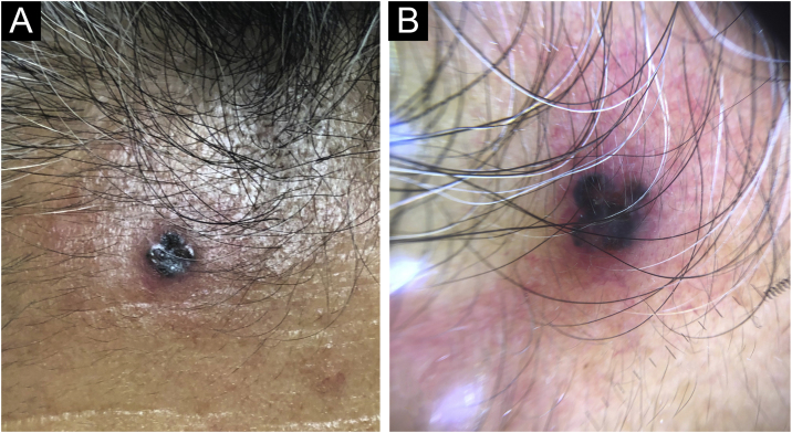 Figure 1
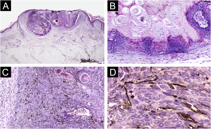 Figure 2
Peer Reviews
No public reviews on file for this paper yet. If you reviewed it on a platform where reviews are public (OpenReview, ICLR, NeurIPS, ICML), you can paste yours below so the community can read it here.
Videos
No videos yet. Explain this paper in a talk, walkthrough, or lecture? Add one.
Taxonomy
TopicsCancer and Skin Lesions · Nonmelanoma Skin Cancer Studies · Cutaneous Melanoma Detection and Management
Dear Editor,
A 74-year-old woman presented to our outpatient dermatology clinic with a 4-year history of an asymptomatic, pigmented tumor located on the forehead. On physical examination, we found a 5 mm, dark brown colored papule, surrounded by an erythematous rim. Past medical history was unremarkable. On dermoscopy, we found blue-gray ovoid nests, ulceration and peripheral telangiectasias (Fig. 1). The tumor was clinically diagnosed as pigmented basal cell carcinoma. A cutaneous biopsy was performed, and the histopathologic study revealed an epithelial, well-circumscribed neoformation that was composed of basophilic cells with hyperchromatic nuclei, a scarce cytoplasm, and prominent nucleoli. Mixed with these basaloid cells, there were multiple cells with basophilic nuclei and eosinophilic cytoplasm that were arranged in small nests. Sparse ghost cells were also found. There were multiple dendritic and pigmented melanocytes as well as areas of compacted keratinization (Fig. 2). Immunohistochemical study with BerEP4 turned positive in basaloid areas. Melanocytic matricoma diagnosis was concluded and a complete tumor resection was performed. The patient remained clinically disease-free during follow-up consultation.Figure 1. Physical examination. (A) A dark brown-black colored papule. (B) Dermoscopy. Blue-gray ovoid nests and ulceration, surrounded by a 3 mm erythematous and elevated rim.Figure 1. Figure 2Histopathology findings (A) Well circumscribed tumor arranged in lobes (Hematoxylin & eosin, 4×). (B) Basaloid and ghost cells with compact keratinization (Hematoxylin & eosin, 10×). (C) Basaloid cells and numerous dendritic melanocytes (Hematoxylin & eosin, 20×). (D) Dendritic pigmented melanocytes (Hematoxylin & eosin, 40×).Figure 2
Melanocytic matricoma is considered an adnexal tumor with matrical differentiation.1, 2 This newly described neoformation predominates in males, and it is strongly associated with sun-damaged skin in elderly patients, with a mean age of 71 years at presentation.3, 4 Only 32 cases have been reported in international literature. This uncommon tumor is predominantly located on the head, particularly in the nose and preauricular area, but it has also been reported on the neck, trunk, and extremities.4 Its classical clinical presentation is described as a small, well-circumscribed, nodular tumor with an asymmetric dark pigmentation.1, 4 Melanocytic matricoma is a biphasic tumor that comprises an epithelial component with matrical differentiation, and a melanocytic component with dendritic melanocytes. The epithelial component consists of basaloid cells with scarce cytoplasm, round nuclei, dotted nuclear chromatin, and prominent nucleoli. These basaloid cells might acquire mild to moderate pleomorphism with a slightly elevated mitotic activity, thus denominated matrical and supramatrical cells. These cells show an abrupt or gradual transition to ghost cells. In contrast, the melanocytic component appears as a well-circumscribed arranged nodule that is composed of melanocytes, intermixed with matrical and supramatrical cells, as well as foci of ghost cells. The epithelial component shows positivity for cytokeratin and beta-catenin, whereas dendritic melanocytes are highlighted by HMB-45, S-100, and Melan-A.5 Most tumors involve superficial to deeper dermis, without an evident epidermal or adnexal connection.1 The histopathologic and immunohistochemical findings suggest that melanocytic matricoma resembles anagen hair growth. Therefore, melanocytic matricoma is currently classified as a cutaneous adnexal tumor with both follicular and matrical differentiation.4
Clinical differential diagnosis includes basal cell carcinoma, melanoma, and hemangioma, but the main clinical differential diagnosis is pilomatrixoma1 (Table 1).6, 7, 8 This benign cutaneous tumor is found predominantly in young females (average 20 years), localized frequently on the neck and extremities, and it is clinically presented as a multilobulated and firm subcutaneous nodule. In contrast, histopathologic differential diagnosis includes tumors with matrical differentiation, such as pilomatrixoma, pigmented pilomatrixoma, and basal cell carcinoma with matrical differentiation (Table 2).9Table 1. Differential diagnosis of melanocytic matricoma.4, 6.Table 1. Melanocytic MatricomaPilomatrixomaBasal Cell CarcinomaMelanomaCommon age groupElderly individualsChildren, younger than 10 years.Adults, elderlyYoung and middle-aged individualsSexM > FF > MM > FF > MSize (cm)0.2 ‒ 1.50.5 ‒ 30.5 ‒ 10VariableClinical FeaturesPigmented nodule, polypoid or exophytic, rarely ulcerated.Solitary, asymptomatic, slow growing, cystic, or firm nodule.Slow growing, ulcerated nodule or plaque.Asymmetric macule or nodule with irregular borders, might present variations in color within the lesionSun Damaged SkinSignificantNot significantSignificantNot significantDermoscopyHomogeneous blue or patched pigmentation. Fork-shaped glassesBlue pigmentation, dilated vessels.Large gray-blue ovoid nests, multiple blue-gray globules, maple leaf-like areasAtypical network, blue whitish veil, atypical vascular pattern, irregular globules, irregular streaks, regression structuresSiteHead and neck, upper extremities.Head and neck, upper extremities.Head and neck, upper extremities (arms and hands).Trunk, limbs, acral regions, headTable 2Histopathological differences between melanocytic matricoma, pigmented pilomatrixoma and pigmented basal cell carcinoma with matrical differentiation.4, 9, 10Table 2. Pigmented PilomatrixomaMelanocytic MatricomaPigmented Basal Cell Carcinoma with Matrical DifferentiationEpidermis involvementAnyAtrophic, or hyperkeratotic with acanthosisAnyHistopathological patternNodular or multinodular with cystic areasSingle solid well-defined nodule, usually no cystic areasNodular pattern accompanied by either superficial pattern, cystic change or an infiltrative patternLocation in the dermisIn deep dermis with frequent extension to adipose tissueIn superficial to medium reticular dermisDermis to subcutaneous tissue in some casesCell typesVariable mixture of basaloid, transitional and ghost cellsMatrix and supra-matrix cells, few occasional ghost cellsBasaloid follicular and germinative cells, ghost cells with basaloid appearing matrical cells in peripheryKeratinization TypePilar and sometimes infundibular keratinizationPilar keratinizationCentral abrupt matrical keratinizationHistopathological alterations of the dermisConnective tissue: Blood vessels, infiltrate of mixed inflammatory cells, giant foreign body cells.Sclerotic stromal response, actinic elastosisActinic elastosisSometimes: hemosiderin, melanin, bone and, rarely, amyloid deposition.Calcium depositionIn 80% of casesInfrequentPresented in some casesForeign body reactionVery frequentInfrequentInfrequentGhost cellsIn the center of the tumor, almost always presentPresent in small foci mixed with pigmented, dendritic melanocytesPresent in most of the tumorMitosisThere may be cytological atypia and mitotic figuresThere may be cytological atypia and mitotic figuresPresent cytologic atypia and high mitotic figuresMelaninMelanin is rare, some dendritic melanocytesPigmented, inside melanophages, and dentritic melanocytes distributed asymmetrically.Tumor cells with melanin in cytoplasm. No prominence of intra tumoral melanocytes
The importance of recognizing this recently described tumor is based on its unknown prognosis and lack of treatment options, besides surgery. Therefore, it is important to consider melanocytic matricoma in elderly, sun-damaged skin patients with a newly discovered pigmented neoplasm, besides pigmented basal cell carcinoma and melanoma, requiring wider surgical margins and a closer follow-up. It is essential to report all melanocytic matricoma cases to establish its clinical course and prognostic features.
Financial support
None declared.
Authors’ contributions
Teresa Alonso-de-León: Writing of the manuscript or critical review of important intellectual content; data collection, analysis, and interpretation; effective participation in the research guidance; critical review of the literature; final approval of the final version of the manuscript.
Carlos Barrera-Ochoa: Critical review of the literature; data collection; analysis and interpretation.
Luis Enrique Cano-Aguilar: Writing of the manuscript or critical review of important intellectual content; data collection, analysis and interpretation; effective participation in the research guidance; intellectual participation in the propaedeutic and/or therapeutic conduct of the studied cases; critical review of the literature.
Katia Lizette Munguia-Galeano: Critical review of the literature.
Jorge Felipe Flores-Ochoa: Critical review of the literature.
Maria Elisa Vega-Memije: Effective participation in the research guidance; intellectual participation in the propaedeutic and/or therapeutic conduct of the studied cases; critical review of the literature.
Conflicts of interest
None declared.
The reference list from the paper itself. Each links out to its DOI / PubMed record.
- 1Carlson J.A.Healy K.Slominski A.Mihm M.C.Jr Melanocytic matricoma: a report of two cases of a new entity Am J Dermatopathol 2119993443491044677510.1097/00000372-199908000-00006 · doi ↗ · pubmed ↗
- 2Rizzardi C.Brollo A.Colonna A.Brutto R.L.Melato M.A tumor with composite pilo-folliculosebaceous differentiation harboring a recently described new entity-melanocytic matricoma Am J Dermatopathol 2420024934971245460210.1097/00000372-200212000-00007 · doi ↗ · pubmed ↗
- 3Williams C.M.Bozner P.Oliveri C.V.Horenstein M.G.Melanocytic matricoma: case confirmation of a recently described entity J Cutan Pathol 3020032752781268096110.1046/j.0303-6987.2003.042.x · doi ↗ · pubmed ↗
- 4Horenstein M.G.Kahn A.G.Pathologic quiz case: a 69-year-old man with a brown-black facial papule. Melanocytic matricoma Arch Pathol Lab Med 1282004 e 16341550408310.5858/2004-128-e 163-PQCAYM · doi ↗ · pubmed ↗
- 5Aranguren-López I.Ibarbia-Oruezabal S.Segués-Merino N.Melanocytic matricoma: a rare tumor that can mimic melanoma An Bras Dermatol 9720228338343608818510.1016/j.abd.2022.08.001PMC 9582867 · doi ↗ · pubmed ↗
- 6Leonardi G.C.Falzone L.Salemi R.ZanghìA.Spandidos D.A.Mccubrey J.A.Cutaneous melanoma: from pathogenesis to therapy (Review)Int J Oncol 522018107110802953285710.3892/ijo.2018.4287 PMC 5843392 · doi ↗ · pubmed ↗
- 7Kibbi N.Kluger H.Choi J.N.Melanoma: clinical presentations Cancer Treat Res 16720161071292660186010.1007/978-3-319-22539-5_4 · doi ↗ · pubmed ↗
- 8Russo T.Piccolo V.Ferrara G.Agozzino M.Alfano R.Longo C.Dermoscopy pathology correlation in melanoma J Dermatol 4420175075142844735510.1111/1346-8138.13629 · doi ↗ · pubmed ↗