Arteriovenous Fistula Caused by Ruptured Abdominal Aortoiliac Aneurysm
Leizhi Ku, Yuhang Wang, Xiaojing Ma

TL;DR
A ruptured abdominal aortoiliac aneurysm leading to an arteriovenous fistula is rare but life-threatening and needs quick diagnosis and treatment.
Contribution
Highlights the rare but severe complication of an arteriovenous fistula from a ruptured aneurysm and emphasizes the need for prompt medical action.
Findings
Ruptured aorto-iliac aneurysms can lead to life-threatening arteriovenous fistulas.
Prompt diagnosis and treatment are crucial for managing this rare complication.
Abstract
Teaching point: A ruptured aorto-iliac aneurysm, complicated by an iliac arteriovenous fistula, is rare but has a possibly fatal outcome and requires prompt diagnosis and appropriate treatment.
Genes, proteins, chemicals, diseases, species, mutations and cell lines named across the full text — each resolved to its canonical identifier and authoritative record.
Click any figure to enlarge with its caption.
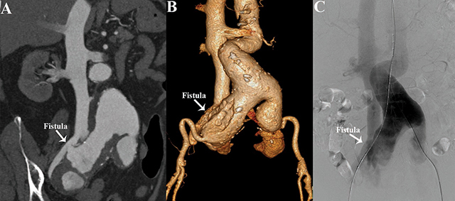 Figure 1
Peer Reviews
No public reviews on file for this paper yet. If you reviewed it on a platform where reviews are public (OpenReview, ICLR, NeurIPS, ICML), you can paste yours below so the community can read it here.
Videos
No videos yet. Explain this paper in a talk, walkthrough, or lecture? Add one.
Taxonomy
TopicsAortic aneurysm repair treatments · Infectious Aortic and Vascular Conditions · Vascular Procedures and Complications
Case History
A 74–year-old female presented with an 8-hour history of sudden-onset lower abdominal pain. The physical examination revealed a blood pressure of 96/52 mmHg. Her symptoms were significantly relieved after she received analgesic therapy and cardiovascular support. A contrast-enhanced computed tomography angiography (CTA) examination was performed. Maximum-intensity projection CTA showed an infrarenal abdominal aortic aneurysm extending into the common iliac artery. The right common iliac artery aneurysm ruptured into the right common iliac vein, with the early appearance of contrast medium in the dilated inferior vena cava, indicative of the presence of an arteriovenous fistula (AVF) (Figure 1A). Three-dimensional volume-rendering CTA identified the aneurysms and the fistula orifice (Figure 1B). Abdominal aortography confirmed that the fistula was located between the right common iliac artery aneurysm and the common iliac vein (Figure 1C and Video S1). The patient underwent an emergency endovascular stent-graft repair.
(A and B) Multiplanar reconstructions (MPR) and 3D volume rendered (3D-VR) CTA images show a large ruptured abdominal aortoiliac aneurysm causing an ilio-iliac arteriovenous fistula. (C) Abdominal aortography Abdominal aortography reveals ilio-iliac AVF with a fistula size of 8.5mm.
Comment
A spontaneous ilio-iliac arteriovenous fistula (AVF) caused by a ruptured abdominal aorto-iliac aneurysm is a rare condition. Patients with this condition may present with manifestations of high-output cardiac failure, vague abdominal pain, hematuria, lower extremity swelling, or multi-organ failure. Early diagnosis enables significant clinical improvement due to the early reversal of multi-organ complications. CTA is thus a key imaging modality to identify the site of the fistula, especially when clinical symptoms are not typical, and provides a road map for endovascular management [1]. Surgical mortality of an AVF is high because of hemorrhagic shock, high-output heart failure, renal failure, and massive blood loss during the operation. Endovascular repair is a less invasive alternative, with less hemorrhagic complications and limited hemodynamic deterioration during the intervention. Several reports show good early results of endoluminal repair of an AVF.
The reference list from the paper itself. Each links out to its DOI / PubMed record.