Characterization of Retinal Microvascular Abnormalities in Birdshot Chorioretinopathy Using OCT Angiography
Aman Kumar, Alexander Zeleny, Sunil Bellur, Natasha Kesav, Enny Oyeniran, Kübra Gul Olke, Susan Vitale, Wijak Kongwattananon, H. Nida Sen, Shilpa Kodati

TL;DR
This study uses OCT angiography to show that birdshot chorioretinopathy causes reduced retinal blood vessel density, which could help track disease severity.
Contribution
The study introduces OCT angiography as a potential biomarker for monitoring retinal microvascular changes in birdshot chorioretinopathy.
Findings
Birdshot chorioretinopathy eyes showed significantly lower vessel density compared to controls in both superficial and deep capillary plexus.
Enlarged foveal avascular zone area was observed in BCR eyes, particularly in the deep capillary plexus.
Longer disease duration and treatment-naive status were associated with decreased vessel density in BCR patients.
Abstract
To characterize changes in the retinal microvasculature in eyes with birdshot chorioretinopathy (BCR) using OCT angiography (OCTA). Retrospective, observational, single center. Twenty-eight patients (53 eyes) with BCR and 59 age-matched controls (110 eyes). En face OCTA images of the superficial capillary plexus (SCP) and deep capillary plexus (DCP) of each eye were assessed for the presence of microvascular abnormalities and used to measure the vessel and foveal avascular zone (FAZ) areas. A longitudinal analysis was performed with a representative cohort of 23 BCR eyes (16 patients) at baseline and at a 2-year time point. Whole-image vessel density (VD, %), extrafoveal avascular zone (extra-FAZ) VD (%), and FAZ area (%) were calculated and compared between control and BCR eyes. The frequency of microvascular abnormalities in BCR eyes was recorded. In the SCP, increased…
Genes, proteins, chemicals, diseases, species, mutations and cell lines named across the full text — each resolved to its canonical identifier and authoritative record.
Click any figure to enlarge with its caption.
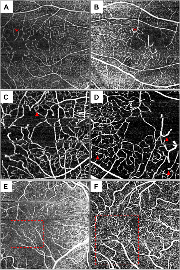 Figure 1
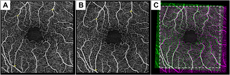 Figure 2
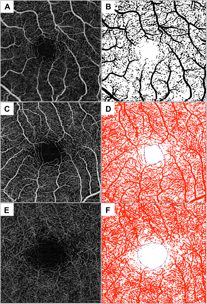 Figure 3
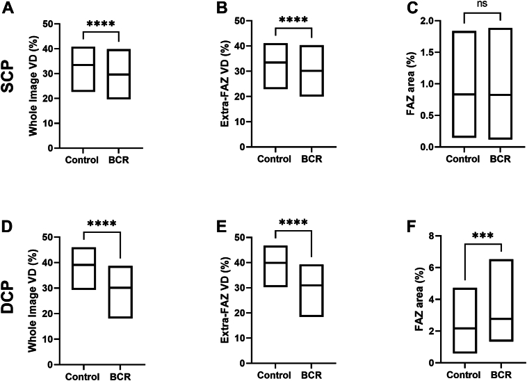 Figure 4
Peer Reviews
No public reviews on file for this paper yet. If you reviewed it on a platform where reviews are public (OpenReview, ICLR, NeurIPS, ICML), you can paste yours below so the community can read it here.
Videos
No videos yet. Explain this paper in a talk, walkthrough, or lecture? Add one.
Taxonomy
TopicsOcular Diseases and Behçet’s Syndrome · Retinal and Optic Conditions · Retinal Diseases and Treatments