Sleep and diurnal alternative polyadenylation sites associated with human APA-linked brain disorders
Carlos C. Flores, Nickolas A. Pasetto, Hongyang Wang, Alexander G. Dimitrov, Jon F. Davis, Zhihua Jiang, Christopher J. Davis, Jason R. Gerstner

TL;DR
This paper explores how sleep and circadian rhythms affect alternative polyadenylation in the brain and links these changes to human brain disorders.
Contribution
The study identifies APA sites in rat brains related to sleep and circadian rhythms and connects them to human brain disorder susceptibility genes.
Findings
Unique APAs in rat brains are associated with time-of-day and sleep deprivation.
Sleep-related PASs are linked to human brain disorder susceptibility genes.
APA changes may influence mRNA function and contribute to neurological diseases.
Abstract
Disruption of sleep and circadian rhythms are a comorbid feature of many pathologies, and can negatively influence many health conditions, including neurodegenerative disease, metabolic illness, cancer, and various neurological disorders. Genetic association studies linking sleep and circadian disturbances with disease susceptibility have mainly focused on changes in gene expression due to mutations, such as single-nucleotide polymorphisms. The interaction between sleep and/or circadian rhythms with the use of Alternative Polyadenylation (APA) has been largely undescribed, particularly in the context of other disorders. APA is a process that generates various transcript isoforms of the same gene affecting its mRNA translation, stability, localization, and subsequent function. Here we identified unique APAs expressed in rat brain over time-of-day, immediately following sleep deprivation,…
Genes, proteins, chemicals, diseases, species, mutations and cell lines named across the full text — each resolved to its canonical identifier and authoritative record.
Click any figure to enlarge with its caption.
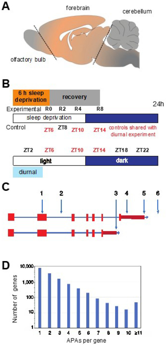 Figure 1
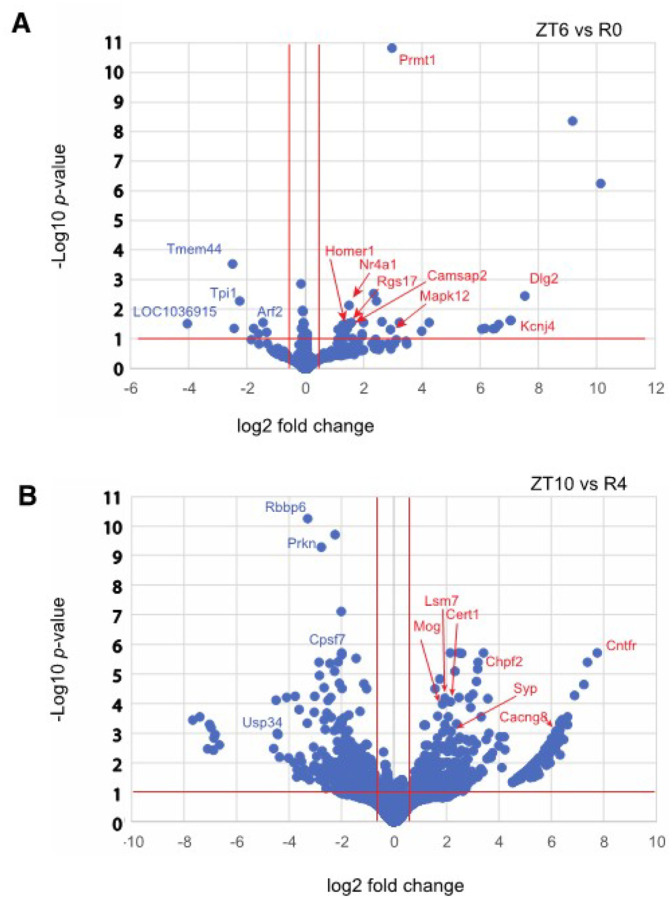 Figure 2
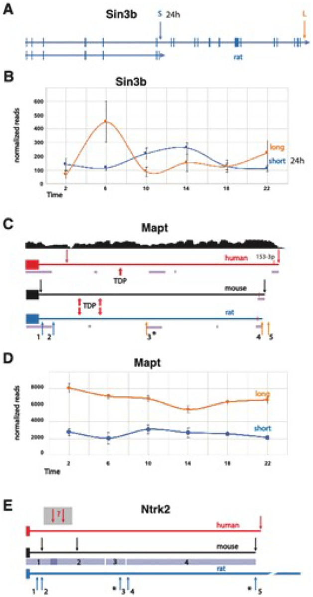 Figure 3
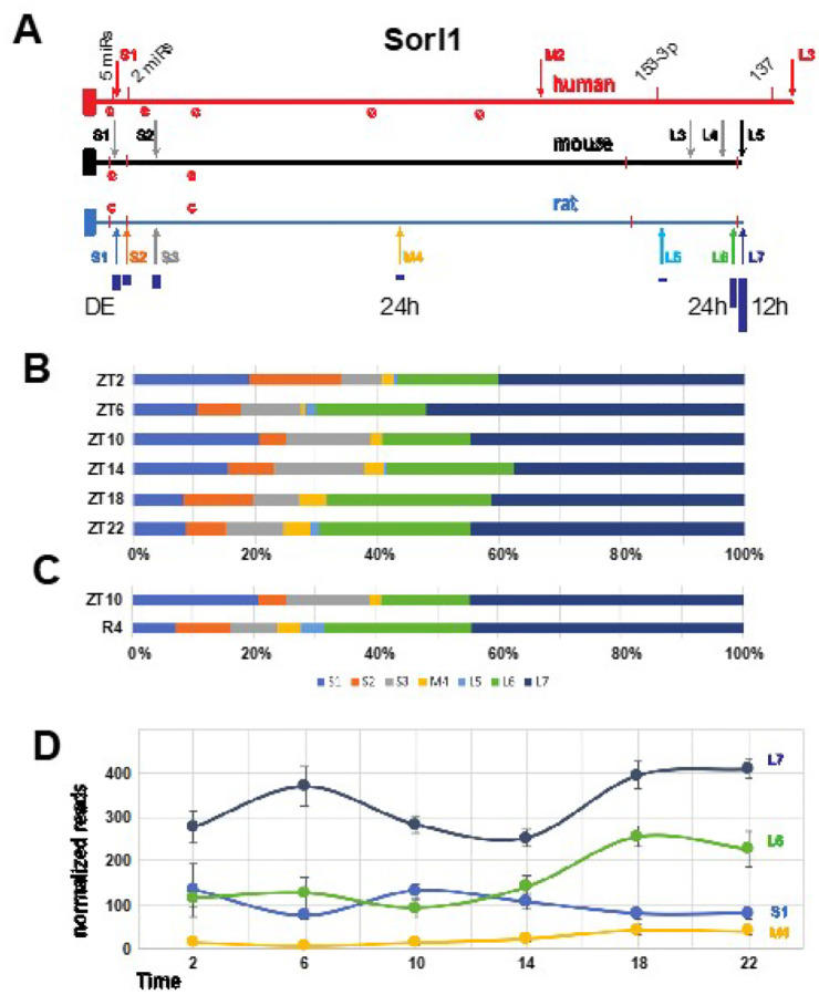 Figure 4
Peer Reviews
No public reviews on file for this paper yet. If you reviewed it on a platform where reviews are public (OpenReview, ICLR, NeurIPS, ICML), you can paste yours below so the community can read it here.
Videos
No videos yet. Explain this paper in a talk, walkthrough, or lecture? Add one.
Taxonomy
TopicsRNA Research and Splicing · Lipid metabolism and biosynthesis · RNA modifications and cancer
Introduction
Dysregulation of sleep and circadian rhythms can profoundly impact human health and compound disease^1,2^. Indeed, sleep disruption is associated with negative outcomes in cardiovascular, metabolic, immunologic, and cognitive health that can have substantial short- and long-term consequences^3^. Alterations in sleep and circadian rhythms are often observed with various brain disorders, including autism spectrum disorder, bipolar disorder, major depression, schizophrenia, Parkinson’s, and Alzheimer’s diseases^4–7^. Complicating the association between sleep and health is the fact that functional aspects of sleep remain largely undefined and inconclusive^8,9^; however, the use of evolutionarily distinct animal models to study sleep has historically offered keen insights^10,11^. For example, studies on circadian- and sleep-dependent gene-regulatory mechanisms in diverse species, including flies, rodents, and humans, have identified important phylogenetically conserved pathways with functional relevance^12–15^. Employing unbiased approaches, such as large-scale metabolomic, transcriptomic, and proteomic analyses, have also greatly aided in the generation of conceptual frameworks for characterizing sleep function in health^14,16^. Therefore, performing such discovery-based studies of sleep and circadian regulatory processes in model organisms will help define the fundamental biological mechanisms underlying sleep function and inform pre-clinical relevance for comorbidities of sleep dysfunction associated with poor health.
Alternative polyadenylation (APA) site usage is an important and often overlooked mechanism of gene regulation, that can affect mRNA stability, mRNA/protein targeting, translational competence, and generate alternative protein isoforms^17,18^. APA sites are common and occur most frequently in the 3’ untranslated region (3’ UTR) of mRNAs across phylogeny, with more than half of human genes having multiple polyadenylation sites (PASs) that generate alternative isoforms^19^. These isoforms can have altered coding sequences or 3’UTRs, resulting in the diversification of cis-regulatory elements (e.g., RNA binding protein sites, microRNA binding sites) that influence transcript abundance, trafficking, stability, and/or translation efficiency^20^. Furthermore, there’s growing evidence of cell-type-specific APA preference^21^. The involvement of APA in the context of sleep and circadian rhythms has been largely unexplored, with the few studies available mostly focused on peripheral organs^22,23^ and cells^24^. Here, we have characterized how APA site usage oscillates based on the time of day as well as how it is altered following acute changes in sleep pressure, specifically in the adult mammalian brain. Multiple methodologies have been developed for transcriptome-wide profiling and mapping of APA sites^25,26^. To complete this study, we performed whole transcriptome termini sequencing (WTTS-seq)^27,28^ analysis to profile the variations in APA usage that occur due to sleep pressure and daily rhythms in the rat forebrain. Over 31,000 PASs were recovered in total, with 45% of the represented genes having multiple APA sites. Interestingly, many of the PASs sequenced were not previously annotated in the rat genome. Moreover, a total of 2,011, (6%) of PASs cycled over the day, and 831 (3%) were homeostatically regulated following sleep loss following sleep loss or during recovery. Over half of all cycling or differentially expressed PASs were APAs, (i.e., in genes with ≥ 2 PASs). Given the importance of sleep^4–7^ and APA in health and disease^25,29,30^, we compared our sequencing results with results from a recent study that determined APA usage in human brain disorder susceptibility^31^. The genes found in both studies warrant further examination and could lead to new preclinical animal models to investigate these disorders.
To the best of our knowledge, the current study represents the first comprehensive, transcriptome-wide mapping of APA sites in adult mammalian brain tissue over the day-night cycle as well as following changes in sleep homeostasis. This global temporal dataset will be useful for future comparative studies that require the determination of baseline APA site usage profiles in the mammalian brain. Furthermore, our study underscores the importance of using alternative-omic approaches to characterize phylogenetically conserved genome-phenome information and reveals another expansive layer of complexity in sleep and circadian gene regulation that has not previously been documented.
Results
Identification of PASs in the rat forebrain.
Given the rat transcriptome is not as extensively annotated as the human or mouse, we first identified all PASs, including novel candidate PASs prior to determining changes in PAS usage. Replicate diurnal (central forebrains) were taken from five rats every four hours starting at two hours after lights on (i.e., ZT2, ZT6, ZT10, ZT14, ZT18 and ZT22) (Fig. 1a, b). RNA was purified from these samples and used to generate WTTS-seq cDNA libraries that were subsequently sequenced. Poly(A)-directed sequence reads were then mapped to the rat genome, giving rise to 31,757 PAS clusters (see Supplementary Table S1). Among the 31,757 PAS clusters identified, a sizable portion mapped to novel unannotated PASs, leaving 26,635 PASs that mapped to named loci (i.e., genes). Many APAs occur at different points within the longest 3’ UTR (Fig. 1c, sites 4 and 5). Some are distal to the longest documented 3’ UTR (site 6), while some occur in internal exons (site 1) or introns (sites 2 and 3) (Fig. 1c). In our data set of all PASs that mapped to genes, 45% mapped to genes with ≥ 2 APA sites, and 19% mapped to genes with ≥ 3 APA sites (Fig. 1d).
Identification of PASs that exhibit a daily cycle.
Periodicity of PAS expression was assessed using meta2d^32^. Diurnal (24 h period) oscillations were demonstrated for 2,011 PASs. Among these, 1,173 were in genes with ≥ 2 total APA sites, including ones in known diurnal transcripts, such as Dbp (diurnal in 2 of 2 APA sites recovered), Nr1d2 (in 1 of 1), Per2 (in 2 of 2), and Ntrk2 (in 2 of 10)^33^ (Table 1 and Supplementary Table S2).
We were interested whether rhythmic PASs might cluster predominantly into certain phases of peak expression, and whether APAs that share a common peak phase might also share some functional relationship. It was evident that some phases had very few APAs relative to other phases and the expression levels of many PASs peaked around ZT18–20 (Supplementary Fig. S1). When diurnal APAs from genes with ≥ 2 total APAs were grouped by phase, GO and pathway analysis on each group found that only phases 2, 10 and 18 had significantly over-represented terms. Phase 18 had the most, with the over-representation of multiple signaling pathways, including ‘neuron to neuron synapse’ and ‘post-synaptic specialization’ (Supplementary Table S4).
There is a growing appreciation that rhythms shorter than 24h are biologically relevant^35–39^. Thus, we evaluated the PASs data for ultradian cycling using meta2d with the period set to 12 h. Overall, 1,502 PASs that cycled with a 12 h period were identified (Supplementary Table S5). Of the 12 h cycling PASs, 1,198 were in genes, and after adjusting for genes with multiple 12h cycling APAs, there were 1,149 unique genes in the set. In total, 827 of the 12 h cycling APA sites were in genes that had ≥ 2 APAs, representing 778 unique genes. Pathway analysis on this set of 778 unique genes (Supplementary Table S6) showed that CREB phosphorylation and circadian entrainment were highly enriched, while GO analysis of this data set resulted in 16 GO terms related to the synapse.
PASs are differentially expressed after sleep deprivation and during recovery sleep.
To investigate changes in APA site usage related to sleep pressure, rats were subjected to SD for 6 h from ZT0 to ZT6, and central forebrain tissue was collected immediately afterwards (R0). Additional animals were allowed to recover for 2, 4, or 8 h after SD (R2, R4 and R8) before tissue was collected. WTTS-seq data from these samples were compared to time-matched controls that were allowed to sleep undisturbed (ZT6, ZT8, ZT10 and ZT14). All groups consisted of 5 biological replicates. Our sequencing data showed that the most significant differences in expression were seen when we compared R0 with its control (ZT6) and R4 with its control (ZT10) (Supplementary Table S7 and Fig. 2). Interestingly, a Homer1a APA isoform is the most abundant at R0, R4 and ZT6, whereas a full-length isoform is dominant at ZT10 (Supplementary Fig. S2 a and b) Also, the expression of one APA isoform of Prmt1, was upregulated with high confidence after 6h of sleep deprivation (Fig. 2). PRMT1 protein regulates multiple stress response pathways^40,41^, which have a roll in acute sleep loss.
The gene names of differentially expressed APA sites from genes with ≥ 2 APAs were used for GO and pathway over-representation analysis (Table 3). ZT6 vs R0 only had significant results for GO while ZT10 vs R4 had significant GO and pathway results.
Discussion
APA site usage is an understudied aspect of gene regulation. Although APA sequencing can reveal changes in overall gene expression, it’s designed to focus on changes in APA usage and cannot reveal differences in splicing or transcription start sites (TSSs). On the other hand, bulk RNA-seq analysis often ignores APA, TSS and splice isoforms to simply assess reads per gene. Currently it would be very difficult to enumerate copies of all the mRNA isoforms for each gene. Yet appreciation is growing for the importance of APA sites in regulating mRNA stability^17,42^, mRNA/protein localization^20,43,44^, and human disease^31,45^.
Rhythmic APA site usage has been uncovered in the mouse liver^22,23,46^, and in temperature-entrained cultured cells, circadian APA usage occurs in many genes and can regulate expression of specific central clock genes^24^. Still, alternative poly(A) site usage hasn’t been given much attention in the sleep and circadian field. We therefore initiated this investigation into the conjunction of APA with sleep and diurnal expression. As far as we are aware, the current study is the first to examine APA sites related to circadian rhythms and sleep pressure in any mammalian brain. There are several, diverse ways in which data from this study can translate into biological relevance as described in the examples below.
Here, we observed that 6% of all PASs cycled with a 24 h period. One of the top pathways identified for the diurnal APA gene set was ‘circadian entrainment’ (Table 2). Since transcription-translation feedback loops are central to circadian regulation, this may not be surprising, but APA site usage suggests a more complex role^24,46^. For example, we find that one Sin3b APA follows a diurnal rhythm (Fig. 3a, b). Sin3b encodes short and long variants conserved in mammals. The short variant binds to CRY1 but cannot bind HDAC1^47^. The long isoform is implicated in regulation of Per1/Per2 transcription^48^, along with many other genes^49^. In our data, long Sin3b APA reads constitute the predominant isoform at ZT6 and ZT22, while the short, diurnal isoform is the most abundant one at ZT10, ZT14 and perhaps ZT2 (Fig. 3b). Sin3b transcript levels in mouse hippocampus have previously been reported to be affected by sleep deprivation^50^, although this effect was not observed using TRAP-seq^51^, suggesting post-transcriptional processing can lead to changes in sleep-dependent differential expression. Together with our work, this example highlights the importance of utilizing various “-omic” approaches to properly decipher the complexity of molecular processing tied to changes in behavioral state in the brain.
Additional significant pathways emerged from the diurnal APAs, such as Oxytocin, Ephrin, and MAPK signaling that have demonstrated links to the circadian clock^52–54^. In the GO analysis of the diurnal genes with multiple PASs, we discovered that terms related to the synapse (12), protein localization (6), and vesicles (7) (Table 2 and Supplementary Table S3) were enriched suggesting APAs are poised to affect neural communication.
A large proportion of diurnal APAs had expression peaks around ZT20 (Supplementary Fig. S1). Considering that rats are nocturnal, this is similar to what has been seen for bulk transcripts in several human tissues, including brain^55^. Interestingly, among the identified diurnal APA sites, 3 were in genes for RNA-binding proteins (Celf2, Elavl3, and Rbfox1) whose expressions correlate with more distal APA usage^47^. Peak expression of these three genes is from ZT21 to ZT1, so it would be interesting to see if transcripts of predicted targets tend to be longer at these times.
In addition to the 24 h circadian rhythm, recent studies have also demonstrated the existence of cell-autonomous ultradian clocks that run independently of the circadian clock to regulate 12 h oscillations in gene expression and metabolism^35–39^. Here we found that 5% of all PASs cycle with a 12 h period. Further analysis of these genes showed enrichment of gene ontology terms and pathways such as “regulation of trans-synaptic signaling” and “protein-protein interactions at synapses” (Supplementary Table S6), indicating that APAs could function to regulate cyclic actions of cell signaling and communication.
Gene expression studies following changes in sleep homeostasis have largely ignored alternative polyadenylation. Of the 31,795 total PASs characterized in rat forebrain in our study, we determined that 2.5% were differentially expressed with sleep deprivation and recovery sleep. We also observed 6 GO terms significantly enriched following 6 hours of sleep loss and 26 following 4 hours of recovery sleep (Table 3).
Human APA isoforms have been linked to many neurological disorders^31^. Among the genes that we identified to have rhythmic expression of APA sites or had APA sites that were affected by sleep pressure, we found that 46 have also been correlated with brain disorder susceptibility (Table 4). For example, the human MAPT/TAU gene produces transcripts containing short or long 3’ UTRs, and a 3’ single-nucleotide polymorphism, (SNP) is associated with both 3’ UTR length and risks for 8 neurological disorders, including Alzheimer’s and Parkinson’s diseases^31^. Homozygosity of the more common SNP variant is associated with short MAPT3’ UTRs, homozygosity of the less common SNP variant is associated with long 3’ UTRs, and heterozygosity is associated with 3’ UTRs of intermediate lengths. In our rat APA data, there were both short and long 3’ UTR forms (5 in total) of the Mapt gene that were identified (Fig. 3c, d). Only two are currently annotated in the rat genome and one of the newly discovered APAs was observed to cycle with time-of-day. In mouse, binding of the ALS-associated protein TDP-43 to two sites in the 3’ UTR of Mapt has been shown to destabilize the mRNA^56^. In Alzheimer’s disease, the expression level of TDP-43 protein is often low, and TAU is overexpressed and eventually forms neurofibrillary tangles. The two TDP-43 binding sites that were experimentally determined in mouse are conserved in sequence and position in the rat gene, implying that transcripts with shorter 3’ UTRs would not be affected by TDP-43, while longer ones could be destabilized^56,57^. The presence of at least one putative TDP-43 binding site in the human MAPT 3’UTR suggests that this may be contributing to the neurological disorder risk.
Ntrk2 is among the APA TWAS genes linked to anxiety^31^ and has been associated with autism in other studies^58^. We found strong time-of-day oscillations of the 2 most abundant APA sites of the short, tyrosine kinase deficient (TK−) Ntrk2 isoform. The TK− isoform of Ntrk2 has several known functions, including a dominant negative effect on the full-length TK + isoform during neuronal proliferation, differentiation, and survival. In addition, the TK− version promotes filopodia and neurite outgrowth; sequesters, translocates, and presents BNDF; and affects calcium signaling and cytoskeletal modifications in glia^59^. Our WTTS-seq data revealed short, medium, and long 3’ UTRs in the rat Ntrk2 TK− isoform (Fig. 3e). In mice, the longer Ntrk2 TK− transcripts are preferentially targeted to apical dendrites^60^. Since the sequence of the rat 3’ UTR is highly conserved with the mouse sequence, it is plausible that an analogous dendritic localization mechanism is also in use in the rat (Fig. 3e). Interestingly, ‘Ntrk signaling’ was one of the pathways over-represented in the diurnal APA genes (Supplementary Table S3). APA sites in Src, Frs2, Atf1, Nras, Sh3gl2, Ntrk3, Mapk1, Grb2, Pik3r1, and Mapk14 contributed to this enrichment.
Four different APAs from the Sorl1 gene exhibited significant changes in our analyses; two diurnal, one cycled with a 12 h period, and one was reduced during recovery from sleep deprivation (Fig. 4). In total, there were seven APAs in the Sorl1 3’UTR, three short, one medium and two long. The longest and most abundant isoform cycles per 12 h, the second longest and medium ones are diurnal and the shortest isoform is differentially expressed after SD (Fig. 4). SORL1 encodes an endosomal recycling receptor^61^, and a deficiency of SORL1 as well as many polymorphisms are strong risk factors for AD^62,63^. The mouse and human 3’ UTRs share extensive similarities including 5 APAs in mouse and 3 in human based on the PolyA_DB v3 (https://exon.apps.wistar.org/polya_db/v2/) and UCSC database^64^. Four microRNA binding sites with high probability of preferential conservation are in good alignment (TargetScanHuman v8.0)^65^. The first motif can be bound by five miRNAs (miR-25–3p, miR-32–5p, miR-92–3p, miR-363–3p, and miR-367–3p), while the second contains overlapping 7mer and 8mer motifs bound by miR-128–3p and miR-27–3p, respectively. The final two more distal sites are recognized by miR-153–3p and mir-137 (Fig. 4a). Sequences matching the consensus binding site for CPEB are present in the 3’ UTRs of all three species, with 2 in very good alignment. Cytoplasmic polyadenylation element binding protein (CPEB) facilitates mRNA trafficking to synapses and local translation^66,67^, and we have previously shown that the core clock-controlled Fabp7 mRNA^68,69^ contains functional CPE sites in its 3’UTR to regulate translation^70^. Since APOE4, an apolipoprotein E variant with increased risk of AD^71^, disrupts FABP7 interaction with sortilin, (an APOE receptor similar to Sorl1), to interfere with neuroprotective lipid signaling^72^, this suggests circadian variation in local translation of CPEB-mediated polyadenylation of target mRNAs may be a generalizable mechanism that modulates AD susceptibility through downstream lipid pathways. Any one or more of these conserved features could lead to conserved functional consequences dependent on APA choice.
One caveat to our approach is that WTTS-seq generates Ion Torrent PGM sequences which may retain more noise compared to Illumina platform reads and since only Illumina has the option of paired-end reads, there can be more uncertainty in mapping Ion Torrent reads. Our strategy was to capture the maximum number of PASs, including the discovery of novel PASs, and the rat genome is not as thoroughly annotated as some other vertebrate species, we therefore included potentially intergenic reads. In our analysis, we found 5,122 PASs and 318 diurnal PASs that mapped outside of known genes, and many APAs within genes mapped to regions in which 3’ ends have yet to be annotated. Based on prior WTTS-seq data sets and other PAS mapping approaches, some portion of our PASs could be method-based artifacts^27,73^, (see Zhou et al.^27^ Figs. 3, 4 and 5). In this, our initial PAS survey, we assayed a large portion of the brain. Therefore, future studies in restricted brain structures or cell types will be required to uncover APAs that cycle or are differentially expressed at a finer scale. Overall, the newly discovered PASs should add valuable insights into regulation of the rat transcriptome and for characterizing PAS usage in the mammalian brain.
Here we used an unbiased discovery-based approach for uncovering novel APA usage following time-of-day or changes in sleep pressure in mammalian brain. These data leverage a call to action for additional work to elucidate the core mechanisms of PAS usage in the brain and to examine the capacity of APA to affect the transcriptomes and proteomes that regulate central brain processes known to be altered by time-of-day and sleep/wake homeostasis. Moreover, it known that PAS usage varies across brain region and cell type^21^ (i.e., substructure-, circuit-, laminar- or nucleus-specific)^74^. These hypothesis-generating data provide an impetus for continued research aimed at delineating how sleep and circadian rhythms impact mental health and neurodegenerative disease.
Methods
Subjects.
All animal procedures were carried out in accordance with the National Institutes of Health Guide for the Care and Use of Laboratory Animals and ARRIVE and OLAW guidelines and approved by the WSU Institutional Animal Care and Use Committee (IACUC; ASAF# 6804). Male Long Evans rats (7–9 weeks old) were housed in pairs at 22 ± 2°C on a 12:12 h light-dark cycle. The rats were acclimated to this light cycle for at least 10 days prior to tissue collection, with water and chow ad libitum. Cages were cleaned weekly (between 8 and 11 AM) unless the rats were being euthanized within 24 h. Thirty rats were randomly assigned to one of six groups (n = 5/group) that were sampled every 4 h, beginning 2 h after light onset (zeitgeber time (ZT)) (i.e., ZT2, 6, 10, 14, 18, and ZT22). For the sleep deprivation (SD) study, twenty rats were randomly assigned to 6 h SD from ZT0–6, wherein rats were kept awake by an automated bedding stir bar (Pinnacle) at the bottom of a cylindrical cage. The bar was set to rotate for 4 s, randomly changing rotation direction, and stopped for a random interval ranging from 10 to 30 s^75,76^. Following SD, rats (n = 5/time point) were euthanized immediately (R0) by live decapitation or were returned to their home cage for 2 h (R2), 4 h (R4), or 8 h (R8) under red light without disruption before sampling. Five additional rats were euthanized at ZT8 as undisturbed, time-matched controls. The other time-matched controls with undisrupted sleep (i.e., ZT6, 10, and 14) were taken from the corresponding time-of-day matched samples described above.
Tissue Collection.
Rats were decapitated by guillotine under normal room light (ZT2–10) or under dim red light (ZT14–22). Following decapitation, forebrains were resected (Fig. 1A), frozen in 2-methylbutane suspended in dry ice, and then stored at −80°C until homogenization for RNA extraction.
RNA isolation.
Just before RNA isolation, forebrains were removed from − 80°C storage and placed on dry ice. Prior to use, a stainless-steel mortar and pestle were cleaned with RNase Zap (Thermo Fisher) and 70% ethanol. The mortar was then partially filled with liquid nitrogen before a forebrain was added, pulverized, and placed in a conical tube. Between each sample, the mortar and pestle were cleaned with 70% ethanol. A small aliquot of sample was removed for RNA isolation using Trizol Reagent (Invitrogen), according to the manufacturer’s instructions. Purified RNA was resuspended in water, and concentration and purity were measured with a Nanodrop spectrophotometer (Thermo Fisher). Samples were stored at −20°C until further processing was performed.
Library preparation.
WTTS-seq libraries were prepared as described by Zhou et al.^27^. Briefly, total RNA (2.5 μg) was incubated at 70°C with 10X Fragmentation buffer (Invitrogen) for 3 min. The fragmentation reaction was halted by the addition of Stop Solution and incubation on ice for at least 2 min. Next, poly(A) + RNA was purified from the fragmented total RNA with Dynabeads Oligo (dT)25 (Invitrogen), according to the manufacturer’s directions, and used for first-strand cDNA synthesis in a 20 μL reaction mixture. First, 1.0 μL of barcode primer (100 μM) and 1.0 μL of a common SMART primer (100 μM) were annealed to the poly(A) + RNA template by heating to 65°C for 5 min and incubating on ice for at least 2 min. Next, 4.0 μL of 5X First-strand buffer (Invitrogen), 1.0 μL of SuperScript III reverse transcriptase (Invitrogen), 1.0 μL of 0.1 M dithiothreitol, 2.5 μL of 10 mM dNTP, and 1.0 μL of RNase OUT (Invitrogen) were added to the mixture. First-strand cDNA was synthesized by incubating the mixture at 40°C for 90 min in the presence of library-specific adaptors. Synthesis was terminated by heating the mixture at 70°C for 15 min. RNases I (100 U/μL; Invitrogen) and H (2 U/μL; Invitrogen) were subsequently added and incubated with the mixture at 37°C for 30 min to hydrolyze the remaining single-stranded RNA molecules and ensure that only single-stranded cDNA remained. RNase activities were terminated by heating the samples at 70°C for 20 min. Following purification with solid-phase reversible immobilization (SPRI) beads, second-strand cDNA was synthesized from first-strand cDNA by asymmetric PCR. In addition to the cDNA, the 50 μL PCR reaction contained 1.0 μL of Phusion Hi-Fidelity DNA polymerase, 10.0 μL of 5X HF buffer, 1.0 μL of 0.4 μM barcode primer, 1.0 μL of 0.8 μM common primer, 1.0 μL of 10 mM dNTP and nuclease-free water. The PCR reaction was carried out by heating at 95°C for 30 sec, followed by 20 cycles of 98°C for 10 s, 50°C for 30 s, and 72°C for 30 s, with a final elongation step at 72°C for 10 min. SPRI beads were used to purify and select 200–500 bp fragments from the final library. After quality control analyses, the size-selected library was sequenced with an Ion PGM Sequencer at the WSU Genomics Core Laboratory.
Data analysis
Raw read processing.
Raw data were obtained from 55 samples and stored in FASTQ format. We filtered raw reads with the FASTQ quality filter in the FASTX Toolkit (v0.0.13), allowing for a minimum score of ≥ 10 for ≥ 50% of bases (http://hannonlab.cshl.edu/fastx_toolkit/). We trimmed T nucleotides or T-rich sequences located at the 5’ ends of the reads using Perl scripts, as described previously^27^. Trimmed reads of at least 16 bp in length were kept for further analysis.
Read mapping and poly(A) site clustering.
For each data set, the processed reads were aligned to the Rattus norvegicus genome (mRatBN7.2/rn7) using the torrent mapping program (TMAP, v3.4.1; http://github.com/iontorrent/tmap) with the unique best hits parameter (-a 0). Raw PASs supported by the uniquely mapped reads were extracted from SAM files and mergeded into a polyadenylation tag (PAT) file with a script previously used for WTTS-seq (freely available by contacting Dr. Zhihua Jiang, Washington State University). The PAT files were merged to determine the final PASs for all samples. PASs within 25 nucleotides of one another were grouped into one polyadenylation site cluster (PAC) using GetPolyaSiteCluster^77^. PACs were filtered taking into account the library size. For libraries that had less than 1.7M reads, PACs were required to have ≥ 1 set of 5 biological replicates had ≥ 3 samples with ≥ 3 reads. For libraries with more than 1.7M reads, at least 3 samples with ≥ 4 reads were required.
Gene annotation and usage of poly(A) sites.
We annotated all the final PACs for PAS_ID, gene symbol, functional region, and other factors, as indicated, using Cuffcompare (v2.2.1)^78^, Perl scripts, and annotation file (GCF_000001895.5_Rnor_6.0_genomic.gtf; https://ftp.ncbi.nlm.nih.gov/). Clusters that mapped to mitochondrial genes were removed, then the number of PAS-covered reads was normalized^79^ to the total number of covered reads within each library and rescaled by a factor of 10^7^.
Diurnal/ultradian PAS discovery.
Using normalized PAS read counts as input, rhythmic patterns were identified using the MetaCycle^32^ R package meta2d, which synthesizes the results of three cycle analysis algorithms (ARSER, JTK_Cycle, and Lomb-Scargle). The analysis was run 5 times with different replicates inserted into each of the appropriate ZT time slots^80^, and the median p-value, median BH.Q, average phase, amplitude, and relative amplitude were calculated. Only the highly corroborated PASs that were significant (p < 0.05) in all 5 trials were used for all analyses. Plots of read counts use normalized reads per 10^7^ and show the SEM of 5 biological replicates.
Detailed mapping of APA sites.
The data supporting all figures depicting APA sites was from rat genome build BN7.2 and the UCSC (http://genome.ucsc.edu) and RDG (https://rgd.mcw.edu/rgdweb/homepage/) genome browsers^64,81^.)
Gene ontology and pathway analysis.
Gene over-representation analysis was performed with the web-based tool WebGestalt^34^. Input gene symbol sets representing genes with cycling APA sites (p < 0.05 in 5 of 5 trials and > 1 PAS) or APA sites that were differentially expressed with sleep pressure (p < 0.01, log2FC > 0.5 and > 1 PAS), were compared to relevantly annotated rat genes using an output threshold of FDR ≤ 0.05. For phase-specific analysis, a sliding window of 5 h centered on each sample collection timepoint was used. For example, for phase ZT6, all PASs with average phase calculations that ranged from 3.5 to 8.5 were grouped.
Differential expression analysis of sleep deprivation/recovery.
To evaluate the expression of PASs in sleep homeostasis experiments, PAS counts from rats recovering from 6 h SD were contrasted with time-matched controls (R0 vs ZT6, R2 vs ZT8, R4 vs ZT10, R8 vs ZT14). We removed high variation from the first principal component systematically, resulting in improved variance estimates for low read counts. Prcomp (in R) was used to perform principal component analysis (PCA) and to find eigenvectors by way of singular value decomposition. DESeq-2 with “Apeglm” Shrinkage^82^ and the Wald Test were used to generate test statistics in R software. The FDRtool was used to determine the Local FDR.
The reference list from the paper itself. Each links out to its DOI / PubMed record.
- 1Abbott S. M., Malkani R. G. & Zee P. C. Circadian disruption and human health: A bidirectional relationship. Eur J Neurosci 51, 567–583, doi:10.1111/ejn.14298 (2020).30549337 PMC 7261021 · doi ↗ · pubmed ↗
- 2Grandner M. A. & Fernandez F. X. The translational neuroscience of sleep: A contextual framework. Science 374, 568–573, doi:10.1126/science.abj 8188 (2021).34709899 PMC 8761057 · doi ↗ · pubmed ↗
- 3Medic G., Wille M. & Hemels M. E. Short- and long-term health consequences of sleep disruption. Nat Sci Sleep 9, 151–161, doi:10.2147/NSS.S 134864 (2017).28579842 PMC 5449130 · doi ↗ · pubmed ↗
- 4Baglioni C. Sleep and mental disorders: A meta-analysis of polysomnographic research. Psychol Bull 142, 969–990, doi:10.1037/bul 0000053 (2016).27416139 PMC 5110386 · doi ↗ · pubmed ↗
- 5Alachkar A. The hidden link between circadian entropy and mental health disorders. Transl Psychiatry 12, 281, doi:10.1038/s 41398-022-02028-3 (2022).35835742 PMC 9283542 · doi ↗ · pubmed ↗
- 6Logan R. W. & Mc Clung C. A. Rhythms of life: circadian disruption and brain disorders across the lifespan. Nat Rev Neurosci 20, 49–65, doi:10.1038/s 41583-018-0088-y (2019).30459365 PMC 6338075 · doi ↗ · pubmed ↗
- 7Musiek E. S. & Holtzman D. M. Mechanisms linking circadian clocks, sleep, and neurodegeneration. Science 354, 1004–1008, doi:10.1126/science.aah 4968 (2016).27885006 PMC 5219881 · doi ↗ · pubmed ↗
- 8Krueger J. M., Frank M. G., Wisor J. P. & Roy S. Sleep function: Toward elucidating an enigma. Sleep Med Rev 28, 46–54, doi:10.1016/j.smrv.2015.08.005 (2016).26447948 PMC 4769986 · doi ↗ · pubmed ↗