Atypical Imaging Features of an Incidentally Discovered Intracranial Meningioma: A Case Report
Nasser Alsharif, Abdulmajeed Alkhathami, Abdullah S Almalki, Shimaa s Elkholy

TL;DR
This case report describes an unusual meningioma with atypical imaging features that led to a more aggressive tumor diagnosis.
Contribution
The study emphasizes the diagnostic importance of atypical imaging features in identifying higher-grade meningiomas.
Findings
The case exhibited atypical imaging characteristics not commonly seen in meningiomas.
Histopathology confirmed a WHO grade II atypical meningioma with increased cellularity and nuclear atypia.
Atypical imaging features may indicate more aggressive tumor behavior and influence management decisions.
Abstract
Intracranial meningiomas are the most common primary brain tumors, typically presenting with well-defined imaging characteristics. This case report focuses on a 56-year-old female patient who was referred due to a history of head trauma and an incidental space-occupying finding to investigate the atypical imaging appearances of intracranial meningiomas, focusing on a specific case with distinct radiological findings. Meningiomas are commonly associated with specific radiological features, such as contrast enhancement, dural tail, and hyperostosis. However, this particular case exhibited atypical imaging characteristics that raised concerns about the underlying tumor type. In-depth analysis and subsequent histopathological examination revealed a World Health Organization (WHO) grade II atypical meningioma. This variant of meningioma demonstrated increased cellularity, nuclear atypia,…
Genes, proteins, chemicals, diseases, species, mutations and cell lines named across the full text — each resolved to its canonical identifier and authoritative record.
Click any figure to enlarge with its caption.
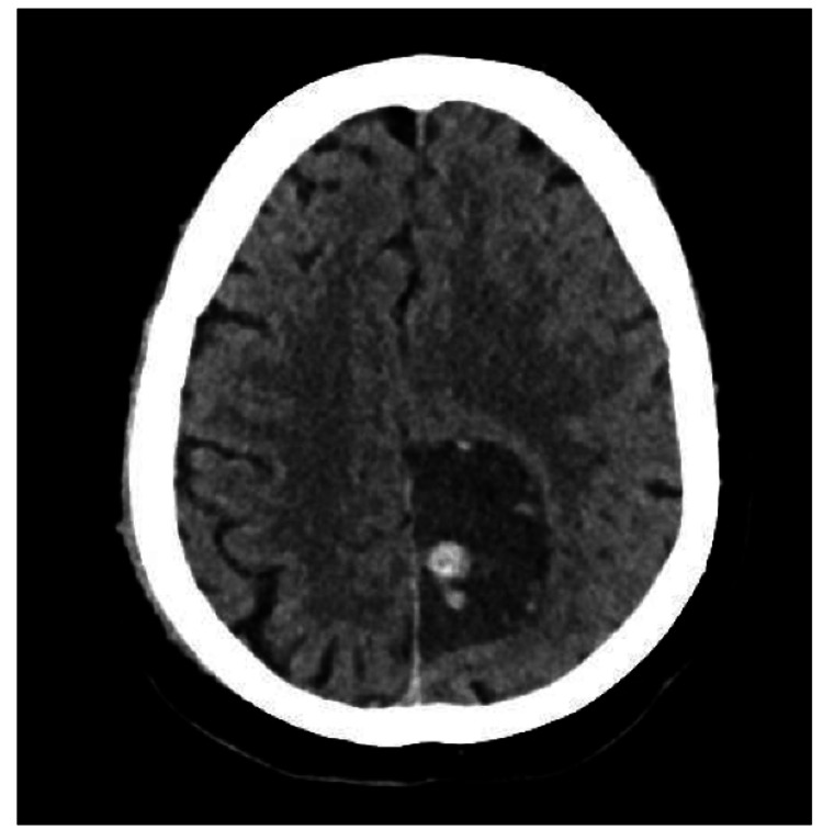 Figure 1
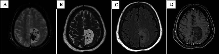 Figure 2
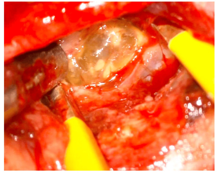 Figure 3
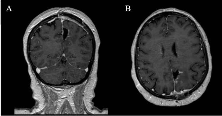 Figure 4
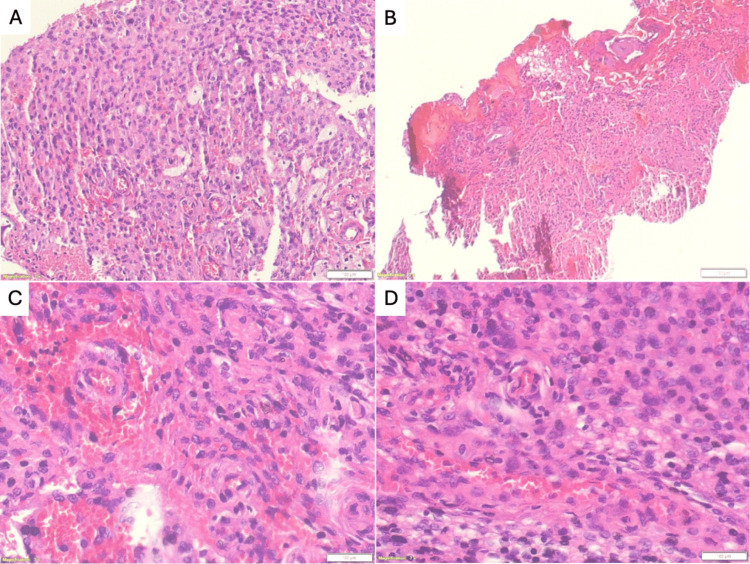 Figure 5
Peer Reviews
No public reviews on file for this paper yet. If you reviewed it on a platform where reviews are public (OpenReview, ICLR, NeurIPS, ICML), you can paste yours below so the community can read it here.
Videos
No videos yet. Explain this paper in a talk, walkthrough, or lecture? Add one.
Taxonomy
TopicsMeningioma and schwannoma management · Neurofibromatosis and Schwannoma Cases · Head and Neck Surgical Oncology