Synthesis of geometrically realistic and watertight neuronal ultrastructure manifolds for in silico modeling
Marwan Abdellah, Alessandro Foni, Juan José García Cantero, Nadir Román Guerrero, Elvis Boci, Adrien Fleury, Jay S Coggan, Daniel Keller, Judit Planas, Jean-Denis Courcol, Georges Khazen

TL;DR
This paper introduces a method to create realistic and watertight 3D models of neurons for detailed simulations of brain cell functions.
Contribution
A robust method for generating realistic and watertight neuronal meshes from morphological data is presented.
Findings
The method successfully generates watertight meshes for various cortical neuron morphologies.
The approach is extended to synthetic astrocytic morphologies with plausible biological detail.
Volumetric meshes are created for scalable in silico reaction-diffusion simulations.
Abstract
Understanding the intracellular dynamics of brain cells entails performing three-dimensional molecular simulations incorporating ultrastructural models that can capture cellular membrane geometries at nanometer scales. While there is an abundance of neuronal morphologies available online, e.g. from NeuroMorpho.Org, converting those fairly abstract point-and-diameter representations into geometrically realistic and simulation-ready, i.e. watertight, manifolds is challenging. Many neuronal mesh reconstruction methods have been proposed; however, their resulting meshes are either biologically unplausible or non-watertight. We present an effective and unconditionally robust method capable of generating geometrically realistic and watertight surface manifolds of spiny cortical neurons from their morphological descriptions. The robustness of our method is assessed based on a mixed dataset of…
Genes, proteins, chemicals, diseases, species, mutations and cell lines named across the full text — each resolved to its canonical identifier and authoritative record.
Click any figure to enlarge with its caption.
 Figure 1
 Figure 2
 Figure 3
 Figure 4
 Figure 5
 Figure 6
 Figure 7
 Figure 8
 Figure 9
 Figure 10
 Figure 11
 Figure 12
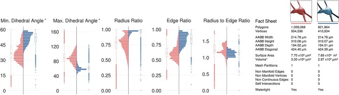 Figure 13
Peer Reviews
No public reviews on file for this paper yet. If you reviewed it on a platform where reviews are public (OpenReview, ICLR, NeurIPS, ICML), you can paste yours below so the community can read it here.
Videos
No videos yet. Explain this paper in a talk, walkthrough, or lecture? Add one.
Taxonomy
TopicsCell Image Analysis Techniques · Advanced Fluorescence Microscopy Techniques · Single-cell and spatial transcriptomics