Detection of Hepatic Metastasis from Early Delayed Images of Modified Dual-Time-Point F-18 FDG PET/CT Images in a Patient with Breast Cancer
Ji Young Lee, Hee-Sung Song

TL;DR
A case study shows how modified PET/CT imaging detected liver metastasis in a breast cancer patient more effectively than standard methods.
Contribution
Modified dual-time-point PET/CT with 80-minute delayed imaging detected metastasis not visible in standard scans.
Findings
Focal FDG uptake in the liver was detected in 80-minute delayed images.
The metastatic lesion was not visible in the usual one-hour post-injection images.
Modified PET/CT reduces scanning time and radiation exposure compared to traditional methods.
Abstract
We present a rare case of focal F-18-2-fluoro-2-deoxyglucose (FDG) uptake in the liver observed during a modified dual-time-point F-18 FDG positron emission tomography (PET)/computed tomography (CT), so-called early delayed scanning, in a 53-year-old woman diagnosed with breast cancer. This metastatic lesion was revealed in 80 min delayed images after FDG injection, but not in the usual one-hour images after injection. Modified dual-time-point F-18 FDG PET/CT is convenient because compared to the 2 h delayed images of dual-time-point PET/CT, it has a shorter scanning time and avoids additional radiation exposure.
Genes, proteins, chemicals, diseases, species, mutations and cell lines named across the full text — each resolved to its canonical identifier and authoritative record.
Click any figure to enlarge with its caption.
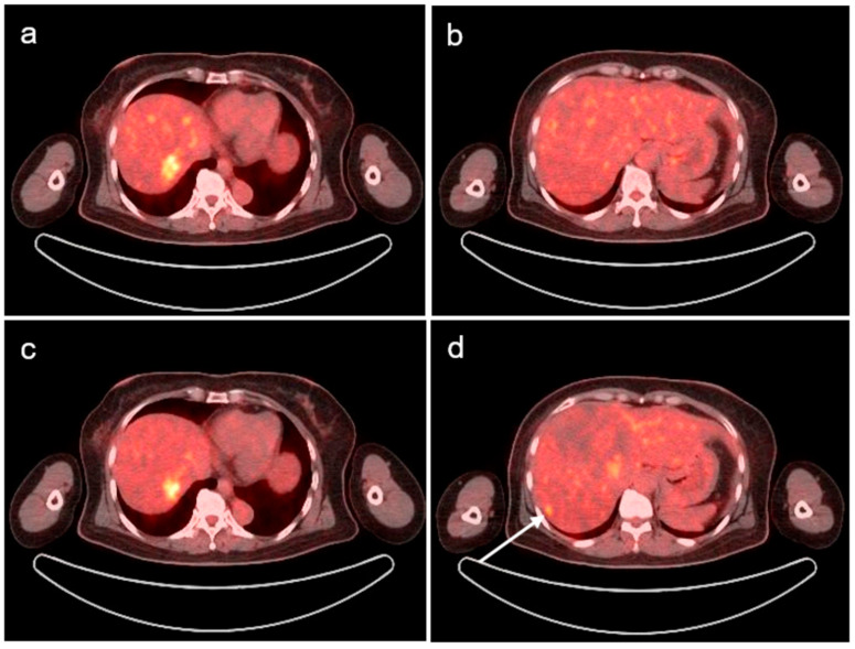 Figure 1
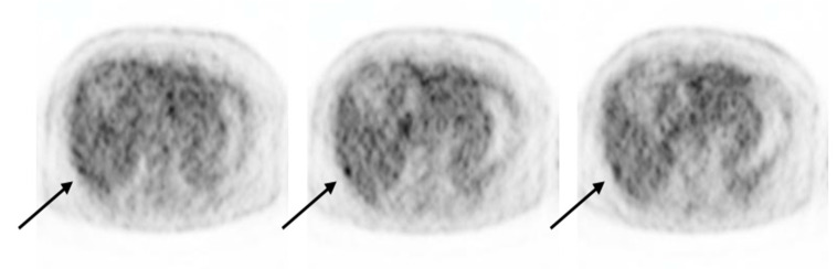 Figure 2
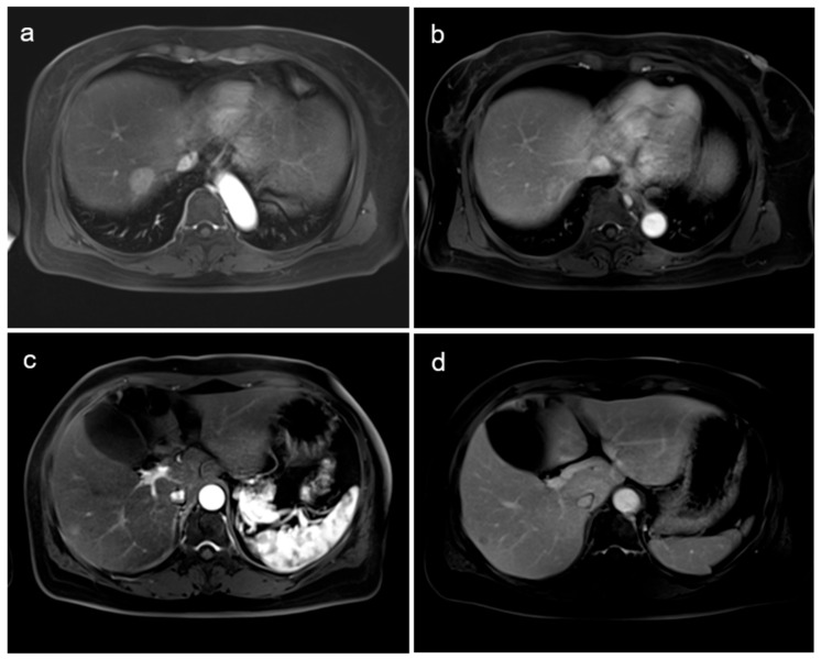 Figure 3
Peer Reviews
No public reviews on file for this paper yet. If you reviewed it on a platform where reviews are public (OpenReview, ICLR, NeurIPS, ICML), you can paste yours below so the community can read it here.
Videos
No videos yet. Explain this paper in a talk, walkthrough, or lecture? Add one.
Taxonomy
TopicsMedical Imaging Techniques and Applications · MRI in cancer diagnosis · Medical Imaging and Pathology Studies
A 53-year-old woman with right-breast cancer underwent F-18-2-fluoro-2-deoxyglucose (FDG) positron emission tomography (PET)/computed tomography (CT) as part of a metastatic workup. She was diagnosed with invasive ductal carcinoma in March 2020 and underwent breast-conserving surgery with axillary dissection. At the time of diagnosis, the pathological stage was IIA (T1N1M0); postoperative chemoradiotherapy was continued until November 2020.
In July 2023, a routine abdominopelvic CT revealed a new ill-defined high-attenuation lesion in segment 7 of the liver. Laboratory test results, including tumor and inflammatory marker levels, were within normal ranges. Consequently, the patient underwent modified dual-time-point (DTP) F-18 FDG PET/CT for further evaluation of the hepatic lesions. A focal hypermetabolic lesion was identified on routine 60 min images post-F-18 FDG injection in segment 7 of the liver, similar to the lesions seen on CT images (Figure 1a). Our department performed a modified, delayed scan 80 min after F-18 FDG injection, and another small increase in focal FDG uptake at the subcapsular portion of segment 6 of the liver (Figure 1d), in addition to the existing segment 7 lesion (Figure 1c), was detected. Although this lesion had a mild degree of FDG uptake, it was observed on several slices, unlike the surrounding heterogeneous hepatic uptake (Figure 2). This additional lesion was not observed on the early PET/CT images (Figure 1b); however, it was suspected to be metastatic. No other abnormal FDG uptakes suggesting metastasis or active inflammation were observed throughout the body.
Subsequent hepatic magnetic resonance imaging (MRI) was performed to assess the hypermetabolic lesions in the liver. On liver MRI, an arterial-enhancing lesion of approximately 2.3 cm with delayed washout in segment 7 of the liver (Figure 3a,b) and a faint arterial-enhancing lesion of approximately 0.8 cm in the subcapsular portion in segment 6 of the liver (Figure 3c,d) were observed; these were probably a hepatocellular carcinoma or metastases. Since the lesion in segment 6 was too small to be observed on liver ultrasound, an ultrasound-guided liver biopsy was performed only on the lesion of segment 8. The hepatic biopsy confirmed a metastatic carcinoma for which the patient received chemotherapy.
Many reports have indicated that DTP F-18 FDG PET may help distinguish benign from malignant lesions and evaluate uncertain metastatic lesions, particularly hepatic metastases; these are based on previous studies showing that FDG uptake increases in malignant tumors for several hours after injection, while uptake in benign lesions decreases or remains stable [1,2,3,4]. Modified DTP F-18 FDG PET/CT images performed 80 min after F-18 FDG injection, so-called early delayed images, have recently been reported in several articles [5,6,7]. This technique is advantageous because compared to the conventional DTP F-18 FDG PET/CT, wherein there is delayed acquisition at 120–180 min after FDG injection, it has a shorter scanning time and avoids additional radiation exposure. This is the first case report in which early delayed DTP F-18 FDG PET/CT was used to detect an additional metastatic lesion, in which only FDG uptake was observed on early delayed images. This conveniently modified DTP FDG PET/CT can help detect metastasis and recurrence in patients with cancer, especially in lesions with physiological FDG uptake such as lesions in the liver.
The reference list from the paper itself. Each links out to its DOI / PubMed record.
- 1Yen Y. Huang W. Chiu C. Tyan Y. Wang J. Wu L. Feng I.J. Lee C.H. Does routine triple-time-point FDG PET/CT imaging improve the detection of liver metastases?Diagnostics 20201060910.3390/diagnostics 1009060932825064 PMC 7554868 · doi ↗ · pubmed ↗
- 2Lin W.Y. Tsai S.C. Hung G.U. Value of delayed 18F-FDG PET imaging in the detection of hepatocellular carcinoma Nucl. Med. Commun.20052631532110.1097/00006231-200504000-0000315753790 · doi ↗ · pubmed ↗
- 3Boanova L.G. Altmayer S. Watte G. Raupp A.A. Francisco M.Z. De Oliveira G.S. Hochhegger B. Andrade R.G.F. Detection of liver lesions in colorectal cancer patients using 18F-FDG PET/CT dual-time-point scan imaging Cancers 202315540310.3390/cancers 1522540338001662 PMC 10670707 · doi ↗ · pubmed ↗
- 4Mao W. Zhou J. Qiu L. Yin H. Tan H. Shi H. The added value of dual-time-point 18F-FDG PET/CT imaging in the diagnosis of colorectal cancer liver metastases Abdom. Radiol.2020451075108110.1007/s 00261-019-02396-331927618 · doi ↗ · pubmed ↗
- 5Pietrzak A. Kazmierska J. Cholewinski W. Sequential 18F-FDG PET/CT imaging parameters for differentiating benign from malignant lymph nodes in head and neck carcinoma Hell. J. Nucl. Med.201720809229324917 · pubmed ↗
- 6Miyake K.K. Nakamoto Y. Togashi K. Dual-time-point 18F-FDG PET/CT in patients with colorectal cancer: Clinical value of early delayed scanning Ann. Nucl. Med.20122649250010.1007/s 12149-012-0599-y 22492392 · doi ↗ · pubmed ↗
- 7Lee J.Y. Song H. Choi J.H. Hyun C.L. Dual-time-point FDG uptake correlates with prognostic factors of invasive breast cancer: Clinical usefulness of early delayed scanning Diagnostics 201994010.3390/diagnostics 902004030970638 PMC 6627602 · doi ↗ · pubmed ↗