Characterization of Bovine Papillomavirus Types Detected in Cattle Rumen Tissues from Amazon Region, Brazil
Paulo Henrique Gilio Gasparotto, Igor Ribeiro dos Santos, Jerônimo Viera Dantas Filho, Mariana Soares da Silva, Fernanda dos Anjos Souza, Jennefer Caroline de Macedo Sousa, David Driemeier, Cláudio Wageck Canal, Flavio Roberto Chaves da Silva, Cíntia Daudt

TL;DR
This study identifies specific bovine papillomavirus types in cattle rumen tissues from the Amazon region of Brazil, contributing to understanding and managing BPV infections.
Contribution
The study reports the first detection of BPV13 and BPV44 in cattle rumen tissues from the Brazilian Amazon region.
Findings
BPV types 2, 13 (Delta PVs), and 44 were identified in rumen samples from cattle in the Amazon region.
One sample was classified as a putative new subtype of BPV44.
Histopathological analysis revealed fibropapilloma and squamous papilloma classifications.
Abstract
Bovine papillomavirus (BPV) infection of the gastrointestinal tract (GIT) can induce the development of masses with high growth, resulting in breathing and eating obstructions leading to animal suffering and death. Beyond this, BPV is related to economic losses worldwide by depressing meat and milk production as well as cattle by-products. Using PCR followed by Sanger sequencing, we were able to identify the high-risk Delta BPVs and the BPV44 on rumen cattle samples collected in slaughterhouses. These results can contribute to future epidemiological studies and vaccine studies regarding BPV infections. The Bos Taurus Papillomavirus, commonly known as bovine papillomavirus (BPV), can cause lesions in the mucosa of the gastrointestinal tract (GIT) in cattle and induce the formation of papillomas in organs such as the pharynx, esophagus, rumen and reticulum. GIT papillomas can lead to…
Genes, proteins, chemicals, diseases, species, mutations and cell lines named across the full text — each resolved to its canonical identifier and authoritative record.
Click any figure to enlarge with its caption.
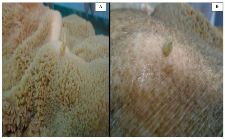 Figure 1
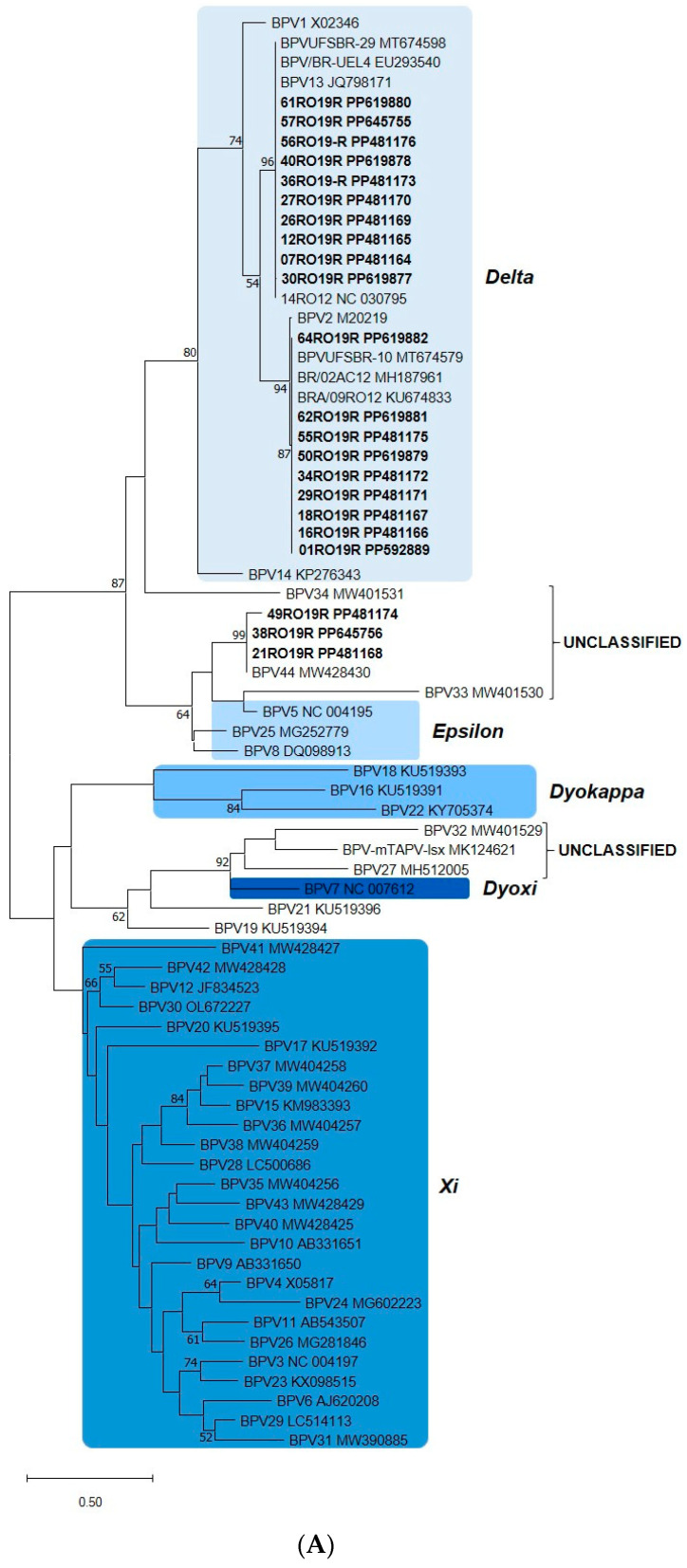 Figure 2
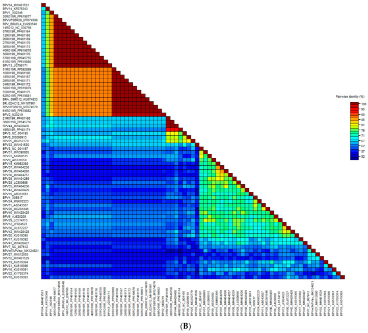 Figure 3
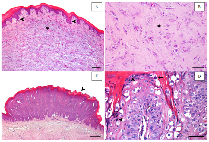 Figure 4
- —Acre State Research Support Foundation (FAPAC)
Peer Reviews
No public reviews on file for this paper yet. If you reviewed it on a platform where reviews are public (OpenReview, ICLR, NeurIPS, ICML), you can paste yours below so the community can read it here.
Videos
No videos yet. Explain this paper in a talk, walkthrough, or lecture? Add one.
Taxonomy
TopicsHistorical Gender and Feminism Studies · Global Public Health Policies and Epidemiology
1. Introduction
Papillomaviruses (PVs) are a large and diverse group of double-stranded circular DNA viruses about 55-60 nm in diameter, lacking lipoprotein envelope and infecting a wide variety of animal species from fish to humans [1,2,3]. Bovine papillomavirus (BPV) induces papillomas and fibropapillomas in both mucosal and cutaneous epithelium, causing vulvovaginal, penile and ocular lesions as well as udder and teats papillomas, urinary bladder and gastrointestinal tract (GIT) cancers [4,5,6,7,8]. Currently, BPVs comprise 44 types classified into five genera (Delta, Xi, Epsilon, Dyokappa and Dyoxipapillomavirus). In addition, the BPV types 19, 21, 27 and tick-associated BPV are still not attributed to a genus [5,9,10,11]. PVs are classified based on the nucleotide sequence of the L1 gene, which differs between each PV genotype [9].
BPV1 and 2 are the most described BPV types in cattle worldwide but are commonly found infecting equids, giraffes, deer and other animal species [12]. In cattle, BPV1 and 2 have been described in different clinical specimens [13,14,15], being mainly associated with skin lesions [12,16,17] and urinary bladder cancer [7,8,12,18,19]. Infection of the gastrointestinal tract (GIT) of cattle by BPV induces the formation of lesions that affect organs such as the pharynx, esophagus, rumen and reticulum [20]. Small, nodular or thin papillomas associated with BPV1, 2 and 5 have already been described in the rumen mucosa [21]. Additionally, GIT papillomas have been primarily associated with BPV4, which can progress to carcinomas [22]. These lesions can lead to clinical signs such as rumen intermittent swelling, wheezing and drooling [22], resulting in feeding and breath difficulties [23]. In cattle and buffaloes, papillomas have been described in the rumen mucosa, ranging from high growth to small nodular, spherical and pedunculated lesions [21,23].
Bovine GIT papillomas are predominantly accidental findings at necropsy or in slaughterhouses. These lesions can progress to squamous cell carcinoma (SCC) frequently associated to the chronic consumption of Pteridium spp. (Dennstaedtiaceae) [8,24]. Benign tumors usually show spontaneous regression; however, they can remain and, in the presence of environmental and/or genetic cofactors, evolve into a malignant lesion [8,24,25]. Despite being a relevant etiological agent in cattle farming, the detection and characterization studies of PV in animals are still deficient, especially regarding cattle rumen lesions [26,27]. BPV infection of the GIT can extend from the mouth, tongue, rumen and reticulum and can present high mass growth, which can result in breathing and eating obstructions and animal suffering and death [21]. Beyond animal suffering, these viruses cause worldwide economic losses by depressing meat and milk production as well as cattle by-products [10,11].
Recently, molecular biology techniques have allowed for the identification and characterization of several new and putative new types (PNTs) of BPV in bovine skin papilloma lesions in the Western Amazon region and in other regions of Brazil [5,6,12]. Herein, we aimed to genetically and histologically characterize papillomatous lesions from the upper gastrointestinal tract of beef cattle raised extensively in the Western Amazon region, Brazil, providing data for future epidemiological studies of cattle GIT lesion etiologies.
2. Material and Methods
2.1. Research Ethics
The approval by the Ethics Committee in the Use of Animals is not required once the specimens were collected from cattle slaughtered in slaughterhouses registered by the Brazilian sanitary inspection (Brazilian Law No 11794/2008). All rumen samples were collected by a veterinarian.
2.2. Sample Collection
All gross neoplastic lesions were collected from 100 animals, totaling 100 samples of papillomatous lesions from the bovine rumen (one sample per animal) (Figure 1) in slaughterhouses in the central region of Rondônia state, Northern Brazil. The samples came from two slaughterhouses in Ji-Paraná and Urupá municipalities, which receive cattle from Ji-Paraná, Alvorada do Oeste, Urupá, Teixeirópolis and Mirante da Serra municipalities, Brazil. The sampling was performed using a sterile scalpel and tweezers for each lesion. Subsequently, half of each sample was conditioned in 10% formalin (according to tissue availability) and the other half was refrigerated and stored at −20 °C.
2.3. PCR, Sequencing and Sequence Analysis
DNA extraction was performed using the commercial Purelink^®^ Genomic DNA Mini kit (Invitrogen, Carlsbad, CA, USA), according to the manufacturer’s instructions. FAP59/FAP64 primer pairs were used for partial amplification of the L1 gene [28]. The PCR reactions were performed with 2 µL of the extracted DNA, 0.2 μM of each primer, 1 unit of Taq DNA polymerase (Invitrogen, Carlsbad, CA, USA), 2.5 μL of 10× PCR buffer, 0.38 mM MgCl_2_, 0.05 mM of each dNTP and sterile ultrapure water, to the final volume of 25 µL. Amplifications were performed in a thermocycler under the following time and temperature conditions: 5 min at 95 °C, followed by 40 cycles of 1 min at 94 °C, 1 min at 50 °C, 1 min at 72 °C and a final extension of 7 min at 72 °C. Afterwards, 5 µL aliquots of the amplification reactions were subjected to electrophoresis in a 2% agarose gel, using Gel Red (Quatro G Biotechnology, Porto Alegre, Brazil), and visualized in a transilluminator UV LTB HE (Loccus, Cotia, Brazil).
The PCR positive samples were purified using the commercial kit NucleoSpin Extract II (Macherey—Nagel, Duëren, Germany) and subsequently sequenced using the automatic sequencer ABI-PRISM 3100 Genetic Analyzer armed with 50 cm capillaries and POP6 polymer (Applied Biosystems, Waltham, MA, USA), with forward and reverse primers. The sequences were edited using Geneious Prime software (version 2023.1.2). BLASTn tool (http://www.ncbi.nlm.nih.gov/BLAST, accessed on 5 April 2024) was used to compare the identity of the sequences obtained in this study with the sequences deposited in public databases (GenBank).
2.4. Phylogenetic Analysis
All BPVs reference genomes, as well as the sequences most similar to those obtained in this study, were retrieved from the NCBI (https://www.ncbi.nlm.nih.gov) for phylogenetic analysis. The alignment was performed with Clustal W [6,28,29] and the phylogenetic tree was built with the maximum likelihood method (ML) with the most suitable nucleotide substitution model, according to the “Find Best DNA/Protein Model” tool available in MEGA X (version 10.2.6) [30,31,32]. The reliability of the tree was tested with 1000 nonparametric bootstrap analyses.
2.5. Histopathological Analysis
Samples of papillomatous lesions from 100 animals were fixed in 10% buffered formalin and routinely processed for histology. For this, the samples were trimmed, dehydrated in increasing concentrations of alcohol, clarified in xylene solution and embedded in paraffin wax. Tissue sections (3–5 μm) were stained with hematoxylin and eosin (HE) and evaluated under light microscopy. Based on histological findings, the tumors were classified as fibropapilloma or squamous papilloma [33].
3. Results
3.1. PCR, Genetic Sequencing and Phylogenetic Analysis
For the 100 rumen samples subjected to PCR with the FAP 59/64 primer pair, papillomavirus (PV) DNA was amplified in 41 samples and not amplified in 59 samples using the well-established PCR with FAP59/FAP64 primer pair (Table 1 and Table 2). Phylogenetic analysis was performed for 22 samples that presented high-quality sanger sequencing (electropherogram showing a single peak to a single nucleotide). In the generated phylogenetic tree, the vast majority of sequences clustered in the genus Deltapapillomavirus (19 sequences) and three sequences grouped in an unclassified genus (Figure 2A,B), in the BPV44 cluster.
The samples belonging to genus Delta PV were classified as BPV2 (40.91%; 9/22) and BPV13 (45.45%; 10/22). Samples classified as BPV2 showed a high degree of identity with each other (100%) and with the reference BPV2 (98.02%). The sequences that clustered with BPV13 (100% identity) also showed 100% identity with each other.
The 21RO19R and 38RO19R sequences (100% similarity between them) have a common ancestor, namely BPV44. Greater phylogenetic distance was observed between the study sequence 49RO19R and BPV44 (only 94.42% of similarity), which can be classified as a possible new viral subtype. Results are shown in Figure 2 and Table 1, Table 2 and Table 3. The average age and weight of the slaughtered animals sampled were 33 months-old and 480 kg, respectively.
3.2. Histopathological Analysis
Histopathological analysis was performed on 22 samples, which had enough material for this purpose. The results show that nineteen (79.10%) can be histologically classified as fibropapilloma and two as squamous papilloma. Additionally, two samples (8.4%) did not show any histopathological alteration and one sample (4.10%) was inconclusive (Table 3). Fibropapillomas were characterized by mild mucosal hyperplasia, which had a wavy surface and formed long epithelial pins (rete pegs) toward the proliferated submucosa (Figure 3A).
The cell proliferation in submucosa was fusiform and formed bundles arranged in random flows, supported by a mild-to-moderate myxoid or collagenous stroma (Figure 3B). There was moderate anisocytosis and anisokaryosis and no mitotic activity. Squamous papilloma consisted of marked mucosal hyperplasia and ortho or parakeratotic hyperkeratosis, with a finger-like or slightly flat surface (Figure 3C). Keratinocytes often had eccentric pyknotic nuclei and a perinuclear halo (consistent with koilocytes). Enlarged keratohyaline granules and intranuclear amphophilic inclusions (Figure 3D) were observed in only two cases.
4. Discussion
Herein, it was possible to amplify PV sequences in 41/100 rumen papillomatous lesions using primer pair FAP59/64. Moreover, we report the occurrence of BPV44 in cattle rumen, which was first identified in bovine teat papilloma lesions using rolling circle amplification followed by high-throughput sequencing [6].
From the 59 negative papillomatous samples, 22 were histologically diagnosed as squamous papilloma or fibropapillomas. This fact may be due to the fact that the FAP primer pair is not able to amplify some types of BPV that were already detected in this region due to the lower affinity for the primers [34,35].
The primer pair FAP59/64 allowed for the amplification of BPV mostly from the Deltapapillomavirus genus (BPV2 and 13). The FAP primer pair has been widely and satisfactorily used in previous BPV screening studies [17,32]. It is important to point that these primers were originally designed to amplify human PVs and have been widely used to amplify PV from a vast range of species worldwide, which made it possible to describe several new PV types from distinct animal species [1,5,12,17,28,29,30,31,32,33,34,35].
Furthermore, two samples were classified as BPV44 and one as a possible new subtype of this recently described viral type. BPV44 was previously isolated in cattle teat lesions in southern Brazil [6], but we describe here the first detection of this viral type in ruminal lesions.
The histological findings of most samples in the current study are consistent with those typically seen in bovine cutaneous papilloma lesions [17,27,36,37]. BPVs of the Deltapapillomavirus genus (BPV1, 2, 13 and 14) usually induce the formation of fibropapilloma, as they infect the epidermis and dermis [5]. In this study, BPV2 and 13 were detected in papillomatous lesions mostly classified as fibropapilloma.
Delta PV have already been detected in papillomatous skin lesions of cattle in Rondônia state [12,17,34], in Southern Brazil [5,7,34,35,36,37,38] and in southeast Brazil [38,39]. Additionally, BPVs 1, 2 and 5 were detected in the mouth, esophagus, rumen and reticulum fibropapillomas of cattle and buffaloes in India, Japan and Brazil [21,39,40], corroborating to the findings of this study. Delta PVs are considered high-risk since they are frequently detected in neoplasms of the upper gastrointestinal tract and urinary bladder, especially when they are associated with the consumption of Pteridium species [7,8,12,41]. In the present study, it was not possible to obtain data on the farms. However, the region presents data on the incidence of the Pteridium species and can be a damaging factor; in addition, the biodiversity of other toxic plants may be related to the incidence [41].
Other studies detected BPV4 (Xi-PV) in GIT papillomatous lesions of cattle in the UK [22] and Italy [42,43]. On the other hand, members of the Xipapillomavirus genus, such as BPV12, were described in a cattle tongue in Japan [44], although these viral types were not detected in this study. BPV4, which has already been found in the GIT, is probably not a predominant viral type in northern Brazil, as it was not detected in any previous study carried out on cattle in the Amazon region, not even using high-throughput sequencing [17,34]. Other studies of GIT papillomas in bovines have already been associated with BPV1, 2, 4 and 5 [21,22,42]. However, regression may not occur in chronically immunodepressed animals and some types of PV have been linked to malignancy, especially in synergism with some chemical or environmental carcinogens [21]. These infections can extend from the mouth and tongue to the esophagus, rumen and reticulum and present low to high growth, with a nodular, spherical and pedunculated shape, which can result in difficulty in feeding and breathing, obstructive bloat and cause the death of the animal [21,22,23,44,45,46,47].
BPV1 DNA is commonly found in skin warts, bovine peripheral blood, placenta, amniotic fluid and bovine colostrum [12,14,19,39,48] and it has been suggested that the bloodstream contributes to viral dissemination [39,49,50,51,52]. Similarly, the identification of BPV2 and BPV13 in the present study could also be explained by bloodstream dissemination. Although the pathogenesis of the PVs involves epitheliotropism, its genetic material can be found in several different tissues in the same animal and also in the peripheral blood of their offspring [46,52].
5. Conclusions
Here, we were able to identify Delta papillomaviruses and the recently identified BPV44 infecting the rumen of cattle from the western Amazon, Brazil. This study shows that ruminal papillomatosis is a post-mortem finding, emphasizing the importance of this type of study for a better understanding of the pathologies caused by BPV.
The reference list from the paper itself. Each links out to its DOI / PubMed record.
- 1Antonsson A. Hansson B.G. Healthy Skin of Many Animal Species Harbors Papillomaviruses Which Are Closely Related to Their Human Counterparts J. Virol.200276125371254210.1128/JVI.76.24.12537-12542.200212438579 PMC 136724 · doi ↗ · pubmed ↗
- 2de Villiers E.-M. Fauquet C. Broker T.R. Bernard H.-U. zur Hausen H. Classification of papillomaviruses Virology 2004324172710.1016/j.virol.2004.03.03315183049 · doi ↗ · pubmed ↗
- 3López-Bueno A. Mavian C. Labella A.M. Castro D. Borrego J.J. Alcami A. Alejo A. Concurrence of Iridovirus, Polyomavirus, and a Unique Member of a New Group of Fish Papillomaviruses in Lymphocystis Disease-Affected Gilthead Sea Bream J. Virol.2016908768877910.1128/JVI.01369-1627440877 PMC 5021401 · doi ↗ · pubmed ↗
- 4Borzacchiello G. Roperto F. Bovine papillomaviruses, papillomas and cancer in cattle Vet. Res.2008394510.1051/vetres:200802218479666 · doi ↗ · pubmed ↗
- 5Bianchi R.M. Alves C.D.B.T. Schwertz C.I. Panziera W. De Lorenzo C. da Silva F.S. de Cecco B.S. Daudt C. Chaves F.R. Canal C.W. Molecular and pathological characterization of teat papillomatosis in dairy cows in southern Brazil Braz. J. Microbiol.20195136937510.1007/s 42770-019-00175-231642003 PMC 7058819 · doi ↗ · pubmed ↗
- 6Sauthier J.T. Daudt C. da Silva F.R.C. Alves C.D.B.T. Mayer F.Q. Bianchi R.M. Driemeier D. Streit R.S.A. Staats C.C. Canal C.W. The genetic diversity of “papillomavirome” in bovine teat papilloma lesions Anim. Microbiome 202135110.1186/s 42523-021-00114-334321106 PMC 8317299 · doi ↗ · pubmed ↗
- 7de Alcântara B.K. Lunardi M. Agnol A.M.D. Alfieri A.F. Alfieri A.A. Detection and Quantification of the E 6 Oncogene in Bovine Papillomavirus Types 2 and 13 From Urinary Bladder Lesions of Cattle Front. Vet. Sci.2021867318910.3389/fvets.2021.67318934055956 PMC 8160092 · doi ↗ · pubmed ↗
- 8Medeiros-Fonseca B. Abreu-Silva A.L. Medeiros R. Oliveira P.A. Gil da Costa R.M. Pteridium spp. and Bovine Papillomavirus: Partners in Cancer Front. Vet. Sci.2021875872010.3389/fvets.2021.75872034796228 PMC 8593235 · doi ↗ · pubmed ↗