Postoperative discovery of molar pregnancy
Haithem Aloui, Hatem Frikha

Abstract
Genes, proteins, chemicals, diseases, species, mutations and cell lines named across the full text — each resolved to its canonical identifier and authoritative record.
Click any figure to enlarge with its caption.
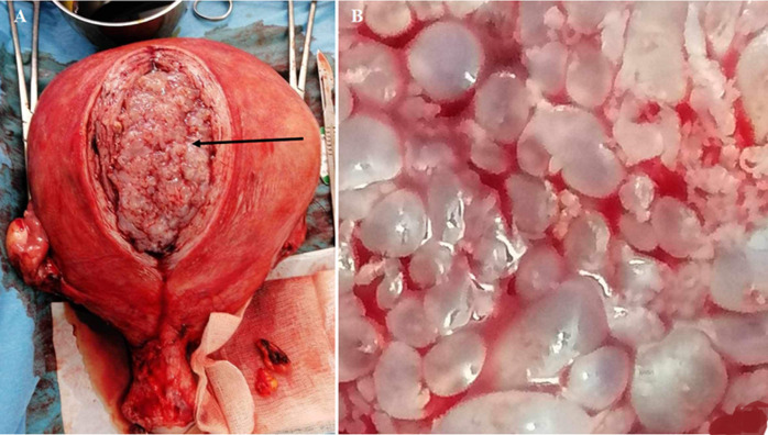 Figure 1
Peer Reviews
No public reviews on file for this paper yet. If you reviewed it on a platform where reviews are public (OpenReview, ICLR, NeurIPS, ICML), you can paste yours below so the community can read it here.
Videos
No videos yet. Explain this paper in a talk, walkthrough, or lecture? Add one.
Taxonomy
TopicsGestational Trophoblastic Disease Studies · Prenatal Screening and Diagnostics
Image in medicine
This concerns a 52-year-old patient presenting with abdominal-pelvic pain and diffuse bloating. Upon gynecological examination, an enlarged uterus is noted. Endovaginal ultrasound reveals a 7 cm intracavitary fibroid with suspected degeneration. The patient underwent a hysterectomy. Intraoperatively, the surgeon observes a soft consistency of the uterus and decides to proceed with bilateral annexectomy. Upon dissection of the operative specimen, the typical appearance of a molar pregnancy is noted. This finding is confirmed by anatomopathological examination. The patient had a good postoperative recovery.
molar pregnancy: A) perioperative cluster of grapes appearance (black arrow); B) macroscopic view