Gastric Adenocarcinoma Masquerading as Cannabis Hyperemesis Syndrome
Charles Altfillisch, Fortune Unegbu, Daniel Buckles

Abstract
Genes, proteins, chemicals, diseases, species, mutations and cell lines named across the full text — each resolved to its canonical identifier and authoritative record.
Click any figure to enlarge with its caption.
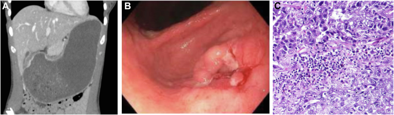 Figure 1
Peer Reviews
No public reviews on file for this paper yet. If you reviewed it on a platform where reviews are public (OpenReview, ICLR, NeurIPS, ICML), you can paste yours below so the community can read it here.
Videos
No videos yet. Explain this paper in a talk, walkthrough, or lecture? Add one.
Taxonomy
TopicsNeuroendocrine Tumor Research Advances · Gastrointestinal disorders and treatments · Cannabis and Cannabinoid Research
A 29-year-old female with a history of marijuana use presented to the Emergency Department due to intractable nausea and vomiting. Symptoms had been present for 3 months and were associated with 30-pound unintentional weight loss. She reported smoking marijuana daily. She had been to various emergency rooms on multiple occasions and was diagnosed with Cannabis Hyperemesis Syndrome.
A computed tomography scan of the abdomen revealed a severely dilated, fluid filled stomach (Figure A). A diagnostic esophagogastroduodenoscopy showed a stomach filled with a large volume of liquid that was suctioned and an ulcerated, friable mass with spontaneous bleeding in the prepyloric region (Figure B) causing gastric outlet obstruction. Biopsies from the mass were positive for invasive adenocarcinoma (Figure C). The patient underwent a partial gastrectomy with Roux-en-Y reconstruction and D2 lymphadenectomy.
While Cannabis Hyperemesis Syndrome is common, this case underscores the importance of a complete diagnostic evaluation to exclude other pathology, especially when red flag symptoms, such as weight loss, are present. The average age of diagnosis of gastric adenocarcinoma is 68 and less than 10% of cases are in patients younger than 40. Given the increasing prevalence with advancing age, the rarity in such a young patient may have additionally contributed to the delay in diagnosis.