Short-Time Development of Esophageal Inflammatory Myofibroblastic Tumor Which Resected by Endoscopic Submucosal Dissection
Sachiyo Onishi, Tsutomu Tanaka, Masahiro Tajika

Abstract
Genes, proteins, chemicals, diseases, species, mutations and cell lines named across the full text — each resolved to its canonical identifier and authoritative record.
Click any figure to enlarge with its caption.
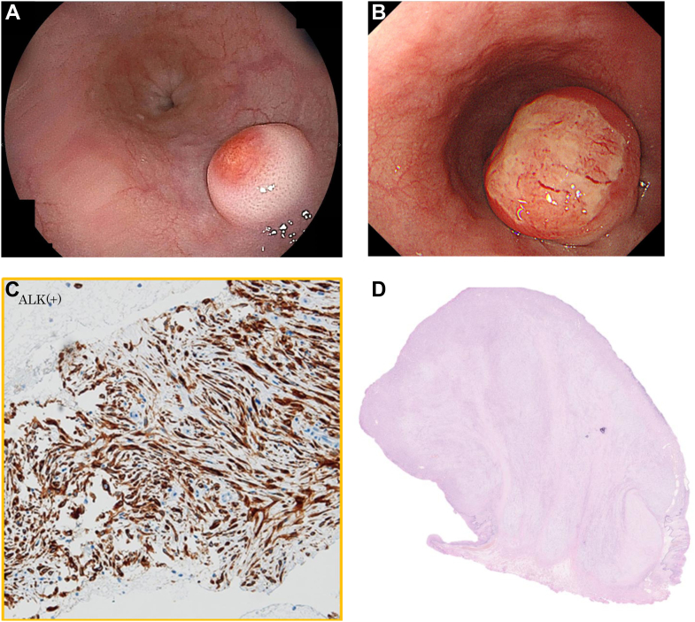 Figure 1
Peer Reviews
No public reviews on file for this paper yet. If you reviewed it on a platform where reviews are public (OpenReview, ICLR, NeurIPS, ICML), you can paste yours below so the community can read it here.
Videos
No videos yet. Explain this paper in a talk, walkthrough, or lecture? Add one.
Taxonomy
TopicsIgG4-Related and Inflammatory Diseases · Neuroendocrine Tumor Research Advances · Gastrointestinal disorders and treatments
A 53-year-old man was pointed out an elevated lesion in the esophagus by screening upper gastrointestinal series. A esophagogastroduodenoscopy showed a submucosal tumor 10 mm in diameter in the lower thoracic esophagus (Figure A). A biopsy specimen taken from the tumor showed no malignancy. One year later, the tumor had more than doubled in size (Figure B). So, we decided to perform endoscopic ultrasonography–guided fine-needle aspiration for this tumor. Endoscopic ultrasonography showed a hypoechoic mass 21 mm in size which come from the second layer. The biopsy specimen showed spindle cell proliferation and actin and anaplastic lymphoma kinase positivity (Figure C), which lead to the diagnosis of inflammatory myofibroblastic tumor (IMT). Because of the strong accumulation on positron emission tomography/computed tomography and the growing trend of submucosal tumor, we decided to perform endoscopic submucosal dissection (ESD). The pathological results were consistent with IMT which complete resected by ESD (Figure D). No recurrence has been observed for a year.
Although IMT is a tumor of mesenchymal origin which occurs systemically, there are few reports of its occurrence in the esophagus. This is the first case in which short-time growth can be observed and resected by ESD.