An Unusual Case of Gastrointestinal Bleeding; Which Is the Bleeding Origin, Upper or Lower Digestive Tract?
Kazuki Natsui, Masaki Maruyama, Shuji Terai

Abstract
Genes, proteins, chemicals, diseases, species, mutations and cell lines named across the full text — each resolved to its canonical identifier and authoritative record.
Click any figure to enlarge with its caption.
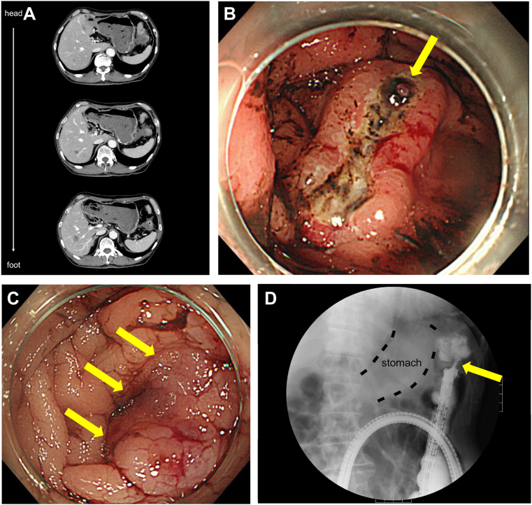 Figure 1
Peer Reviews
No public reviews on file for this paper yet. If you reviewed it on a platform where reviews are public (OpenReview, ICLR, NeurIPS, ICML), you can paste yours below so the community can read it here.
Videos
No videos yet. Explain this paper in a talk, walkthrough, or lecture? Add one.
Taxonomy
TopicsGastrointestinal Bleeding Diagnosis and Treatment · Gastrointestinal disorders and treatments · Biliary and Gastrointestinal Fistulas
A 75-year-old man with no medical history presented to our emergency room with a massive bloody stool. His vital signs on arrival indicated shock, and blood tests showed decreased hemoglobin (9.2 g/dL; reference range, 12–16 g/dL). Computed tomography showed the stomach being full of high-density contents, a wall break in a part of the descending colon, a soft shadow from the wall break to the gastric side, and multiple liver tumors (Figure A, head side at the top). Urgent esophagogastroduodenoscopy showed massive blood clots and an ulcer of approximately 40 mm in size at the fornix (Figure B) with an exposed blood vessel (yellow arrow). Endoscopic coagulation was performed. Subsequently, urgent colonoscopy and lower gastrointestinal series showed a semicircular submucosal tumor (yellow arrows, Figure C) and scope-impassible stenosis (Figure D). Pathological findings for the gastric ulcer and colonic submucosal tumor were muc, tub1, and tub2. The patient was diagnosed with gastric cancer, and the histological type was poorly differentiated adenocarcinoma with colonic metastasis via peritoneal dissemination. He planned to undergo nivolumab/S-1/oxaliplatin chemotherapy.
As colonic metastasis of gastric cancer is extremely rare and presents difficult image finding to diagnose, intestinal findings in the presence of gastric cancer should be noted.