Endoscopic ultrasound-guided hepaticojejunostomy for complete biliary anastomotic stricture: the echo-free space technique for scope insertion in surgically altered anatomy
Michihito Kono, Shunsuke Omoto, Mamoru Takenaka, Akito Furuta, Shunsuke Ogata, Taro Inoue, Wataru Ono

Abstract
Genes, proteins, chemicals, diseases, species, mutations and cell lines named across the full text — each resolved to its canonical identifier and authoritative record.
Click any figure to enlarge with its caption.
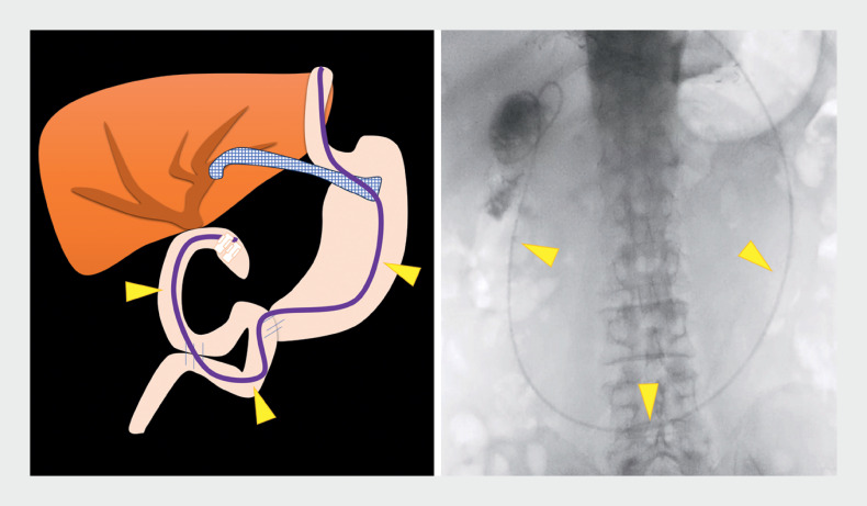 Fig. 1
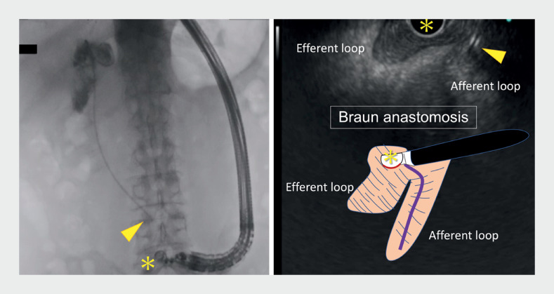 Fig. 2
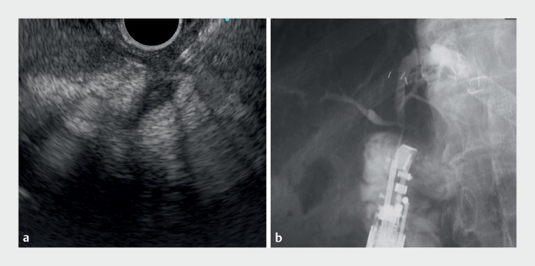 Fig. 3
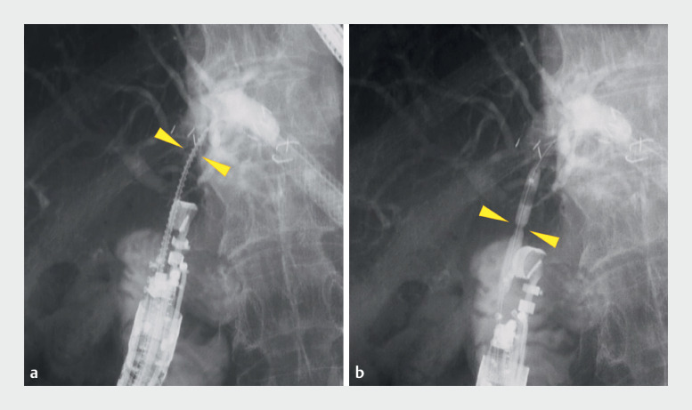 Fig. 4
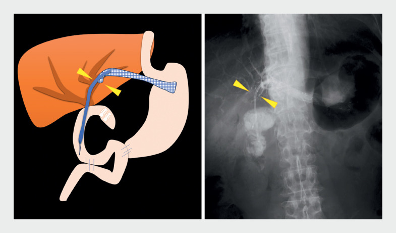 Fig. 5
Peer Reviews
No public reviews on file for this paper yet. If you reviewed it on a platform where reviews are public (OpenReview, ICLR, NeurIPS, ICML), you can paste yours below so the community can read it here.
Videos
No videos yet. Explain this paper in a talk, walkthrough, or lecture? Add one.
Taxonomy
TopicsGallbladder and Bile Duct Disorders · Pancreatic and Hepatic Oncology Research · Cholangiocarcinoma and Gallbladder Cancer Studies
Postoperative biliary strictures are estimated to occur in 2.6% of patients. When endoscopic treatment is difficult, they can be treated with endoscopic ultrasound-guided hepaticojejunostomy (EUS-HJS) using a forward-viewing linear endoscope 1 2 3 4 . However, in many institutions, the forward-viewing scope is not readily available, making immediate intervention difficult. We have developed a safe and reliable method for inserting a side-viewing linear endoscope using the “echo-free space” technique 5 .
We present the case of a 71-year-old man who underwent total pancreatectomy and choledochojejunostomy for pancreatic cancer. After 8 months, he developed cholangitis due to an anastomotic stricture and was referred to our department. Single-balloon endoscopic retrograde cholangiopancreatography (ERCP) and EUS-guided hepaticogastrostomy (EUS-HGS) were attempted, but the patient continued to have recurrent cholangitis. We therefore decided to perform EUS-HJS from the anastomotic site.
The single-balloon enteroscope was first inserted up to the anastomosis, which was marked with a clip; a 6-Fr endoscopic nasobiliary drainage (ENBD) catheter was placed near the anastomotic stricture ( Fig. 1 ). The scope was switched to a side-viewing linear endoscope (GF-UCT260) and the Braun anastomosis was identified on careful observation of the echo image, with the ENBD catheter used as a guide to reach the HJS ( Fig. 2 ; Video 1 ). The bile duct was punctured through the anastomosis with an 19G EZ Shot 3 Plus (Olympus) and a guidewire was placed ( Fig. 3 ). The stenosis was dilated with a spiral drill dilator (Tornus ES; Olympus) and then with a tapered-tip balloon catheter (REN; Kaneka) up to 4 mm ( Fig. 4 ), and the procedure was completed with the placement of a 7-Fr, 9-cm inside stent ( Fig. 5 ).
Schematic diagram and fluoroscopic image showing a 6-Fr endoscopic nasobiliary drainage catheter (arrowheads) placed near the anastomotic stricture after a single-balloon enteroscope had been inserted up to the anastomosis, which was marked with a clip.
A side-viewing linear endoscope (asterisk) is used to identify the Braun anastomosis, relying on careful observation of the echo image, the endoscopic nasobiliary drainage catheter (arrowhead) is used as a guide to reach the choledochojejunostomy.
Endoscopic ultrasound-guided hepaticojejunostomy is performed in a patient with complete biliary anastomotic stricture using the echo-free space technique to insert the scope into the choledochojejunostomy site.Video 1
Endoscopic ultrasound and fluoroscopic images showing: a the bile duct being punctured through the anastomosis using a 19G needle; b the appearance after the injection of contrast medium.
Fluoroscopic images showing the stenosis being dilated with a spiral drill dilator and tapered-tip balloon catheter.
Schematic diagram and fluoroscopic image showing a 7-Fr, 9-cm inside stent (arrowhead) placed to complete the procedure.
This case suggests that the echo-free space technique using a side-viewing linear endoscope can be useful in postoperative patients and represents a new option for EUS-HJS in the treatment of complete biliary anastomotic stricture.
Endoscopy_UCTN_Code_TTT_1AS_2AH
The reference list from the paper itself. Each links out to its DOI / PubMed record.
- 1House MG Fong Y Arnaoutakis DJ Preoperative predictors for complications after pancreaticoduodenectomy: impact of BMI and body fat distribution J Gastrointest Surg 20081227027810.1007/s 11605-007-0421-718060467 · doi ↗ · pubmed ↗
- 2Itoi T Ikeuchi N Tonozuka REUS-guided choledochojejunostomy with a lumen-apposing metal stent in a post-Whipple patient Gastrointest Endosc 2015811259126025440682 10.1016/j.gie.2014.08.033 · doi ↗ · pubmed ↗
- 3Kida M Yamauchi H Okuwaki K Endoscopic ultrasound-guided choledochojejunostomy with a forward-viewing echoendoscope for severe benign bilioenteric stricture in a patient with Child’s resection Endoscopy 2015471 E 303E 30426099105 10.1055/s-0034-1392208 · doi ↗ · pubmed ↗
- 4Koizumi K Masuda S Shionoya K Endoscopic ultrasound-guided hepaticojejunostomy using forward-viewing echoendoscope for transected aberrant right posterior hepatic duct in Roux-en-Y hepaticojejunostomy Endoscopy 202254 E 933E 93410.1055/a-1881-406835835153 PMC 9736790 · doi ↗ · pubmed ↗
- 5Omoto S Takenaka M Fukunaga T The “echo-free space” technique: a safe and reliable method for endoscopic ultrasound scope insertion Endoscopy 202355 E 698E 69937142249 10.1055/a-2062-5718 PMC 10159774 · doi ↗ · pubmed ↗