Transluminal opening technique for multiple septa in liver abscess using novel cholangioscope
Takeshi Ogura, Yuki Uba, Nobuhiro Hattori, Kimi Bessho, Hiroki Nishikawa

Abstract
Genes, proteins, chemicals, diseases, species, mutations and cell lines named across the full text — each resolved to its canonical identifier and authoritative record.
Click any figure to enlarge with its caption.
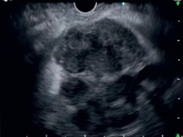 Fig. 1
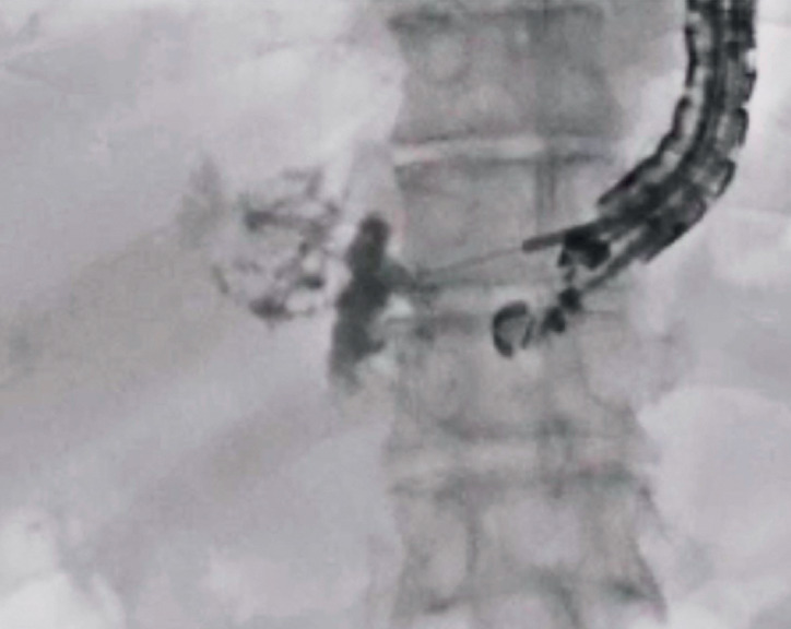 Fig. 2
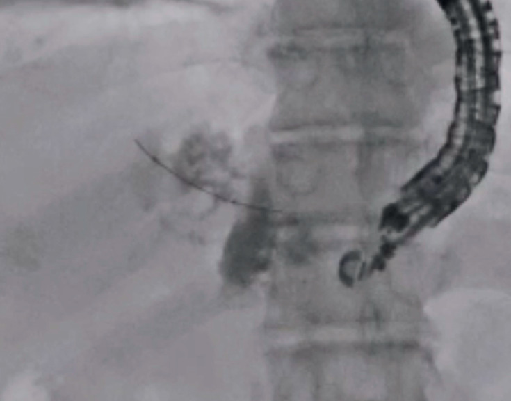 Fig. 3
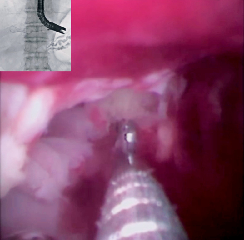 Fig. 4
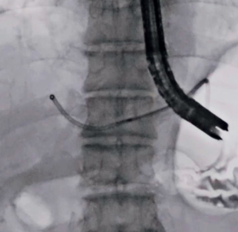 Fig. 5
Peer Reviews
No public reviews on file for this paper yet. If you reviewed it on a platform where reviews are public (OpenReview, ICLR, NeurIPS, ICML), you can paste yours below so the community can read it here.
Videos
No videos yet. Explain this paper in a talk, walkthrough, or lecture? Add one.
Taxonomy
TopicsAmoebic Infections and Treatments · Gallbladder and Bile Duct Disorders · Pancreatic and Hepatic Oncology Research
The gold standard technique for liver abscess drainage is the percutaneous transhepatic approach 1 ; however, endoscopic ultrasound-guided liver abscess drainage (EUS-LAD) has been reported 2 3 4 5 to overcome several disadvantages of percutaneous transhepatic liver abscess drainage, including external drainage or the risk of self-tube removal. However, if the liver abscess has multiple septa, the drainage effect after EUS-LAD might be limited. Several techniques, such as guidewire manipulation, can be used to divide the septa. However, breaking the septum might be challenging in cases with thick-walled septa. In addition, if a blood vessel is present in the septal wall, the procedure carries the risk of bleeding.
Recently, a novel cholangioscope (EyeMAX; Micro-Tech Co., Ltd, Nanjing, China), with a large working channel, has been developed. This scope has several benefits, such as allowing favorable visualization because of strong injection and aspiration functions due to the large working channel. Herein, we describe a novel technique, called the “transluminal opening technique,” for managing multiple septa in a liver abscess using the novel cholangioscope.
A 90-year-old man was admitted for the treatment of liver abscess. EUS imaging demonstrated a liver abscess with multiple septa ( Fig. 1 ). As the patient had dementia, a transluminal approach to the abscess was selected. After liver abscess puncture and contrast medium injection ( Fig. 2 ), a balloon catheter was inserted. Then, division of the septa was performed as much as possible ( Fig. 3 ). Finally, EUS-LAD using a metal stent was performed.
Multiple septa were observed within the liver abscess.
The liver abscess was punctured using a 19-G needle and contrast medium was injected.
The septa were broken, as much as possible, using a catheter and guidewire.
Owing to inadequate clinical effects, a transluminal opening technique was attempted after 7 days. First, an endoscopic retrograde cholangiopancreatography catheter was inserted within the EUS-LAD stent, and the contrast medium was injected. However, the cavity of the liver abscess was small. To break the septa, the novel cholangioscope was inserted within the liver abscess via a fistula. The septa were broken using biopsy forceps under direct visualization ( Fig. 4 ), and the cavity of the liver abscess was opened. A double-pigtail plastic stent was deployed without any adverse events ( Fig. 5 , Video 1 ). Following this procedure, the patient’s clinical course was excellent and he was discharged after 10 days.
The septa were broken using biopsy forceps under cholangioscopic guidance.
A plastic stent was deployed.
The septa were broken using biopsy forceps under cholangioscopic guidance.Video 1
In conclusion, the present technique might be useful for liver abscesses with multiple septa.
Endoscopy_UCTN_Code_TTT_1AS_2AG
The reference list from the paper itself. Each links out to its DOI / PubMed record.
- 1Ahmed S Chia CL Junnarkar SP Percutaneous drainage for giant pyogenic liver abscess – is it safe and sufficient?Am J Surg 20162119510110.1016/j.amjsurg.2015.03.00226033361 · doi ↗ · pubmed ↗
- 2Noh SH Park DH Kim YREUS-guided drainage of hepatic abscesses not accessible to percutaneous drainage (with videos)Gastrointest Endosc 2010711314131920400078 10.1016/j.gie.2009.12.045 · doi ↗ · pubmed ↗
- 3Chandra S Chandra U Endoscopic ultrasound-guided transgastric drainage of radiologically inaccessible left lobe liver abscess involving segment 4, caudate lobe, and left lateral segments using a modified technique Endosc Int Open 20219 E 35E 4010.1055/a-1293-774633403234 PMC 7775807 · doi ↗ · pubmed ↗
- 4Ogura T Sano T Onda S Endoscopic ultrasound-guided biliary drainage for right hepatic bile duct obstruction: novel technical tips Endoscopy 201547727510.1055/s-0034-137811125264761 · doi ↗ · pubmed ↗
- 5Tonozuka R Itoi T Tsuchiya TEUS-guided drainage of hepatic abscess and infected biloma using short and long metal stents (with videos)Gastrointest Endosc 2015811463146925843615 10.1016/j.gie.2015.01.023 · doi ↗ · pubmed ↗