Removal of obstructed pancreatic stent tube and biliary stones in patient with Roux-en-Y anatomy under direct endoscopic view
Jingjing Yao, Shengxue Pan, Hongbo Li, Kunpeng Liu, Guangyao Zhao, Jindong Fu

Abstract
Genes, proteins, chemicals, diseases, species, mutations and cell lines named across the full text — each resolved to its canonical identifier and authoritative record.
Click any figure to enlarge with its caption.
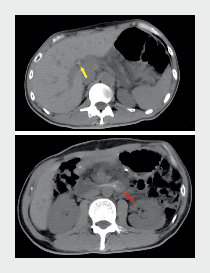 Fig. 1
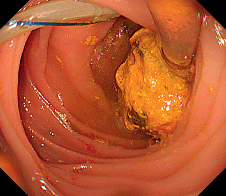 Fig. 2
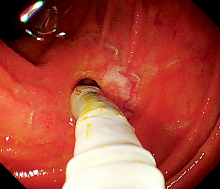 Fig. 3
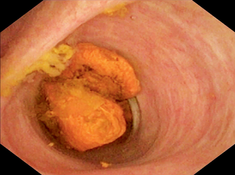 Fig. 4
Peer Reviews
No public reviews on file for this paper yet. If you reviewed it on a platform where reviews are public (OpenReview, ICLR, NeurIPS, ICML), you can paste yours below so the community can read it here.
Videos
No videos yet. Explain this paper in a talk, walkthrough, or lecture? Add one.
Taxonomy
TopicsPancreatic and Hepatic Oncology Research · Gallbladder and Bile Duct Disorders · Esophageal and GI Pathology
A 42-year-old man was admitted due to upper abdominal pain for 2 hours. He had undergone a pancreaticoduodenectomy for a pancreatic tumor with a Roux-en-Y anastomosis 3 years earlier. Over the past year, he had experienced recurrent episodes of acute pancreatitis. Abdominal computed tomography scans revealed high-density images at the terminus of the pancreatic stent tube and the hilar bile duct ( Fig. 1 ). Stent obstruction was suspected and endoscopic removal was performed ( Video 1 ).
Abdominal computed tomography scans revealed high-density images at the terminus of the pancreatic stent tube (red arrow) and the hilar bile duct (yellow arrow).
Endoscopic removal of the obstructed pancreatic stent tube and biliary stones.Video 1
We inserted an Olympus PCF-260 enteroscope (Olympus, Tokyo, Japan) through the oral cavity into the jejunal input loop, reaching the pancreaticojejunal anastomosis site. Here we observed a long pancreatic stent tube affixed to the intestinal wall with sutures. An adhesive stone was found at the distal end, completely obstructing the lumen ( Fig. 2 ). Endoscopic scissors were employed to cut the sutures and the stent to facilitate stent removal. A snare device was then used to sequentially extract the stent and the attached stone. The choledochojejunal anastomosis was identified adjacent to the pancreatic anastomosis. Upon insertion of a cholangioscope (Eye-Max CDS11001, 9 Fr; Micro-Tech, Nanjing, China) for direct visualization ( Fig. 3 ), two calculi were revealed at the bile duct convergence ( Fig. 4 ). Following anastomosis dilation with a balloon catheter, the calculi were successfully extracted using a stone retrieval basket under direct visual guidance from the cholangioscope ( Video 1 ). Finally, a hemostatic clip was applied to constrict the dilated anastomosis, preventing reflux cholangitis.
An adhesive stone was found at the distal end of the pancreatic stent tube, completely obstructing the lumen.
A cholangioscope was inserted into the bile duct through the choledochojejunal anastomosis.
Two calculi were visible at the bile duct convergence under direct visualization with the cholangioscope.
The patient was kept fasting for 48 hours. No complications were reported postoperatively. His abdominal symptoms resolved. He was discharged 4 days postoperatively.
Following pancreatic surgery, stent tubes are commonly used to reduce the risk of pancreatic fistula formation 1 2 . In this rare case, the stent was completely obstructed by a stone, leading to recurrent episodes of acute pancreatitis. However, with the aid of endoscopic scissors, the obstructed stent was successfully removed endoscopically. Furthermore, for this patient with Roux-en-Y anatomy presenting with bile stones, the utilization of the cholangioscope was more intuitive and accurate, offering a direct and radiation-free approach.
Endoscopy_UCTN_Code_TTT_1AR_2AG
The reference list from the paper itself. Each links out to its DOI / PubMed record.
- 1Kawai M Yamaue H Jang JY Propensity score-matched analysis of internal stent vs external stent for pancreatojejunostomy during pancreaticoduodenectomy: Japanese-Korean cooperative project Pancreatology 20202098499132680728 10.1016/j.pan.2020.06.014 · doi ↗ · pubmed ↗
- 2Shrikhande SV Sivasanker M Vollmer CM Pancreatic anastomosis after pancreatoduodenectomy: a position statement by the International Study Group of Pancreatic Surgery (ISGPS)Surgery 20171611221123410.1016/j.surg.2016.11.02128027816 · doi ↗ · pubmed ↗