Comparison of the Application Value of Transthoracic Echocardiography in Diagnosing Patent Foramen Ovale Under Different States of Stimulation: A Retrospective Study
Jianwei Shi, Haijuan Gu, Wenjun Fan, Jiesheng Xia, Huanhuan Gu

TL;DR
This study shows that using contrast-enhanced transthoracic echocardiography during specific physical maneuvers improves the accuracy of diagnosing patent foramen ovale.
Contribution
The study introduces the clinical benefit of using contrast-enhanced TTE under different stimulation states for more accurate PFO diagnosis.
Findings
Contrast-enhanced TTE during Valsalva maneuvers significantly improves PFO detection sensitivity.
Microbubble count increases under stimulation, especially during prolonged Valsalva maneuvers.
PFO diagnosis accuracy is enhanced by combining TTE with different physiological testing conditions.
Abstract
This study aims to evaluate the application value of contrast‐enhanced transthoracic echocardiography (cTEE) in the diagnosis of patent foramen ovale (PFO) under different states of stimulation, with the goal of enhancing the accuracy and efficiency of PFO diagnosis. This research consecutively enrolled patients suspected of having PFO from October 2022 to February 2024, presenting primary clinical symptoms such as unexplained syncope, headache, dizziness, and stroke. Patients underwent standard transthoracic echocardiography (TTE) and cTEE under three different states of stimulation (resting state, coughing, and Valsalva maneuver). Based on the presence of microbubbles in the left heart and their initial appearance time, patients were classified into PFO and control groups, with further diagnostic confirmation via transesophageal echocardiography (TEE) or foramen ovale closure…
Genes, proteins, chemicals, diseases, species, mutations and cell lines named across the full text — each resolved to its canonical identifier and authoritative record.
Click any figure to enlarge with its caption.
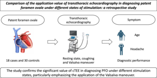 Figure 1
| Category | Number of cases | PFO group | Control group |
|
|---|---|---|---|---|
| Gender | ||||
| Male | 28 | 10 (35.7%) | 18 (64.3%) | 0.243 |
| Female | 20 | 8 (40.0%) | 12 (60.0%) | |
| Age ( ± s) | 47.4 ± 14.6 | 49.4 ± 17.7 | 0.034 | |
| Symptoms | ||||
| Syncope | 1 | 1 (100.0%) | 0 (0.0%) | 0.342 |
| Headache | 19 | 17 (89.5%) | 2 (10.5%) | < 0.001 |
| Dizziness | 24 | 15 (62.5%) | 9 (37.5%) | 0.342 |
| Stroke | 10 | 8 (80.0%) | 2 (20.0%) | 0.050 |
| TTE positive examination | ||||
| Resting state | 7 | 6 (85.7%) | 1 (14.3%) | 0.004 |
| Coughing action | 11 | 9 (81.8%) | 2 (18.2%) | < 0.001 |
| Valsalva maneuver of different durations | ||||
| 5 s | 12 | 10 (83.3%) | 2 (16.7%) | < 0.001 |
| 10 s | 16 | 13 (81.3%) | 3 (18.8%) | < 0.001 |
| 15 s | 21 | 16 (76.2%) | 5 (23.8%) | < 0.001 |
| Number of cases | PFO group (total of 18 cases) | |||
|---|---|---|---|---|
| Item | < 3 | 3–5 | > 5 | |
| Resting state | 7 | 2 | 5 | 0 |
| Coughing action | 11 | 3 | 7 | 1 |
| Valsalva maneuver of different durations (5/10/15 s) | 10 | 3 | 7 | 0 |
| 13 | 3 | 9 | 1 | |
| 16 | 5 | 9 | 2 | |
| Category | Number of cases | Cardiac cycle (number) |
|
|---|---|---|---|
| Resting state | 7 | 3.43 | |
| Coughing action | 11 | 2.86 | < 0.01 |
| Valsalva maneuver of different durations | 10 | 3.17 (5 s) | < 0.05 |
| 13 | 2.82 (10 s) | < 0.01 | |
| 16 | 1.54 (15 s) | < 0.001 |
| Category | Number of cases | Microbubble quantity |
|
| |||
|---|---|---|---|---|---|---|---|
| < 5 | 5–10 | 10–30 | > 30 | ||||
| Resting state | 7 | 4 (57.1%) | 2 (28.6%) | 1 (14.3%) | 0 | 50.32 | < 0.001 |
| Coughing action | 11 | 3 (27.3%) | 4 (36.4%) | 2 (18.2%) | 2 (18.2%) | 37.43 | < 0.001 |
| Valsalva maneuver of different durations | 10 (5 s) | 1 (10%) | 3 (30%) | 4 (40%) | 2 (20%) | 61.32 | < 0.001 |
| 13 (10 s) | 2 (15.4%) | 3 (23.1%) | 4 (30.7%) | 4 (30.8%) | 65.32 | < 0.001 | |
| 16 (15 s) | 1 (6.3%) | 3 (18.7%) | 6 (37.5%) | 6 (37.5%) | 71.32 | < 0.001 | |
- —This study was supported by Nantong Science and Technology Bureau (MSZ2023164).
Peer Reviews
No public reviews on file for this paper yet. If you reviewed it on a platform where reviews are public (OpenReview, ICLR, NeurIPS, ICML), you can paste yours below so the community can read it here.
Videos
No videos yet. Explain this paper in a talk, walkthrough, or lecture? Add one.
Taxonomy
TopicsCardiovascular and Diving-Related Complications · Cardiovascular Syncope and Autonomic Disorders · Trauma Management and Diagnosis
Introduction
1
Patent Foramen Ovale (PFO) represents a common cardiac anatomical anomaly present in approximately 25% of adults [1]. While PFO is benign in most instances, it has been associated with certain medical conditions, such as cryptogenic stroke, migraines, and potential thromboembolic diseases [2]. Consequently, the accurate diagnosis of PFO is crucial for assessing the risk of these conditions and formulating appropriate treatment strategies. Contrast‐enhanced transthoracic echocardiography (cTEE) serves as a widely utilized noninvasive diagnostic approach for detecting PFO, involving the use of ultrasound waves and contrast agents to observe cardiac structures and functions [3]. However, the diagnostic process of cTEE can vary depending on the patient's physiological state, such as their state of stimulation. Common stimuli include the resting state, coughing, and the Valsalva maneuver (VM)—a technique involving an attempt to exhale against a closed airway [4]. While cTEE provides baseline data in the resting state without additional stimuli, it may not reveal all instances of PFO. In contrast, the VM, by increasing intrathoracic pressure, significantly enhances the sensitivity of detecting right‐to‐left shunts (RLS) in the diagnosis of PFO [5]. Under this stimulus, cTEE shows greater diagnostic accuracy. A study found that during the VM, a significant percentage of patients exhibited right‐to‐left cardiac shunting in transcranial Doppler (TCD) assessments, while a similar percentage showed shunting in cTEE. These findings underscore the crucial role of the VM in revealing shunts across both diagnostic modalities [6].
This study aims to compare the application value of cTEE in diagnosing PFO under different states of stimulation. By assessing the resting state, coughing, and varying durations of the VM, the research will explore the sensitivity and specificity of PFO detection under these conditions. This investigation holds significant importance for improving the diagnostic methods for PFO, contributing to enhanced diagnostic accuracy and efficiency.
Materials and Methods
2
Study Participants
2.1
This study consecutively recruited adult patients from October 2022 to February 2024, who exhibited unexplained clinical symptoms such as syncope, headaches, dizziness, and strokes, with a clinical suspicion of PFO. All participants underwent standard transthoracic echocardiography (TTE) and cTTE across three different states of stimulation. Based on the detection of microbubbles in the left atrium within three to six cardiac cycles post‐contrast injection, patients were categorized into the PFO group (microbubbles observed) and the control group (no microbubbles observed) [7]. Patients diagnosed with PFO underwent further evaluation with transesophageal echocardiography (TEE) or underwent a foramen ovale closure procedure. The study groups were divided as follows: (1) resting state, coughing, VM for 5 s, VM for 10 s, and VM for 15 s groups; (2) transthoracic echocardiography group and transesophageal echocardiography group. This is a retrospective study and has received approval from the ethics committee. All patients and some requiring TEE examinations were informed about the purpose, methodology, potential risks, and mitigation measures of the echocardiographic studies and they signed informed consent forms.
Inclusion and Exclusion Criteria
2.2
Inclusion criteria: (1) Patients without contraindications to right heart catheterization, able to complete transthoracic and TEE examinations and standard VMs; (2) Patients agreeing to the examinations and signing informed consent forms. Exclusion criteria: (1) Patients with contraindications to right heart catheterization; (2) Patients with poor echocardiographic window, making it difficult to accurately observe shunt bubbles (e.g., patients with chronic obstructive pulmonary disease, morbid obesity); (3) Patients unable to cooperate with the examination or whose family did not agree to participate; (4) Patients with unclear transthoracic echocardiographic views; (5) Patients with structural heart diseases other than PFO; (6) Patients with cognitive impairments or unable to perform VMs and coughing; (7) Patients in the acute phase of infection, with severe heart failure, renal failure, pulmonary failure, or coagulation disorders.
Methodology and Criteria for Judgment
2.3
TTE and TEE
2.3.1
Patients were positioned in either the left or right lateral decubitus position for transthoracic and/or transesophageal echocardiographic examinations through right heart contrast imaging. TTE utilized the standard four‐chamber view, while TEE employed the bicameral view to clearly display the foramen ovale flap. The right heart contrast procedure involved the rapid injection of a mixture comprising 8 mL of saline, 1 mL of air, and 1 mL of blood, agitated more than 10 times before rapid administration via the median cubital vein. Echocardiographic imaging storage [8]: Dynamic videos of right heart contrast echocardiography during 15 cardiac cycles were stored at rest, during coughing, and during VMs at 5, 10, and 15 s. Outcome interpretation: Observation and recording of the presence and quantity of microbubbles in the left atrium within six cardiac cycles post‐contrast through video playback.
Transthoracic and Transesophageal Right Heart Contrast Echocardiography
2.3.2
A venous pathway was established in the patient's median cubital vein, connected to a three‐way stopcock; two 10 mL syringes were prepared, one with 8 mL of saline and the other with 1 mL of air, connected via the stopcock. After ensuring patency of the venous pathway and retracting 1 mL of blood, saline, blood, and air were agitated between the two syringes thirty times before evenly paced injection. The apical four‐chamber view was selected to observe the presence and quantity of microbubbles in the left heart after right heart contrast imaging at rest and during the VM for three to five cardiac cycles. The positive quantification criteria for contrast‐enhanced TTE (C‐TTE) followed the method of Calvert et al.: Grade I, < 5 microbubbles in the left heart chamber; Grade II, 5–25 microbubbles; Grade III, > 25 microbubbles but with lower density; Grade IV, > 25 microbubbles, dense, causing opacification of the heart chamber [9]. For C‐TTE, a 20G catheter was placed in the patient's left median cubital or antecubital vein, connected to a three‐way stopcock with two 10 mL syringes attached, filled with 8 mL of saline, 1 mL of air, and 1 mL of the patient's blood [10]. The mixture was agitated at least 20 times to prepare a saline solution rich in microbubbles, which was then rapidly injected during the patient's resting state, coughing, VM, and modified VM (mVM), with at least a 5‐min interval between injections. Observation focused on the cardiac cycle when left heart microbubbles first appeared and the quantity of microbubbles. For transesophageal right heart contrast echocardiography (C‐TEE), if PFO presence was uncertain, the agitation method for saline preparation was identical to that of TTE, with rapid intravenous injection after preparation. The appearance of microbubbles in the left atrium within three cardiac cycles post right heart bubble opacification indicated PFO. This procedure was performed up to three times, with at least a 5‐min interval between attempts [11].
VM
2.3.3
Patients were instructed to pinch their nostrils, close their mouth, and forcefully attempt to exhale, engaging the abdomen without allowing air to escape, and suddenly releasing the breath after 5, 10, and 15 s.
Statistical Methods
2.4
Data analysis was performed using SPSS software version 25.0. Count data were presented as case numbers and percentages, with TEE serving as the gold standard for the diagnostic test of C‐TTE. The methods used for intergroup comparisons included the following:
The χ ^2^ test was employed to compare the gender distribution differences between the PFO group and the control group. Independent samples t‐tests were used to compare age differences between these groups. Analysis of Variance (ANOVA) was conducted to compare the initial appearance time of microbubbles and the quantity of microbubbles under different stimulation states (resting state, coughing, VMs of 5, 10, and 15 s). The K‐value was used to compare the distribution differences in microbubble quantities under various stimulation states. Statistical significance (p values) was determined to identify significant differences between groups, such as the differences in microbubble quantity and appearance time.
Results
3
Clinical Case Data
3.1
In this study, we compared the differences between the PFO group and the control group in terms of gender, age, clinical symptoms, and the positivity rate of TTE examinations. The results indicated no significant difference in gender distribution between the two groups (p = 0.243). However, there was a statistically significant difference in age between the PFO group and the control group (mean ages were 47.4 ± 14.6 years and 49.4 ± 17.7 years, respectively; p = 0.034). Analysis of clinical symptoms revealed that headaches were significantly more prevalent in the PFO group compared to the control group (89.5% vs. 10.5%, p < 0.001), while other symptoms such as syncope, dizziness, and cerebral infarction did not show significant differences, although the incidence of cerebral infarction was higher in the PFO group (80% vs. 20%, p = 0.05). The positivity rate of TTE examinations was higher in the PFO group both at rest and during coughing, reflecting the correlation between PFO and specific clinical manifestations, as shown in Table 1.
Distribution of Initial Appearance Time of Left Heart Microbubbles Under Different States of Stimulation
3.2
In this study, we analyzed the performance of 18 PFO patients under various physiological testing conditions, including at rest, during coughing, and during VMs of varying durations (5, 10, 15 s). The results showed that at rest, 7 PFO patients were identified, with two scoring less than 3 and five falling within the 3–5 range, with no patients scoring above 5. During the coughing test, out of 11 PFO patients, 3 scored less than 3, 7 scored within the 3–5 range, and 1 scored above 5. For VMs of different durations, the distribution of scores among patients performing 10‐ and 15‐s maneuvers indicated that the number of patients scoring above 5 gradually increased (from 0 to 2) as the duration of the maneuver increased (Table 2).
Comparison of Microbubble Appearance Time Under Different States of Stimulation in CTTE
3.3
We explored the differences in the initial cardiac cycle of microbubble appearance indicating RLS under various states of stimulation (resting state, coughing, and VMs of varying durations). The average cardiac cycle at rest was 3.43, during coughing it was 2.86, and during VMs of different durations, the cardiac cycles were 3.17 (5 s), 2.82 (10 s), and 1.54 (15 s), respectively. This demonstrates a significant reduction in cardiac cycles as the duration of the VM increases (p < 0.001, indicating a significant difference compared to the resting state) (Table 3).
Comparison of RLS Microbubble Quantities Under Different States of Stimulation in TTE
3.4
By comparing the quantity of RLS microbubbles at rest, during coughing, and during VMs of varying durations, we observed that at rest, among 7 patients, 57.1% had fewer than 5 microbubbles, 28.6% had between 5 and 10 microbubbles, and 14.3% had between 10 and 30 microbubbles, with no patients exceeding 30 microbubbles. The K‐value for this state was 50.32, with p < 0.001, indicating statistically significant differences compared to other states. During coughing, among 11 patients, 27.3% had fewer than 5 microbubbles, 36.4% had between 5 and 10, and 18.2% had between 10 and 30 and over 30 microbubbles. The K‐value was 37.43, with p < 0.001. VMs of varying durations (5, 10, 15 s) showed an increasing trend in the quantity of microbubbles with the duration of the maneuver. For instance, at 15 s, 6.3% of patients had fewer than 5 microbubbles, 18.7% had between 5 and 10, and 37.5% had between 10 and 30 and over 30 microbubbles. The corresponding K‐values were 61.32, 65.32, and 71.32, with p < 0.001, indicating a significant increase compared to the resting state (Table 4).
Discussion
4
PFO is a common cardiac structural anomaly observed in approximately 25% of adults, which is mostly harmless but has been linked to conditions such as cryptogenic stroke, migraines, and certain types of thromboembolic diseases [12, 13]. Despite the prevalence of PFO, its diagnosis remains challenging, especially in asymptomatic cases. Traditional diagnostic methods include cTEE and TEE, whose effectiveness may be influenced by the patient's state, such as resting, coughing, and performing VMs [14, 15]. This study, by comparing the application value of cTEE in diagnosing PFO under different states of stimulation, revealed the correlation between age and headache symptoms with PFO and highlighted that the significant increase in microbubble quantity during VMs can enhance the sensitivity and specificity of PFO detection. The results underscore the clinical importance of considering cTEE examinations under different states of stimulation to improve the accuracy and efficiency of PFO diagnosis.
Our findings indicate significant differences in age between the PFO and control groups, with a notably higher incidence of headaches in the PFO group. These observations align with the study by Seward et al., which also reported a connection between PFO and migraines [16]. This could be due to PFO allowing microemboli in the blood to bypass pulmonary filtration and directly enter the brain, triggering headaches. Additionally, our study found that VMs significantly increase the sensitivity of PFO detection, consistent with the findings of Brisson et al., who noted that VMs can increase RLS [17], thereby enhancing PFO detection sensitivity. This result emphasizes the importance of incorporating VMs in cTEE examinations. The significant increase in microbubble quantity during VMs and its positive correlation with the duration of the maneuver provides a new perspective for PFO diagnosis. Van Laecke and Pan et al. also reflected this, finding an increase in microbubble quantity positively correlated with the size of PFO, which might facilitate more refined risk stratification in PFO diagnosis [18, 19]. These data reveal the differential response of PFO patients under various physiological tests, particularly during longer durations of VMs, reflecting potential cardiovascular regulatory abnormalities. This variability may have clinical significance for the diagnosis and management of PFO, suggesting that further research is needed to understand the physiological response characteristics of PFO patients more deeply [20].
Our results also indicate that the state of stimulation significantly affects the initial cardiac cycle of RLS microbubble appearance, especially during the VM. With an increase in the duration of the maneuver, there is a significant decrease in cardiac cycles, which may reflect changes in cardiac physiological responses and the sensitivity of RLS detection. This finding is of significant importance for understanding the clinical assessment and diagnosis of RLS, highlighting the necessity to consider changes in cardiac cycles under different states of stimulation.
A major limitation of this study is the relatively small sample size, which might affect the generalizability of the results [21]. Additionally, the retrospective analysis design could introduce selection bias [22]. Future studies should validate these findings through prospective, multicenter research and explore the differential responses of different PFO sizes and shapes under various states of stimulation.
In conclusion, this study highlights the application value of cTEE in diagnosing PFO under different states of stimulation, particularly with the use of VMs. These findings provide important insights for clinical practice regarding PFO diagnosis, aiding in enhancing diagnostic accuracy and efficiency. Further research is needed on a larger sample size to validate these results and explore other factors that may influence PFO diagnosis, to further optimize diagnostic and treatment strategies for PFO.
Conflicts of Interest
The authors declare no conflicts of interest.
The reference list from the paper itself. Each links out to its DOI / PubMed record.
- 1A. Turley , J. Thambyrajah , P. Finn , et al., “Transthoracic Contrast Echocardiography in the Detection of Patent Foramen Ovale,” Critical Care 10 (2006): 1–2.
- 2T. Wang , Z. Li , M. Huang , et al., “Echo CP: An Echocardiography Dataset in Contrast Transthoracic Echocardiography for Patent Foramen Ovale Diagnosis,” in Medical Image Computing and Computer Assisted Intervention–MICCAI: 24th International Conference, Strasbourg, France, September 27–October 1, 2021, Proceedings, Part VI, eds. M. C. Erasmus , P. C. Cattin , S. Cotin , N. Padoy , S. Speidel , Y. Zheng and C. Essert (Springer International Publishing, 2021), 506–515.
- 3S. Wang , G. Zhu , Z. Liu , J. Zhou , and W. Zang , “Only Transesophageal Echocardiography Guided Patent Foramen Ovale Closure: A Single‐Center Experience,” Frontiers in Surgery 9 (2022): 977959.36303848 10.3389/fsurg.2022.977959 PMC 9592899 · doi ↗ · pubmed ↗
- 4J. Zhu , A. Chen , L. Zhu , et al., “Calf Muscle Pump Tensing as a Novel Maneuver to Improve the Diagnostic Performance of Detecting Patent Foramen Ovale During Transesophageal Echocardiography,” Frontiers in Neurology 14 (2023): 1116764.36761345 10.3389/fneur.2023.1116764 PMC 9905729 · doi ↗ · pubmed ↗
- 5A. M. Lanzone , E. V. Castelluccio , P. Della Pina , et al., “Comparative Diagnostic Accuracy of Transcranial Doppler and Contrast‐Enhanced Transthoracic Echocardiography for the Diagnosis of Patent Foramen Ovale and Atrial Septal Defect,” Panminerva Medica 66, no. 2 (2024): 124–130, https://pubmed.ncbi.nlm.nih.gov/38563605.38563605 10.23736/S 0031-0808.24.05123-1 · doi ↗ · pubmed ↗
- 6A. Chen , J. Zhu , L. Zhu , et al., “Neglected Intrapulmonary Arteriovenous Anastomoses: A Comparative Study of Pulmonary Right‐to‐Left Shunts in Patients With Patent Foramen Ovale,” Frontiers in Cardiovascular Medicine 10 (2023): 1111818.37089892 10.3389/fcvm.2023.1111818 PMC 10117845 · doi ↗ · pubmed ↗
- 7Y. Niu , J. Pan , S. Fan , L. Wang , and X. Tang , “The Value of Right Heart Contrast Echocardiography Combined With Migraine Rating Scale in Evaluating the Efficacy of Patent Foramen Ovale Closure,” BMC Cardiovascular Disorders 23, no. 1 (2023): 390.37558988 10.1186/s 12872-023-03411-8PMC 10410887 · doi ↗ · pubmed ↗
- 8J. Chen , R. Li , J. Chen , et al., “Acute Cerebral Infarction With Acute Myocardial Infarction Due to Patent Foramen Ovale: A Case Report,” Medicine 99, no. 19 (2020): e 20054.32384468 10.1097/MD.0000000000020054 PMC 7220755 · doi ↗ · pubmed ↗