The genome sequence of a barkfly, Mesopsocus fuscifrons Meinander, 1966
Duncan Sivell, Jae Hak Son, Kosuke Kataoka

TL;DR
This paper reports the genome sequence of the barkfly Mesopsocus fuscifrons, including a detailed assembly of its chromosomes and mitochondrial DNA.
Contribution
The novel contribution is the first genome assembly for the species Mesopsocus fuscifrons, including chromosomal scaffolding and mitochondrial genome.
Findings
The genome assembly spans 184.3 megabases and is scaffolded into 9 chromosomal pseudomolecules.
The mitochondrial genome is 20.13 kilobases in length.
Abstract
We present a genome assembly from an individual female Mesopsocus fuscifrons (barkfly; Arthropoda; Insecta; Psocodea; Mesopsocidae). The genome sequence is 184.3 megabases in span. Most of the assembly is scaffolded into 9 chromosomal pseudomolecules. The mitochondrial genome has also been assembled and is 20.13 kilobases in length.
Genes, proteins, chemicals, diseases, species, mutations and cell lines named across the full text — each resolved to its canonical identifier and authoritative record.
Click any figure to enlarge with its caption.
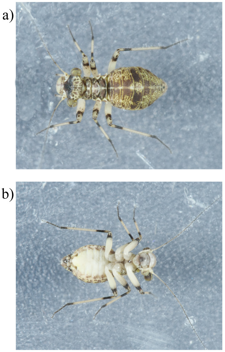 Figure 1
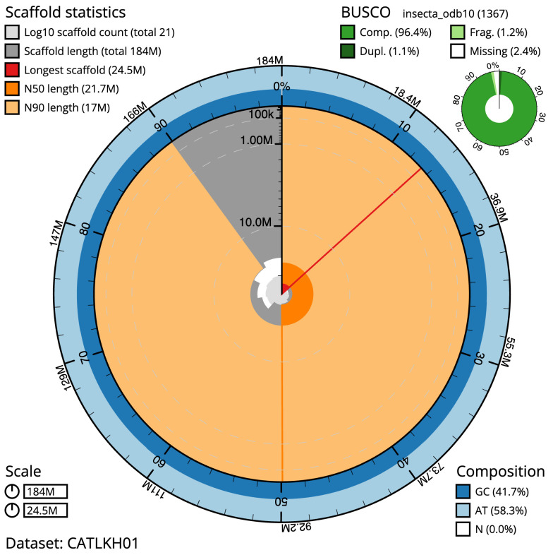 Figure 2
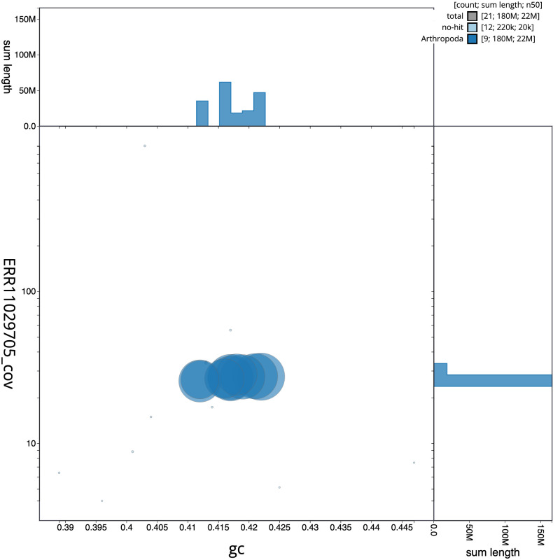 Figure 3
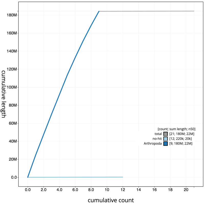 Figure 4
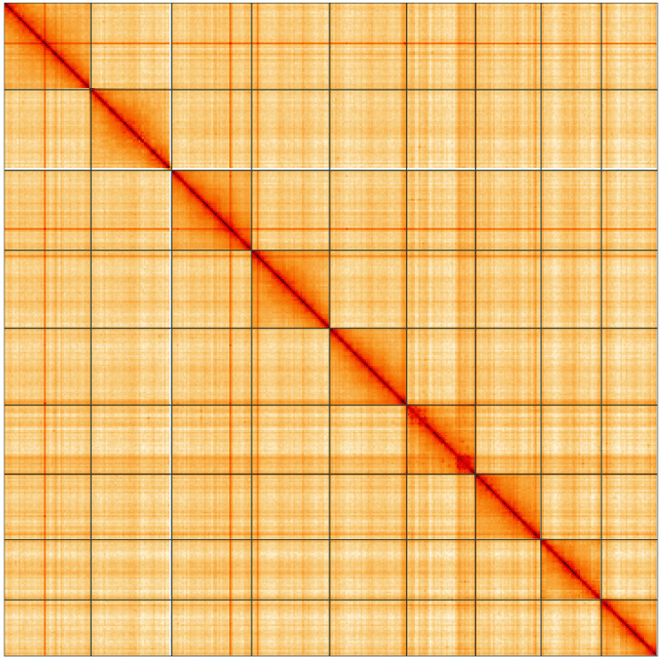 Figure 5
| Project accession data | ||
|---|---|---|
| Assembly identifier | iuMesFusc1.1 | |
| Species |
| |
| Specimen | iuMesFusc1 | |
| NCBI taxonomy ID | 2866285 | |
| BioProject | PRJEB60722 | |
| BioSample ID | SAMEA10241719 | |
| Isolate information | iuMesFusc1, female: whole organism (DNA
| |
| Assembly metrics
|
| |
| Consensus quality (QV) | 58.2 |
|
|
| 100.0% |
|
| BUSCO
| C:96.4%[S:95.3%,D:1.1%],
|
|
| Percentage of assembly
| 99.9% |
|
| Sex chromosomes | Not identified |
|
| Organelles | Mitochondrial genome: 20.13 kb |
|
| Raw data accessions | ||
| PacificBiosciences SEQUEL II | ERR11029705 | |
| Hi-C Illumina | ERR11042967 | |
| Genome assembly | ||
| Assembly accession | GCA_950004255.1 | |
|
| GCA_949987595.1 | |
| Span (Mb) | 184.3 | |
| Number of contigs | 276 | |
| Contig N50 length (Mb) | 1.2 | |
| Number of scaffolds | 20 | |
| Scaffold N50 length (Mb) | 21.7 | |
| Longest scaffold (Mb) | 24.53 | |
| INSDC accession | Chromosome | Length (Mb) | GC% |
|---|---|---|---|
| 1 | 24.53 | 42.0 | |
| 2 | 22.71 | 41.5 | |
| 3 | 22.5 | 42.0 | |
| 4 | 21.98 | 41.5 | |
| 5 | 21.66 | 42.0 | |
| 6 | 19.48 | 41.0 | |
| 7 | 18.44 | 42.0 | |
| 8 | 16.98 | 41.5 | |
| 9 | 15.86 | 41.0 | |
| MT | 0.02 | 40.5 |
| Software tool | Version | Source |
|---|---|---|
| BlobToolKit | 4.1.7 |
|
| BUSCO | 5.3.2 |
|
| Hifiasm | 0.16.1-r375 |
|
| HiGlass | 1.11.6 |
|
| Merqury | MerquryFK |
|
| MitoHiFi | 3 |
|
| PretextView | 0.2 |
|
| purge_dups | 1.2.5 |
|
| sanger-tol/genomenote | v1.0 |
|
| sanger-tol/readmapping | 1.1.0 |
|
| YaHS | 1.2a.2 |
|
- —Wellcome Trust
Peer Reviews
No public reviews on file for this paper yet. If you reviewed it on a platform where reviews are public (OpenReview, ICLR, NeurIPS, ICML), you can paste yours below so the community can read it here.
Videos
No videos yet. Explain this paper in a talk, walkthrough, or lecture? Add one.
Taxonomy
TopicsGenomics and Phylogenetic Studies · Environmental DNA in Biodiversity Studies · Insect symbiosis and bacterial influences
Species taxonomy
Eukaryota; Metazoa; Eumetazoa; Bilateria; Protostomia; Ecdysozoa; Panarthropoda; Arthropoda; Mandibulata; Pancrustacea; Hexapoda; Insecta; Dicondylia; Pterygota; Neoptera; Paraneoptera; Psocodea; Psocoptera; Psocomorpha; Homilopsocidea; Peripsocoidea; Mesopsocidae; Mesopsocus; Mesopsocus fuscifrons (Meinander, 1966) (NCBI:txid2866285).
Background
Mesopsocus fuscifrons is a large, 3.5–5.0 mm long, barkfly (Psocodea: ‘Psocoptera’) that can be recognised by its very dark frons and distinctive eye pattern ( Lienhard, 1998). The wings are fully formed in males, but reduced to vestigial wing buds in females. Female M. fuscifrons also have dark rings near the base of the tibiae that are characteristic of this species. The eye pattern can be described as a dark chevron that lies horizontally across the eye. In practice the eyes appear dark with a paler grey line running horizontally across their middle. Either interpretation is clearly different from the eyes of other British Mesopsocus, which can be identified using New (2005).
Mesopsocus fuscifrons is a recent addition to the British fauna with a population in the Wildlife Garden of the Natural History Museum in South Kensington ( McCarter et al., 2021). Specimens captured in a Malaise trap in the summer of 2013 are the earliest British records we are aware of. This species has also been found at three sites in Richmond and Ham ( Cook, 2023), and undoubtedly occurs elsewhere in the Greater London area.
This barkfly is a Mediterranean species known from Morocco, Algeria, France, Italy, Macedonia and Greece ( Lienhard, 1998) that has expanded its range into northern Europe in the past decade. Mesopsocus fuscifrons is now recorded in Germany, northern France, Belgium, the Netherlands and Denmark as well as the UK ( Schuch et al., 2021). There were also records from southern Sweden in 2006 but it is not clear if a population persists there ( Svensson & Hall, 2010).
Mesopsocus fuscifrons has been found on a wide variety of broadleaved trees and shrubs that may or may not have lichens growing on the bark ( Cook, 2023; Schuch et al., 2021). Lienhard (1998) also reports M. fuscifrons living on conifers. Across its geographic range M. fuscifrons adults may be found in any month of the year, though are more likely to be seen from May to October ( Schuch et al., 2021). British records so far have been made from May to August ( Cook, 2023).
A single female observed on the trunk of a young ash tree ( Fraxinus excelsior) in the NHM Wildlife Garden was captured and submitted to the Darwin Tree of Life project on 8 June 2021. It has been suggested that Mesopsocus fuscifrons may be synonymous with M. ypsilon Ball, 1937, a species known from Morocco and Greece ( Lienhard, 1998). The publication of this genome sequence will help to answer this question.
Genome sequence report
The genome was sequenced from one female Mesopsocus fuscifrons (NHMUK014043153, Figure 1) collected from Natural History Museum Wildlife Garden, England (51.5, –0.18). A total of 93-fold coverage in Pacific Biosciences single-molecule HiFi long reads was generated. Primary assembly contigs were scaffolded with chromosome conformation Hi-C data. Manual assembly curation corrected 204 missing joins or mis-joins and removed 106 haplotypic duplications, reducing the assembly length by 4.60% and the scaffold number by 75.29%, and increasing the scaffold N50 by 13.37%.
Photographs of the Mesopsocus fuscifrons (iuMesFusc1, NHMUK014043153) specimen used for genome sequencing a) dorsal view, b) ventral view.
The final assembly has a total length of 184.3 Mb in 20 sequence scaffolds with a scaffold N50 of 21.7 Mb ( Table 1). The snailplot in Figure 2 provides a summary of the assembly statistics, while the distribution of assembly scaffolds on GC proportion and coverage is shown in Figure 3. The cumulative assembly plot in Figure 4 shows curves for subsets of scaffolds assigned to different phyla. Most (99.9%) of the assembly sequence was assigned to 9 chromosomal-level scaffolds, representing 9 autosomes. Chromosome-scale scaffolds confirmed by the Hi-C data are named in order of size ( Figure 5; Table 2). Sex chromosomes were not identified due to the lack of comparators within this order. Psocodea have multiple sex determination mechanisms ( Hodson et al., 2017). While not fully phased, the assembly deposited is of one haplotype. Contigs corresponding to the second haplotype have also been deposited. The mitochondrial genome was also assembled and can be found as a contig within the multifasta file of the genome submission.
Table 1.: Genome data for Mesopsocus fuscifrons, iuMesFusc1.1.
Genome assembly of Mesopsocus fuscifrons, iuMesFusc1.1: metrics.The BlobToolKit Snailplot shows N50 metrics and BUSCO gene completeness. The main plot is divided into 1,000 size-ordered bins around the circumference with each bin representing 0.1% of the 184,352,988 bp assembly. The distribution of scaffold lengths is shown in dark grey with the plot radius scaled to the longest scaffold present in the assembly (24,526,141 bp, shown in red). Orange and pale-orange arcs show the N50 and N90 scaffold lengths (21,655,844 and 16,975,629 bp), respectively. The pale grey spiral shows the cumulative scaffold count on a log scale with white scale lines showing successive orders of magnitude. The blue and pale-blue area around the outside of the plot shows the distribution of GC, AT and N percentages in the same bins as the inner plot. A summary of complete, fragmented, duplicated and missing BUSCO genes in the insecta_odb10 set is shown in the top right. An interactive version of this figure is available at https://blobtoolkit.genomehubs.org/view/CATLKH01/dataset/CATLKH01/snail.
Genome assembly of Mesopsocus fuscifrons, iuMesFusc1.1: BlobToolKit GC-coverage plot. Scaffolds are coloured by phylum. Circles are sized in proportion to scaffold length. Histograms show the distribution of scaffold length sum along each axis. An interactive version of this figure is available at https://blobtoolkit.genomehubs.org/view/CATLKH01/dataset/CATLKH01/blob.
Genome assembly of Mesopsocus fuscifrons, iuMesFusc1.1: BlobToolKit cumulative sequence plot.The grey line shows cumulative length for all scaffolds. Coloured lines show cumulative lengths of scaffolds assigned to each phylum using the buscogenes taxrule. An interactive version of this figure is available at https://blobtoolkit.genomehubs.org/view/CATLKH01/dataset/CATLKH01/cumulative.
Genome assembly of Mesopsocus fuscifrons, iuMesFusc1.1: Hi-C contact map of the iuMesFusc1.1 assembly, visualised using HiGlass.Chromosomes are shown in order of size from left to right and top to bottom. An interactive version of this figure may be viewed at https://genome-note-higlass.tol.sanger.ac.uk/l/?d=I7ipiU4sRtSk_uJrqsXM3Q.
Table 2.: Chromosomal pseudomolecules in the genome assembly of Mesopsocus fuscifrons, iuMesFusc1.
The estimated Quality Value (QV) of the final assembly is 58.2 with k-mer completeness of 100.0%, and the assembly has a BUSCO v5.3.2 completeness of 96.4% (single = 95.3%, duplicated = 1.1%), using the insecta_odb10 reference set ( n = 1,367).
Metadata for specimens, barcode results, spectra estimates, sequencing runs, contaminants and pre-curation assembly statistics are given at https://links.tol.sanger.ac.uk/species/2866285.
Methods
Sample acquisition and nucleic acid extraction
A female Mesopsocus fuscifrons (specimen ID NHMUK014043153, ToLID iuMesFusc1) was observed on the trunk of a young ash tree in the Natural History Museum Wildlife Garden, England, UK (latitude 51.5, longitude –0.18) on 2021-06-08. The specimen was collected and identified by Duncan Sivell (Natural History Museum) and then dry frozen at –80 °C.
The workflow for high molecular weight (HMW) DNA extraction at the Wellcome Sanger Institute (WSI) includes a sequence of core procedures: sample preparation; sample homogenisation, DNA extraction, fragmentation, and clean-up. The sample was prepared in the WSI Tree of Life Core laboratory: the iuMesFusc1 sample was weighed and dissected on dry ice ( Jay et al., 2023). Tissue from the whole organism was homogenised using a PowerMasher II tissue disruptor ( Denton et al., 2023a).
HMW DNA was extracted in the WSI Scientific Operations core using the Automated MagAttract v2 protocol ( Oatley et al., 2023). HMW DNA was sheared into an average fragment size of 12–20 kb in a Megaruptor 3 system with speed setting 31 ( Bates et al., 2023). Sheared DNA was purified by solid-phase reversible immobilisation ( Strickland et al., 2023): in brief, the method employs a 1.8X ratio of AMPure PB beads to sample to eliminate shorter fragments and concentrate the DNA. The concentration of the sheared and purified DNA was assessed using a Nanodrop spectrophotometer and Qubit Fluorometer and Qubit dsDNA High Sensitivity Assay kit. Fragment size distribution was evaluated by running the sample on the FemtoPulse system.
Protocols developed by the Wellcome Sanger Institute (WSI) Tree of Life core laboratory are publicly available on protocols.io ( Denton et al., 2023b).
Sequencing
Pacific Biosciences HiFi circular consensus DNA sequencing libraries were constructed according to the manufacturers’ instructions. DNA sequencing was performed by the Scientific Operations core at the WSI on a Pacific Biosciences SEQUEL II (HiFi) instrument. Hi-C data were also generated from remaining tissue of iuMesFusc1 using the Arima2 kit and sequenced on the Illumina NovaSeq 6000 instrument.
Genome assembly, curation and evaluation
Assembly was carried out with Hifiasm ( Cheng et al., 2021) and haplotypic duplication was identified and removed with purge_dups ( Guan et al., 2020). The assembly was then scaffolded with Hi-C data ( Rao et al., 2014) using YaHS ( Zhou et al., 2023). The assembly was checked for contamination and corrected using the gEVAL system ( Chow et al., 2016) as described previously ( Howe et al., 2021). Manual curation was performed using gEVAL, HiGlass ( Kerpedjiev et al., 2018) and Pretext ( Harry, 2022). The mitochondrial genome was assembled using MitoHiFi ( Uliano-Silva et al., 2023), which runs MitoFinder ( Allio et al., 2020) or MITOS ( Bernt et al., 2013) and uses these annotations to select the final mitochondrial contig and to ensure the general quality of the sequence.
A Hi-C map for the final assembly was produced using bwa-mem2 ( Vasimuddin et al., 2019) in the Cooler file format ( Abdennur & Mirny, 2020). To assess the assembly metrics, the k-mer completeness and QV consensus quality values were calculated in Merqury ( Rhie et al., 2020). This work was done using Nextflow ( Di Tommaso et al., 2017) DSL2 pipelines “sanger-tol/readmapping” ( Surana et al., 2023a) and “sanger-tol/genomenote” ( Surana et al., 2023b). The genome was analysed within the BlobToolKit environment ( Challis et al., 2020) and BUSCO scores ( Manni et al., 2021; Simão et al., 2015) were calculated.
Table 3 contains a list of relevant software tool versions and sources.
Wellcome Sanger Institute – Legal and Governance
The materials that have contributed to this genome note have been supplied by a Darwin Tree of Life Partner. The submission of materials by a Darwin Tree of Life Partner is subject to the ‘Darwin Tree of Life Project Sampling Code of Practice’, which can be found in full on the Darwin Tree of Life website here. By agreeing with and signing up to the Sampling Code of Practice, the Darwin Tree of Life Partner agrees they will meet the legal and ethical requirements and standards set out within this document in respect of all samples acquired for, and supplied to, the Darwin Tree of Life Project.
Further, the Wellcome Sanger Institute employs a process whereby due diligence is carried out proportionate to the nature of the materials themselves, and the circumstances under which they have been/are to be collected and provided for use. The purpose of this is to address and mitigate any potential legal and/or ethical implications of receipt and use of the materials as part of the research project, and to ensure that in doing so we align with best practice wherever possible. The overarching areas of consideration are:
• Ethical review of provenance and sourcing of the material
• Legality of collection, transfer and use (national and international)
Each transfer of samples is further undertaken according to a Research Collaboration Agreement or Material Transfer Agreement entered into by the Darwin Tree of Life Partner, Genome Research Limited (operating as the Wellcome Sanger Institute), and in some circumstances other Darwin Tree of Life collaborators.
The reference list from the paper itself. Each links out to its DOI / PubMed record.
- 1Abdennur N Mirny LA : Cooler: Scalable storage for Hi-C data and other genomically labeled arrays. Bioinformatics. 2020;36(1):311–316. 10.1093/bioinformatics/btz 540 31290943 PMC 8205516 · doi ↗ · pubmed ↗
- 2Allio R Schomaker-Bastos A Romiguier J : Mito Finder: Efficient automated large-scale extraction of mitogenomic data in target enrichment phylogenomics. Mol Ecol Resour. 2020;20(4):892–905. 10.1111/1755-0998.13160 32243090 PMC 7497042 · doi ↗ · pubmed ↗
- 3Bates A Clayton-Lucey I Howard C : Sanger Tree of Life HMW DNA Fragmentation: Diagenode Megaruptor ®3 for LI Pac Bio. protocols.io. 2023. 10.17504/Protocols.Io.81wgbxzq 3lpk/V 1 · doi ↗
- 4Bernt M Donath A Jühling F : MITOS: Improved de novo metazoan mitochondrial genome annotation. Mol Phylogenet Evol. 2013;69(2):313–319. 10.1016/j.ympev.2012.08.023 22982435 · doi ↗ · pubmed ↗
- 5Challis R Richards E Rajan J : Blob Tool Kit - interactive quality assessment of genome assemblies. G 3 (Bethesda). 2020;10(4):1361–1374. 10.1534/g 3.119.400908 32071071 PMC 7144090 · doi ↗ · pubmed ↗
- 6Cheng H Concepcion GT Feng X : Haplotype-resolved de novo assembly using phased assembly graphs with hifiasm. Nat Methods. 2021;18(2):170–175. 10.1038/s 41592-020-01056-5 33526886 PMC 7961889 · doi ↗ · pubmed ↗
- 7Chow W Brugger K Caccamo M : g EVAL — a web-based browser for evaluating genome assemblies. Bioinformatics. 2016;32(16):2508–2510. 10.1093/bioinformatics/btw 159 27153597 PMC 4978925 · doi ↗ · pubmed ↗
- 8Cook P : Annual Exhibition 2022, Psocoptera. British Journal of Entomology and Natural History. 2023;36:130–131.