Superficial Spreading Cervical Squamous Cell Carcinoma Manifesting as Intrauterine Mural Nodules on Magnetic Resonance Imaging
Hiroki Takahashi, Risa Ide, Yuri Narusawa, Toshitaka Maejima, Hideyoshi Matsumura

TL;DR
A rare case of cervical cancer spreading into the uterus was identified using MRI, showing how the disease can appear as nodules in imaging.
Contribution
This case highlights the MRI features of superficially spreading cervical SCC, aiding in its early recognition.
Findings
MRI showed intrauterine mural nodules with restricted diffusion in a patient with cervical SCC.
DWI imaging was effective in identifying the tumor's spread into the uterine lumen.
Histopathology confirmed the superficial extension of cervical SCC into the uterus.
Abstract
Superficially spreading cervical squamous cell carcinoma (SCC) is the superficial extension of SCC of the cervix into the uterine lumen, replacing the endometrium. Here, we report a case of superficially spreading cervical SCC manifesting as intrauterine mural nodules with restricted diffusion on magnetic resonance imaging (MRI). A 76-year-old woman with a history of conization presented with a pelvic mass. MRI revealed a large cystic lesion with mural nodules and wall thickening. The nodular lesions and thickened walls showed high signal intensity on diffusion-weighted imaging (DWI) and low signal intensity on apparent diffusion coefficient (ADC) maps. We performed a laparotomy for diagnosis and treatment and suspected that the tumor was of uterine origin. Hysterectomy and bilateral adnexectomy were performed. Histopathological examination revealed superficial spreading of the cervical…
Genes, proteins, chemicals, diseases, species, mutations and cell lines named across the full text — each resolved to its canonical identifier and authoritative record.
Click any figure to enlarge with its caption.
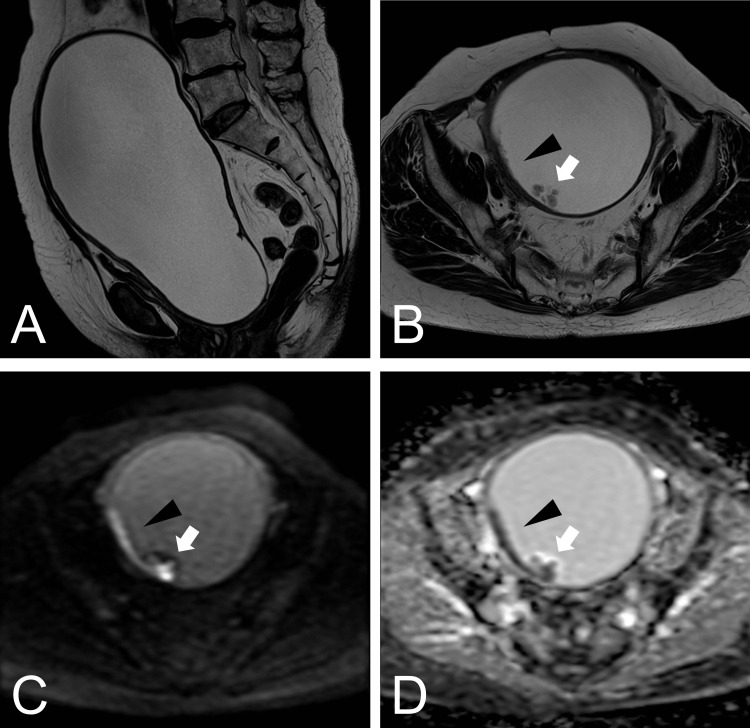 Figure 1
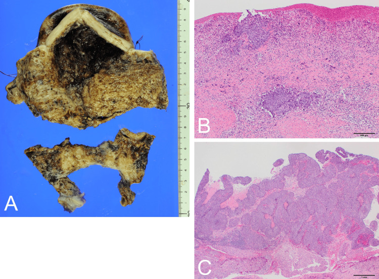 Figure 2
| Reference | Clinical presentation | Mural nodules | Irregular or thickened endometrium | Intrauterine fluid retention | Cervical tumor | DWI/ADC | Contrast-enhanced MRI |
| Narui et al. [ | Abdominal mass, abdominal pain | Multiple | + | + | + | High/low | NA |
| Adler et al. [ | Dysuria, urinary incontinence, pelvic mass | - | + | + | + | NA | + |
| Bagde et al. [ | Discharge | - | + | + | + | NA | NA |
| Dokić et al. [ | Bleeding | - | - | - | + | NA | + |
| Shu et al. [ | No symptoms | - | - | + | - | NA | - |
| Mannan et al. [ | Vaginal discharge, bleeding | - | - | + | - | NA | NA |
| Current study | Abdominal mass | Multiple | + | + | - | High/low | NA |
Peer Reviews
No public reviews on file for this paper yet. If you reviewed it on a platform where reviews are public (OpenReview, ICLR, NeurIPS, ICML), you can paste yours below so the community can read it here.
Videos
No videos yet. Explain this paper in a talk, walkthrough, or lecture? Add one.
Taxonomy
TopicsEndometrial and Cervical Cancer Treatments · Cervical Cancer and HPV Research · Ovarian cancer diagnosis and treatment
Introduction
Cervical cancer is one of the most common malignancies in women, and squamous cell carcinoma (SCC) accounts for 70-80% of cases [1]. Superficial spreading is a rare form of cervical SCC that extends superficially to the inner surface of the uterus with endometrial replacement [2]. Reports on the magnetic resonance imaging (MRI) findings for this form of SCC are limited [3-8]. There are no reports of diffusion-weighted imaging (DWI) in the English literature. Therefore, the characteristics and usefulness of the MRI findings remain unclear. Here, we report a case of superficially spreading cervical SCC that manifested as intrauterine mural nodules on MRI. DWI is useful for delineating lesions.
Case presentation
A 76-year-old Japanese woman presented to our hospital with a chief complaint of a lower abdominal mass for the past three months. Ten years ago, the patient had undergone cervical conization and was diagnosed with cervical intraepithelial neoplasia 3 with positive surgical margins. On physical examination, a mass was palpated on the abdomen. The cervix was indistinct and difficult to expand. The cervical and endometrial cytology results were negative for atypical cells. Transvaginal and transabdominal ultrasound failed to identify the uterus and left ovary and showed a large cystic lesion. MRI showed a 200 mm unilocular cystic lesion extending from the pelvis to the umbilical level (Figure 1A). The wall of the cystic lesion was partially thickened. Mural nodules were observed within the cystic lesion (Figure 1B). The thickened and mural nodular areas were hyperintense on DWI and hypointense on the apparent diffusion coefficient (ADC) maps (Figures 1C, 1D).
MRI findings(A) Sagittal, T2-weighted MRI section showed an unilocular cystic lesion.(B) Axial T2-weighted MRI.(C) DWI and (D) ADC maps showed a thickened wall (black arrowhead) and mural nodule (white arrow).MRI, magnetic resonance imaging; DWI, diffusion-weighted imaging; ADC, apparent diffusion coefficient
Laparotomy was performed for diagnosis and treatment. Intraoperative examination revealed that the uterus enlarged to the size of an adult head. No apparent abnormalities in the serosal surface or adnexa were observed. We punctured the uterine body and aspirated 1300 mL of brown serous fluid. We suspected that the tumor was of uterine origin and performed a hysterectomy and bilateral adnexectomy (Figure 2A). Histopathological findings showed that atypical epithelial cells had formed foci and infiltrated the stroma of the uterine cervix (Figure 2B). Most of the endometrial epithelium detached from the endometrium. Intra-epithelial carcinoma components were also observed in some areas. The outward proliferation almost coincided with the site of the mural nodule on MRI (Figure 2C). Immunohistochemistry revealed that the atypical epithelial cells were positive for p16 and p63. No malignancy was observed bilaterally in the adnexa. We diagnosed cervical SCC with superficial spread to the uterus based on a history of conization, cervical stromal invasion, and p16 positivity. Subsequently, computed tomography (CT) showed lung metastasis, and the patient was scheduled for chemotherapy for stage IVB cervical cancer (the 2018 International Federation of Gynecology and Obstetrics staging system).
Histopathological findings(A) Macroscopic findings of the uterus. The uterine lumen was dilated, and the wall was slightly thinned. The endometrial surface was generally rough. The cervix was separated from the body by surgical manipulation.(B) Histopathological findings of the cervical mucosa showed atypical epithelial cells in the superficial layer, infiltrating the stroma.(C) Histopathological findings of the endometrium in the uterus showed the outward proliferation of atypical epithelial cells.
Discussion
This case report highlights two important clinical findings. Superficially spreading cervical SCC can manifest as intrauterine mural nodules on MRI. DWI is useful for delineating this disease.
First, a superficially spreading cervical SCC can manifest as intrauterine mural nodules on MRI. There have been six previous reports of MRI findings of superficially spreading cervical SCC (Table 1) [3-8]. Cervical mass lesions, luminal fluid retention, and endometrial thickening have been reported previously. The Japanese literature also showed irregular and intermittent raised lesions in the endometrium of the uterine body [3]. However, intrauterine mural nodules have not been reported in the English literature. In this patient, a stratified area of atypical cells was histologically found to almost coincide with a mural nodule on MRI. The presence of intrauterine mural nodules may be a novel indicator of superficially spreading cervical SCC. Cervical stenosis is a late complication of conization that results in false-negative cervical cytology [2,9]. It is common for MRI to fail to detect lesions in early-stage cervical cancer because of the small tumor volume [10]. In previous reports of six cases with MRI findings, mass lesions were found in the cervix in four cases [3-6]. Cervical cytology was negative, and MRI showed no cervical abnormalities. Intrauterine mural nodules and wall thickening may be useful for diagnosing and detecting recurrence in these cases.
Second, DWI is useful for delineating superficially spreading cervical SCC. DWI is particularly useful for the evaluation of cervical cancers [11]. It can be used to differentiate benign and malignant uterine lesions [11]. Cervical cancer shows high signal intensity on DWI and low signal intensity on ADC maps, reflecting a high cell density [11]. DWI in superficial spreading cervical SCC has only been reported in the Japanese literature [3] and not in the English literature. In this patient, the nodular lesion and thickened wall showed high signal intensity on DWI and low signal intensity on the apparent ADC map. This finding suggests a superficial extension of cervical cancer to the uterine body. Similar to other gynecological tumors, DWI and ADC maps are useful for the assessment of superficially spreading cervical SCC.
Superficially spreading cervical SCC can manifest as intrauterine mural nodules on MRI. DWI is useful for delineating this disease. If mural nodules or endometrial thickening with restricted diffusion are found in the uterine lumen, clinicians should consider the possibility of the superficial spread of cervical SCC, especially in patients with a history of conization or suspected cervical cancer. MRI findings in superficial spreading cervical SCC are limited. The characteristics and usefulness of MRI findings, including those of DWI and contrast-enhanced MRI, should be further explored. Whether an MRI should be performed to detect the superficial spread of cervical SCC after conization or early-stage cervical cancer should also be investigated.
Conclusions
Superficially spreading cervical SCC can manifest as intrauterine mural nodules on MRI. DWI is useful for delineating this disease. If mural nodules or endometrial thickening with restricted diffusion are found in the uterine lumen, clinicians should consider the possibility of the superficial spread of cervical SCC.
The reference list from the paper itself. Each links out to its DOI / PubMed record.
- 1Cervical cancer: ESMO Clinical Practice Guidelines for diagnosis, treatment and follow-up Ann Oncol Marth C Landoni F Mahner S Mc Cormack M Gonzalez-Martin A Colombo N 029201810.1093/annonc/mdy 16029741577 · doi ↗ · pubmed ↗
- 2Superficial spreading cervical squamous cell carcinoma in situ involving the endometrium: a case report and review of the literature J Med Case Rep Martín-Vallejo J Laforga JB Molina-Bellido P Clemente-Pérez PA 1961620223559033510.1186/s 13256-022-03433-4PMC 9121615 · doi ↗ · pubmed ↗
- 3Superficial spreading squamous cell carcinoma of the uterine cervix involving the endometrium and fallopian tubes―a case report J Jpn Soc Clin Cytol Narui C Sakamoto M Fukushima S Umayahara K Iwaya K Okamoto A 286292612022 https://www.researchgate.net/publication/363049031_Superficial_spreading_squamous_cell_carcinoma_of_the_uterine_cervix_involving_the_endometrium_and_fallopian_tubeszigongtibutoluanguannibiaocengjinzhanshitazigongjingaino_1_li_-A_case_report-
- 4Superficial endometrial spread of squamous cell cervical carcinoma: a diagnostic challenge at magnetic resonance imaging J Comput Assist Tomogr Adler F Rabban JT Yeh BM Qayyum A Chen LM Coakley FV 2472503120071741476210.1097/01.rct.0000238006.19258.74 · doi ↗ · pubmed ↗
- 5A review and case report of enigmatic superficial endometrial spread of cancer of the uterine cervix: need for vigilance in the primary care setting J Family Med Prim Care Bagde MN Bagde NK Hussain N Thangaraju P 350535101020213476078210.4103/jfmpc.jfmpc_39_21PMC 8565160 · doi ↗ · pubmed ↗
- 6Curious case of superfitial spreading cervical squamocellular carcinoma with adnexal involvement Medicina (Kaunas) DokićM MilenkovićS JovanovićL MiloševićB AndrićL Šaponjski D KesićV 16555820223642219410.3390/medicina 58111655 PMC 9694448 · doi ↗ · pubmed ↗
- 7Endometrial squamous cell carcinoma originating from the cervix: a case report World J Clin Cases Shu XY Dai Z Zhang S Yang HX Bi H 878287871020223615782210.12998/wjcc.v 10.i 24.8782 PMC 9453359 · doi ↗ · pubmed ↗
- 8Superficial spreading, microinvasive CIN 3 of cervix: report of an unusual pattern of endometrial involvement J Obstet Gynaecol India Mannan KA Rao M Yadav G Yadav T 4484517220223645744810.1007/s 13224-022-01667-2PMC 9701269 · doi ↗ · pubmed ↗