Intraplantar aminoglutethimide, a P450scc inhibitor, reduced the induction of mechanical allodynia in a rat model of thrombus-induced ischemic pain
Soon-Gu Kwon, Hoon-Seong Choi, Seo-Yeon Yoon, Dae-Hyun Roh, Jang-Hern Lee

TL;DR
This study shows that blocking a specific enzyme with a drug reduces the development of pain in a rat model of ischemic pain, but only during the early phase.
Contribution
The study reveals a new peripheral mechanism involving P450scc and Sig-1Rs in ischemic pain and suggests a potential early treatment window.
Findings
Inhibiting P450scc with aminoglutethimide reduced mechanical allodynia during the early phase of ischemic pain.
A sigma-1 receptor agonist reversed the analgesic effects of aminoglutethimide during the induction phase.
P450scc expression increased in the ischemic hind paw skin of the rat model.
Abstract
Neuroactive steroids (NASs) directly affect neuronal excitability. Despite their role in the nervous system is intimately linked to pain control, knowledge is currently limited. This study investigates the peripheral involvement of NASs in chronic ischemic pain by targeting the cytochrome P450 side-chain cleavage enzyme (P450scc). Using a rat model of hind limb thrombus-induced ischemic pain (TIIP), we observed an increase in P450scc expression in the ischemic hind paw skin. Inhibiting P450scc with intraplantar aminoglutethimide (AMG) administration from post-operative day 0 to 3 significantly reduced the development of mechanical allodynia. However, AMG administration from post-operative day 3 to 6 did not affect established mechanical allodynia. In addition, we explored the role of the peripheral sigma-1 receptor (Sig-1R) by co-administering PRE-084 (PRE), a Sig-1R agonist, with AMG.…
Genes, proteins, chemicals, diseases, species, mutations and cell lines named across the full text — each resolved to its canonical identifier and authoritative record.
Click any figure to enlarge with its caption.
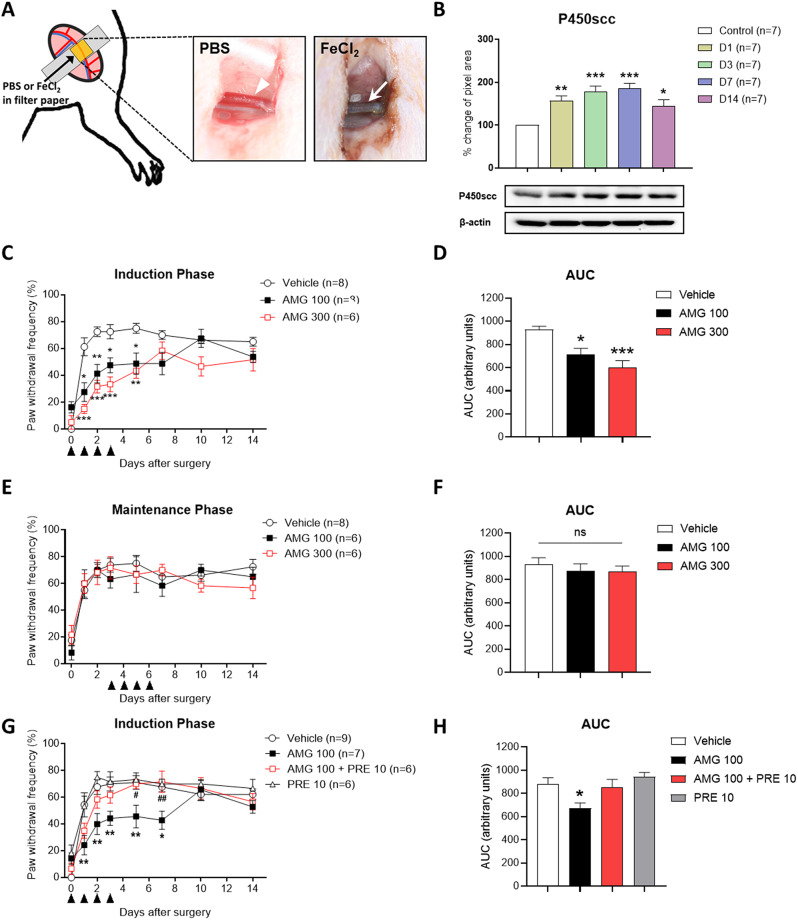 Figure 1
- —http://dx.doi.org/10.13039/501100014188Ministry of Science and ICT, South Korea
- —http://dx.doi.org/10.13039/501100003621Ministry of Science, ICT and Future Planning
Peer Reviews
No public reviews on file for this paper yet. If you reviewed it on a platform where reviews are public (OpenReview, ICLR, NeurIPS, ICML), you can paste yours below so the community can read it here.
Videos
No videos yet. Explain this paper in a talk, walkthrough, or lecture? Add one.
Taxonomy
TopicsPain Mechanisms and Treatments · Pharmacological Receptor Mechanisms and Effects · Pharmacological Effects of Natural Compounds
Main text
Neuroactive steroids (NASs) are a class of steroids that can modify neuronal excitability in both the central and peripheral nervous systems [1, 2]. The initial rate-limiting step in NAS biosynthesis is regulated by the cytochrome P450 cholesterol side-chain cleavage enzyme (P450scc), which catalyzes the conversion of cholesterol to pregnenolone (PREG). Both NASs and P450scc are involved in the normal functioning of central nervous systems as well as in various pathological conditions, including chronic neuropathic pain induced by peripheral nerve injury [3]. However, the role of P450scc in the peripheral nervous system, where the initial cause of pain originates, remains underexplored.
Peripheral ischemia is common causes of pain in the lower extremities [4]. Previously, we developed a peripheral ischemic pain model named the “thrombus-induced ischemic pain (TIIP) model,” which mimicked the pathogenesis of peripheral ischemic pain in humans [5]. Using this model, we focused on the pain mechanism in context of peripheral sensitization and examined related receptors, such as acid-sensing ion channels, ATP receptors, and sigma-1 receptors (Sig-1Rs), which are known as primary target receptors of NASs [5, 6]. In current study, based on the fact that NASs have been reported to control ischemic damage in the nervous system [7], we hypothesized that intraplantar injection of P450scc inhibitor could modulate ischemic pain at the peripheral site. Therefore, the present study was designed to investigate the potential role of the P450scc in the peripheral ischemic pain and to explore the involvement of Sig-1Rs at the peripheral site.
Male Sprague–Dawley rats (250–350 g) were anesthetized with 3% isoflurane in a N_2_O/O_2_ gas mixture, and small incision was made in the femoral triangle of the left hind limb. Femoral artery was carefully separated from the vein and nerve, and a filter paper disc (0.5 × 0.5 cm) soaked in a 20% FeCl_2_ in sterile PBS was applied to the femoral artery for 20 min. We defined the time periods in the TIIP model during which mechanical sensitivity increases or is sustained as the ‘induction phase’ (Day 0 to 3) and the ‘maintenance phase’ (Day 4 to 14), respectively (Fig. 1A). 30 µl of either aminoglutethimide (AMG, a P450scc inhibitor) or PRE-084 (PRE, a Sig-1R agonist) were subcutaneously injected daily into the ischemic paw during induction or maintenance phase. A von Frey filament (4.0 g) was applied 10 times to sole of ischemic paw, and results are presented as a percentage of the paw withdrawal frequency (PWF %). Western blot analysis was performed with the ipsilateral skin and temporal expression pattern of P450scc was quantified with Metamorph software (Molecular Devices, Sunnyvale, CA, USA). Statistical analysis was conducted using Prism 9.5 software (GraphPad, San Diego, CA, USA). A one-way ANOVA or two-way ANOVA was used to analyze the data as shown in the figure legend. A p-value of less than 0.05 was considered statistically significant.
Fig. 1. Schematic diagram for surgery of thrombus-induced ischemic pain (TIIP) injury and the effect of intraplantar aminoglutethimide (AMG) and PRE-084 (PRE) on TIIP induced mechanical allodynia. (A) Schematic diagram for surgery of thrombus-induced ischemic pain (TIIP) injury. In femoral arteries, normal conditions are observed with PBS treatment (arrowhead), whereas FeCl2-treatment in animals results in the development of thrombus formation or occlusion (arrow). (B) Representative Western blots images of P450scc (top) and β-actin (bottom) expression and the graph depicts the P450scc expression levels in the ipsilateral skin on post-operative days 1, 3, 7, and 14. Compared to the sham control group, significant differences were observed from postoperative day 1, and P450scc expression gradually increased until day 7 and then showed decreased trend on day 14. (C) During the induction phase, repeated AMG (100 and 300 nmol) treatments inhibited the increase in PWF (%) compared to vehicle-treated TIIP rats. After treatment, the inhibitory effect of AMG on mechanical allodynia gradually recovered. On day 7 after surgery, there was no significant difference between the AMG-treated and vehicle-treated groups. (D) The area under curve (AUC) data analysis demonstrated a dose-dependent antiallodynic effect of AMG treated on induction phase of TIIP. (E) During the maintenance phase, daily AMG (100 and 300 nmol) treatment did not alleviate the ischemia-induced increase in PWF (%) to mechanical stimuli. (F) The AUC data analysis also showed that the treatment of AMG on maintenance phase did not effective on mechanical allodynia in TIIP. (G) In TIIP rats, co-administration of AMG (100 nmol) and PRE (10 nmol) reversed AMG’s analgesic effect during the induction phase of mechanical allodynia. A single daily injection of AMG (100 nmol) inhibited the induction of mechanical allodynia in TIIP rats while there was no significant difference between AMG (100 nmol) + PRE (10 nmol) compared to the vehicle-treated group. Repeated injections of PRE (10 nmol) had no significant effect on mechanical allodynia during the induction phase. (H) The area under curve (AUC) data analysis showed diminished anti-allodynic effect of AMG (100 nmol) by co-administration of AMG (100 nmol) and PRE (10 nmol) on induction phase of TIIP (n = 6 ~ 8 per group). *p < 0.05, and **p < 0.01, and ***p < 0.001 compared to the vehicle-treated group, #p < 0.05, and ##p < 0.01 compared to the AMG 100 group), All values are expressed as mean ± standard error of the mean (SEM)
The temporal expression level of P450scc on the ipsilateral skin exhibited a gradual increase following ischemia. Immunoblot analysis revealed that P450scc levels peaked on post-operative day 7. Although there was still a significant difference compared to the control group, P450scc expression showed a decreasing trend by post-operative day 14 (Fig. 1B). To investigate the role of P450scc in the ischemic pain, pharmacological intervention was applied by administering AMG (100 and 300 nmol) directly into the ischemic hind paw during induction or maintenance phase of TIIP. During the induction phase, repeated AMG treatments significantly inhibited the increase in PWF (%) in TIIP rats (Fig. 1C). The AUC analysis also showed that AMG treatment dose-dependently inhibited the increase in mechanical allodynia (Fig. 1D). The inhibitory effect of AMG on mechanical allodynia gradually diminished after treatment. However, during the maintenance phase, AMG had no effect on the established mechanical allodynia in TIIP rats (Fig. 1E and F).
To investigate the role of peripheral Sig-1Rs in AMG-induced pain inhibition in induction phase, PRE (10 nmol) was co-administered with AMG (100 nmol) during the induction phase. Co-administration of PRE with AMG reversed the analgesic effect observed in the AMG-treated group on post-operative days 5, and 7 (Fig. 1G). Additionally, AUC analysis demonstrated that the combined treatment of PRE and AMG reversed the anti-allodynic effect of AMG in TIIP (Fig. 1H).
Skin is one of the main extra-gonadal steroidogenic organs that contains the P450scc enzyme [8]. Previous studies have shown that P450scc expression in skin can be regulated by external irritants [9]. Our current research further indicates increased P450scc levels following peripheral ischemic insult. It is noteworthy that peripheral nerves express various NASs and enzymes, including P450scc, which function as physiological regulators and protective agents for the peripheral nerve [7]. Consequently, the elevated peripheral P450scc observed in TIIP rats may be part of a physiological mechanism that provides protection against nerve damage resulting from ischemia.
Sig-1Rs are primary target receptors for NAS such as PREG and dehydroepiandrosterone (DHEA) [10]. We previously reported that peripherally administered PREG sulfate (PREGS) and DHEA sulfate (DHEAS) enhance pain sensitivity and it is closely related with peripheral Sig-1Rs activation [11]. Thus, it is plausible that PREGS and DHEAS contribute to mechanical allodynia during the induction phase of TIIP. Intraplantar administrated AMG inhibits the activation of P450scc, consequently reducing peripheral PREGS and DHEAS levels, which results in a decrease in mechanical allodynia by attenuating peripheral Sig-1R activity. Regarding the role of Sig-1R in TIIP, we previously demonstrated that inhibition of peripheral Sig-1Rs alleviated the mechanical allodynia only during the induction phase of TIIP [6]. Moreover, peripheral Sig-1Rs expression was increased peaking on day 3 and returning to baseline levels by day 7 [6]. These findings suggest that peripheral Sig-1Rs play a limited role in the induction phases of TIIP. Therefore, considering the role of Sig-1R in ischemic pain, which is specific to the induction phase, despite a significant increase in the expression of P450scc from day 1 to 14, pharmacological inhibition of P450scc could only impact the initial induction phase of TIIP.
Overall, our study demonstrated that peripheral P450scc level was increased in ischemic damage and intraplantar injection of AMG significantly alleviated mechanical allodynia during the induction of TIIP through Sig-1Rs activation. These findings indicate that peripheral NASs plays a pivotal role in peripheral sensitization and could represent a novel therapeutic target for chronic ischemic pain.
Electronic supplementary material
Below is the link to the electronic supplementary material.
Supplementary Material 1
Supplementary Material 2
Supplementary Material 3
The reference list from the paper itself. Each links out to its DOI / PubMed record.