High charge of cerebroid nests in nodular melanomas predicts tumor aggressiveness and high mutational tumoral burden: a pilot study
Stefania Caramaschi, Alessandro Mangogna, Laura Bertoni, Marco Manfredini, Francesca Farnetani, Paola Parente, Vito Attino, Gerardo Cazzato, Tiziana Salviato, Giovanni Pellacani, Luca Reggiani Bonetti

TL;DR
This study shows that the number of cerebroid nests in nodular melanomas is linked to more aggressive tumor behavior and higher mutation rates.
Contribution
The study introduces cerebroid nests as a potential biomarker for tumor aggressiveness and BRAF mutation status in nodular melanomas.
Findings
Melanomas with ≥10 cerebroid nests are more likely to have ulceration and BRAF V600E mutations.
Cerebroid nests correlate with epithelioid cell morphology, deeper tumor levels, and vascular invasion.
CNM count is a predictive marker for tumor aggressiveness independent of tumor size.
Abstract
Even today, melanoma is a highly aggressive neoplasm with a high mortality rate. The nodular type is very aggressive and has cerebroid nests of melanocytes (CNMs) at the growth edge, morphologically similar to the poorly differentiated neoplastic epithelial cell clusters described in colorectal, breast, and endometrioid endometrial cancers. We selected 25 nodular melanomas (NMs) with known molecular profiles, of which the entire paraffin-embedded lesion was available. We counted CNMs under a microscopic at a magnification of 20x (i.e., a microscopic field with a major axis of 1 mm). Based on the number of CNMs in the area, melanomas were classified into three groups: G1 (CNMs ranging from 0 to 4), G2 (CNMs ranging from 5 to 9), and G3 (CNMs ≥ 10). The presence of CNMs and their counts were compared with molecular and histopathological data. Seventeen (NMs) were grouped as G1 (68%), 5…
Genes, proteins, chemicals, diseases, species, mutations and cell lines named across the full text — each resolved to its canonical identifier and authoritative record.
Click any figure to enlarge with its caption.
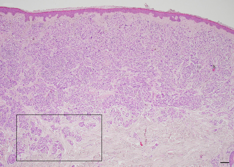 Figure 1
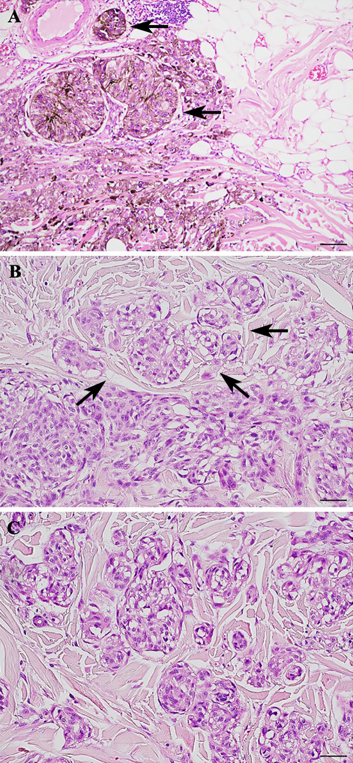 Figure 2
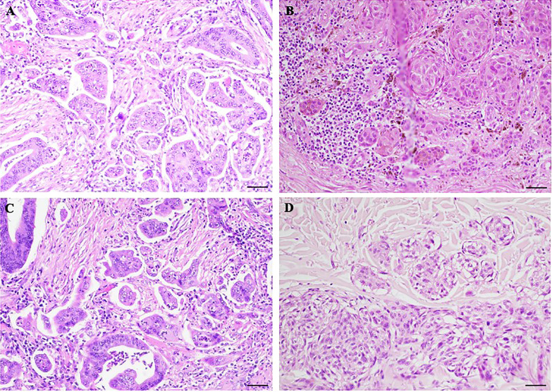 Figure 3
Peer Reviews
No public reviews on file for this paper yet. If you reviewed it on a platform where reviews are public (OpenReview, ICLR, NeurIPS, ICML), you can paste yours below so the community can read it here.
Videos
No videos yet. Explain this paper in a talk, walkthrough, or lecture? Add one.
Taxonomy
TopicsCutaneous Melanoma Detection and Management · Cancer Cells and Metastasis · Melanoma and MAPK Pathways