Trigeminal neuralgia due to Varicella-zoster virus reactivation
Denise Jourdan Oliveira, Diogo Goulart Corrêa, Sérgio Ferreira Alves

Abstract
Genes, proteins, chemicals, diseases, species, mutations and cell lines named across the full text — each resolved to its canonical identifier and authoritative record.
Click any figure to enlarge with its caption.
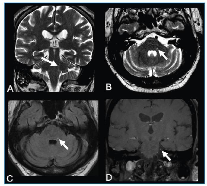 Figure 1
Peer Reviews
No public reviews on file for this paper yet. If you reviewed it on a platform where reviews are public (OpenReview, ICLR, NeurIPS, ICML), you can paste yours below so the community can read it here.
Videos
No videos yet. Explain this paper in a talk, walkthrough, or lecture? Add one.
Taxonomy
TopicsTrigeminal Neuralgia and Treatments · Facial Nerve Paralysis Treatment and Research · Herpesvirus Infections and Treatments
A 61-year-old man presented with left facial pain that worsened with digital touch and was associated with cutaneous vesicles in the left periorbital region for two weeks. Brain magnetic resonance imaging (MRI) revealed a longitudinal lesion with a hyperintense signal on T2-weighted imaging in the left posterolateral portion of the pons and medulla oblongata along the left spinal nucleus of the trigeminal nerve, associated with gadolinium enhancement in the cisternal segment (Figure 1). Cerebrospinal fluid (CSF) analysis revealed mild pleocytosis. Polymerase chain reaction revealed positivity for varicella zoster virus (VZV). Trigeminal postherpetic neuralgia was diagnosed, and the treatment comprised acyclovir, amitriptyline, and gabapentin, which reduced pain intensity and led to the progressive disappearance of the cutaneous lesions.
FIGURE 1:Trigeminal neuralgia due to varicella zoster virus reactivation. Brain MRI demonstrated a longitudinal lesion with hyperintense signal on T2-weighted imaging in the left spinal nucleus of the trigeminal nerve, in the coronal (arrow in A) and axial (arrow in B) planes and on an axial FLAIR (arrow in C), associated with gadolinium enhancement in the cisternal segment of the left trigeminal nerve (arrow in D). PCR of a cerebrospinal fluid sample revealed VZV positivity.
VZV is a herpesvirus whose primary infection causes varicella (chickenpox). Following primary infection, the virus becomes latent in the sensory neurons of the trigeminal ganglia and/or dorsal root ganglia along the neuraxis1. VZV reactivation is a common complication that occurs spontaneously or in response to several triggers, usually associated with aging and immunosuppression and, in some patients, may cause postherpetic neuralgia2. MRI may demonstrate gadolinium-enhancement of the trigeminal nerve or ganglion due to ganglionitis, and a hyperintense lesion on T2-weighted imaging along the trigeminal nuclei and intra-axial portion, including its entry zone, in patients with postherpetic neuralgia3. The spinal trigeminal nucleus extends from the inferior pons to the medulla and cervical spinal cord and receives sensory information regarding facial pain and temperature4. Thus, its involvement in VZV reactivation may explain the symptoms of postherpetic neuralgia.
The reference list from the paper itself. Each links out to its DOI / PubMed record.
- 1Niemeyer CS Harlander-Locke M Bubak AN Rzasa-Lynn R Birlea M Trigeminal Postherpetic Neuralgia: From Pathophysiology to Treatment Curr Pain Headache Rep 202428429530610.1007/s 11916-023-01209-z 38261232 PMC 10940365 · doi ↗ · pubmed ↗
- 2Skripuletz T Pars K Schulte A Schwenkenbecher P YildizÖ Ganzenmueller T Varicella zoster virus infections in neurological patients: a clinical study BMC Infect Dis 201818123823810.1186/s 12879-018-3137-229801466 PMC 5970536 · doi ↗ · pubmed ↗
- 3Cordano C Caverzasi E Spigno L De Maria A Bandini F MRI Findings in Varicella Zoster Trigeminal Neuritis Without Rash Headache 201858576476510.1111/head.1326429399793 · doi ↗ · pubmed ↗
- 4Boeddinghaus R Whyte A Imaging of Trigeminal Neuralgia and Other Facial Pain Neuroimaging Clin N Am 202131448550810.1016/j.nic.2021.05.00834689929 · doi ↗ · pubmed ↗