Actinomycosis mimicking malignancy
Buğra Kerget, Alperen Aksakal, Sevilay Özmen

Abstract
Genes, proteins, chemicals, diseases, species, mutations and cell lines named across the full text — each resolved to its canonical identifier and authoritative record.
Click any figure to enlarge with its caption.
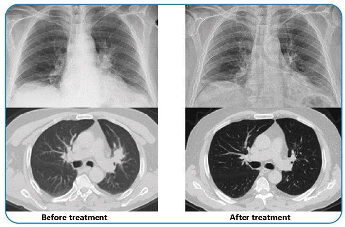 Figure 1
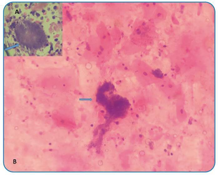 Figure 2
Peer Reviews
No public reviews on file for this paper yet. If you reviewed it on a platform where reviews are public (OpenReview, ICLR, NeurIPS, ICML), you can paste yours below so the community can read it here.
Videos
No videos yet. Explain this paper in a talk, walkthrough, or lecture? Add one.
Taxonomy
TopicsActinomycetales infections and treatment · Infective Endocarditis Diagnosis and Management · Fungal Infections and Studies
A 70-year-old male patient was admitted to the emergency department with persistent cough, hemoptysis, and back pain that had lasted for three weeks. Although his physical examination in the emergency department revealed no significant findings, a chest radiograph displayed a consolidated area with spicular extension in the left hilar region. Subsequent thoracic computed tomography (CT) showed a mass lesion measuring 38x25x36 mm with a standard uptake value (SUV) of 7.44 adjacent to the hilar region (Figure 1). Suspecting lung malignancy, the medical team performed endobronchial ultrasonography (EBUS), which identified a heterogeneous hypoechoic intramural lesion on the anterior wall of the left upper lobe entrance. Cytological samples were taken from the lesion twice, which tested negative for malignancy. However, they revealed bacterial clusters consistent with Actinomyces (Figure 2). The patient was initially treated with 4 x 2 g of intravenous ampicillin for eight weeks. A follow-up CT scan showed complete regression of the mass in the upper lobe (Figure 1). Treatment continued with amoxicillin plus clavulanic acid, administered orally at a dosage of 3 x 1 g for an additional eight weeks, during which he was closely monitored. Pulmonary actinomycosis is known to mimic a range of lung pathologies, from benign infections to metastatic tumors1, and 25% of thoracic actinomycosis cases are initially misdiagnosed as malignancy2. The patient achieved complete recovery with appropriate treatment over a sufficient duration.
FIGURE 1:Comparison of radiological findings before and after treatment.
FIGURE 2:Basophilic filamentous bacterial aggregates stained with hematoxylin and eosin dye (A, B).
The reference list from the paper itself. Each links out to its DOI / PubMed record.