Imaging of labile Fe2+ and Fe3+ in living Arabidopsis thaliana roots
Carine Alcon, Arnaud Comte, Catherine Curie, Tou Cheu Xiong
TL;DR
This paper introduces a method to image iron in living plant roots, showing how iron exists in different forms in different parts of the plant.
Contribution
The study adapts fluorescent imaging to visualize labile Fe2+ and Fe3+ in living Arabidopsis roots.
Findings
Fluorescent imaging reveals heterogeneous iron redox status in plant tissues.
Labile Fe2+ and Fe3+ pools are spatially distinct in Arabidopsis roots.
Abstract
Adapting fluorescent iron imaging to living plants enables visualizing labile Fe2+ and Fe3+ pools, revealing the heterogeneous distribution of iron redox status at the tissue and cellular levels.
Genes, proteins, chemicals, diseases, species, mutations and cell lines named across the full text — each resolved to its canonical identifier and authoritative record.
Click any figure to enlarge with its caption.
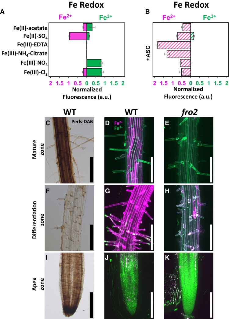 Figure 1
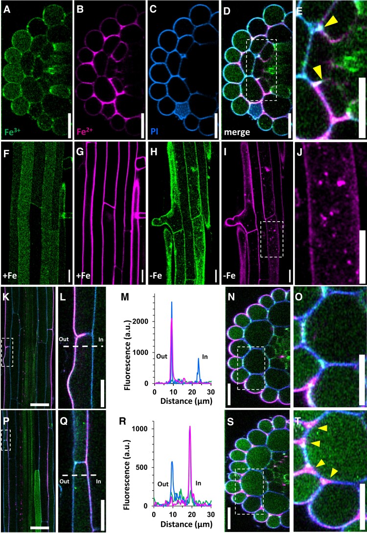 Figure 2
Peer Reviews
No public reviews on file for this paper yet. If you reviewed it on a platform where reviews are public (OpenReview, ICLR, NeurIPS, ICML), you can paste yours below so the community can read it here.
Videos
No videos yet. Explain this paper in a talk, walkthrough, or lecture? Add one.
Taxonomy
TopicsMilitary Technology and Strategies · Legal and Regulatory Analysis · Linguistic, Cultural, and Literary Studies