Multiple Mental Foramina: A Rare Anatomical Variation Detected by Cone-Beam Computed Tomography (CBCT)
Maryam Mohebiniya, Mobina Kamani

TL;DR
This paper discusses a rare anatomical variation where multiple mental foramina are found in the mandible, detected using CBCT, and highlights its clinical importance.
Contribution
The study emphasizes the detection of accessory mental foramina using CBCT and their clinical implications.
Findings
Multiple mental foramina can be identified using cone-beam computed tomography.
Accessory mental foramina are clinically significant due to their neurovascular content.
Identifying these foramina helps prevent post-surgical complications like paraesthesia and pain.
Abstract
The mental foramen is a single anatomical structure that can be seen bilaterally in the body of the mandible and generally in the lower area of the premolars. Sometimes, the mental foramen can have accessory foramina that should be considered. Clinical evaluation of the accessory mental foramina is critical because of its neurovascular fibers. Identifying the secondary mental foramen reduces the possibility of paraesthesia and pain after surgery.
Genes, proteins, chemicals, diseases, species, mutations and cell lines named across the full text — each resolved to its canonical identifier and authoritative record.
Click any figure to enlarge with its caption.
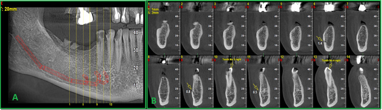 Figure 1
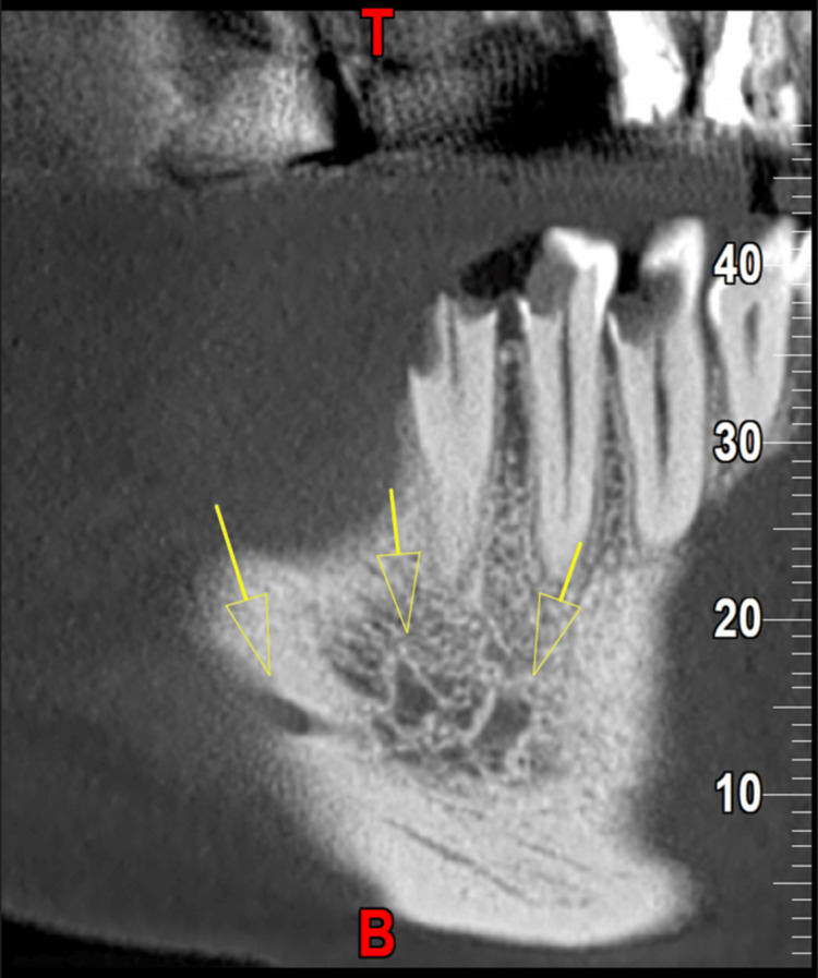 Figure 2
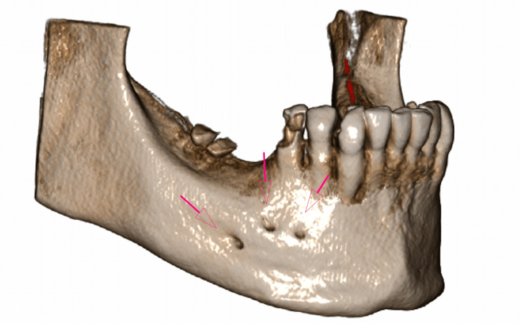 Figure 3
 Figure 4
Peer Reviews
No public reviews on file for this paper yet. If you reviewed it on a platform where reviews are public (OpenReview, ICLR, NeurIPS, ICML), you can paste yours below so the community can read it here.
Videos
No videos yet. Explain this paper in a talk, walkthrough, or lecture? Add one.
Taxonomy
TopicsDental Radiography and Imaging · Oral and Maxillofacial Pathology · Sinusitis and nasal conditions